Introduction
Ubiquitination, the process through which the
ubiquitin protein is covalently attached to another protein, can
alter the functional status of proteins or label them for
degradation through either mono- or poly-ubiquitination.
Functionally, ubiquitination is instrumental in, among other
things, regulating the circadian rhythm, silencing genes and
opening ion channels (1). The
regulation of this process is often disrupted in cancer cells, and
in particular, the polyubiquitin tail that marks proteins for
proteasomal degradation can be removed by deubiquinating enzymes
(DUBs), allowing the buildup of oncogenic proteins (2). Both ubiquitination and
deubiquitination are regulated through a specific set of enzymes.
Of interest is the deubiquitinating enzymes (DUBs) as they have
been identified as emerging therapeutic targets in cancers.
DUBs activate ubiquitin prior to conjugation, as
well as remove it from the ubiquitinated proteins (1). The 79 DUBs tentatively identified in
the human genome can be broadly classified as either cysteine
proteases or Zn2+ metalloproteases. The DUBs are further
divided into 5 subclasses based on the structure of the catalytic
domain: Ubiquitin C-terminal hydrolases (UCH), ubiquitin-specific
processing proteases (USP or UBP), Machado-Joseph disease protein
domain proteases (MJDs), ovarian tumor proteases (OTUs) and
(JAB1/MPN/Mov34 metalloenzyme) JAMM motif proteases (3).
Mechanistically, the active sites of UCH and UBP
DUBs are reminiscent of the papain proteases; however, these
enzymes must undergo a conformational change in the presence of
ubiquitin to become catalytically competent. This requirement
prevents UCH and UBP DUBs from off target effects when ubiquitin is
not present. OTU family DUBs have an almost identical geometry to
UCH and UBP family DUBS within their active sites. The primary
difference is that while the active site of the OTUs is fully
functional even before binding ubiquitin, it remains unable to act
due to being sterically blocked by an α-helix. The enzyme can
perform its function once ubiquitin binds and moves the α-helix
away from the catalytic site. The JAMM motif DUBs have a very
similar active site structure to that of a cytidine deaminase. In
each case, Zn2+ polarizes a water molecule, which then
performs a nucleophilic attack on an isopeptide bond (4).
DUBs are upregulated in several types of cancer,
including non-small cell lung cancer (NSCLC) and mesothelioma, and
thus represent attractive therapeutic targets (5-7).
Several DUB inhibitors have been proposed as anticancer agents
based on promising results in cell lines and in vivo models
(8). However, highly specific
compounds have not yet been developed and none of the current
inhibitors have entered the clinic as yet (9). The present study evaluated 3 DUB
inhibitors, PR-619, RA-9 and LDN-91946, with different DUB target
profiles. These inhibitors exerted differential effects on cell
migration and proliferation between the lung carcinoma and
mesothelioma cell lines. PR-619 is a wide-spectrum ubiquitin/UbL
isopeptidase inhibitor (10). RA-9
is a non-specific inhibitor that irreversibly inhibits DUBs by
exposing its carbonyl group to a nucleophilic attack from the SH-
group of cysteine (11). In
contrast to the two previously described compounds, LDN-91946 is a
specific inhibitor of ubiquitin C-terminal hydrolase-L1 (UCH-L1),
binding to the complex of the enzyme and the substrate (12).
It was hypothesized that DUB inhibition utilizing
these compounds at concentrations both comparable to other studies
and in line with concentration recommendations from Selleckchem
would affect the growth and/or motility of cancer cell lines
(11). The data of the present
study indicated that while LDN-91946 and RA-9 attenuated the
proliferation of both lung cancer and mesothelioma cell lines, they
had no effect on their migration. By contrast, PR-619 attenuated
both the proliferation and migration of the mesothelioma cell line.
PR-619 also inhibited the proliferation of the lung cancer cell
line; surprisingly, however, PR-619 augments the migration of lung
cancer cells. Moreover, the effects of these 3 drugs on the kinase
activity of the NSCLC cell line, A549, and the malignant pleural
mesothelioma (MPM) cell line, H2373, were examined using PamGene's
peptide-based microarray platform. A549 cells were selected for
analysis along with 3 DUB inhibitors based on previous publications
(13,14) that used siRNA library screening to
identify 12 DUBs in A549 cells. These DUBs are involved in the
activation of MET receptor tyrosine kinase by HGF agonist and the
scattering response of A549 cells. As a second target, MPM H2373
cells we selected, which expressed only a single nuclear
deubiquitinase, BAP1. Protein kinetic profiles of TRKs showed fewer
instances of phosphorylated peptides in H2373 as compared to A549,
indicating that the DUBs inhibitors could have a more specific
impact on cell function in H2373 cells. The variety of potential
interactions between these drugs and a complex set of
ubiquitination pathways demonstrated in the present study
emphasizes the need to explore the effects of these drugs in
further detail in future studies.
Materials and methods
Cell culture and reagents
The mesothelioma cell line, H2373, and the NSCLC
cell line, A549, (ATCC) were maintained in RPMI-1640 (Corning Inc.)
with 10% fetal bovine serum (Sigma-Aldrich; Merck KGaA) as
previously described (15).
PR-619, RA-9 and LDN-91946 were purchased commercially (Selleck
Chemicals) and used at concentrations of 10, 5 and 10 µM for
treatment for 24 h, or unless otherwise noted.
Cell adhesion and proliferation
assay
The resistance of cells in culture was measured
using ECIS (16). Briefly, cells
(7.5×10⁴) were plated in the chambers of the 96W1E+ well
plate of single-electrode ECIS arrays (Applied Biophysics) that
were pre-coated with fibronectin. Cells were treated either with
PR-619, RA-9 and LDN-91946, or dimethyl sulfoxide (DMSO), control.
Data were collected at 30 min post-inoculation. Data collection was
initiated by passing a small fixed-amplitude alternating current
between the electrodes and measuring the potential across them and
cell resistance was measured at a frequency of 40 kHz. As cells
grew and replicated, an increase in the electrical potential
between the electrodes was observed. Therefore, measuring cellular
impedance served as a readout for cell attachment and growth
(17). Cell attachment, adhesion
and proliferation were measured for 24-30 h.
Cell migration assay
Cells were seeded on a 96-well ImageLock (Essen
BioScience) plate to reach 90% confluency by the following day.
Following cell adherence, 96 uniform wounds were created
simultaneously using the WoundMaker (Essen BioScience) tool. Cells
were washed once with serum-free medium and replenished with 2% FBS
medium with vehicle or drug at a concentration of 10 µM. Low
serum medium inhibited cell proliferation as the assay was intended
to measure only cell migration. To monitor wound healing, the plate
was placed in the IncuCyte S3 Live-Cell Analysis System (Essen
BioScience) and images were acquired every hour for 48 h. Data
analysis was generated by IncuCyte software using a set confluence
mask to measure relative wound density over time.
Protein phosphorylation kinetics
For protein phosphorylation kinetics the PamGene
tyrosine kinase array (PamGene) was used along with a 4-array
semi-automated system (PamStation® 12, PamGene) designed
for processing PamChip®-4 arrays. The assay contains 144
phospho-peptides representing tyrosine kinase substrates. The
peptides are immobilized on a porous microarray surface through the
N-terminus. Briefly, the array was blocked with 0.2% bovine serum
albumin (BSA) for 30 cycles of 30 sec each following which each
array was washed thrice for 30 sec with 1X ABL protein tyrosine
kinase reaction (PK) buffer solution (New England Biolabs).
Thereafter, the arrays were incubated at 30°C with the reaction
mix, containing 30 µg cell lysate, 1X PK Buffer, 0.4
µl 1 M dithiothreitol (Sigma-Aldrich; Merck KGaA), 0.4
µl 100X BSA (New England Biolabs), 1 µl of 4 mM ATP
(Sigma-Aldrich; Merck KGaA), and 0.3 µl of 1 mg/ml
monoclonal anti-phosphotyrosine FITC conjugate (clone PY20, Exalpha
Biologicals), adjusted to 40 µl with distilled
H2O. The sample was pulsed through the array for 60
cycles, and every fifth pump cycle, a 16-bit TIFF image was
acquired with a built-in CCD camera. Spot intensities were
normalized to local background signal by subtracting the median
background signal and the data were log-transformed to normalize
the distribution of intensities. Fold changes were calculated by
subtracting mean log control values from mean log treatment
values.
Western blot analysis
Cell lines were treated with LD91946 (10 µM),
RA9 (5 µM) and PR619 (10 µM) for 24 h, followed by
lysing with RIPA buffer. Protein concentration for each sample was
measured with Bradford protein assay (Bio-Rad Laboratories, Inc.).
A total of 50 µg of protein was loaded into each well of an
SDS-polyacrylamide gel (4-20% Mini-Protein TGX precast protein gel,
Bio-Rad Laboratories, Inc.) and transferred onto a PVDF membrane
using the Bio-Rad Mini Trans-Blot apparatus. The membrane was
blocked with 5% milk solution for 1 h while rocking gently. Primary
anti-ubiquitin antibody 1:1.000 (ab7780; Abcam) was diluted in 5%
milk and applied to a PVDF membrane overnight at 4°C and then
incubated with secondary antibodies (HRP-labeled antibody 1:10,000
(ab205718; Abcam) for 1 h at room temperature. β-actin (1:3,000)
was used as a loading control (A5441; Sigma-Aldrich; Merck KGaA).
To visualize protein bands, PVDF membranes were treated with ECL
Western Blotting Detection Reagents and then exposed to X-ray
Medical Film before being developed.
Statistical analysis
Mathematica software package (Wolfram Research,
Inc.) was used to perform statistical computation. ANOVA followed
by Tukey's post hoc test were used to evaluate significant
differences in stabilized resistance values of the adhesion and
proliferation assay. The same methodology was used for the slopes
of the migration assay curves, as well as for the phosphorylation
assay. In all three assays the drugs were the factors. P<0.05
was considered to indicate a statistically significant
difference.
Results and Discussion
Effect of DUB inhibitors on cell adhesion
and proliferation
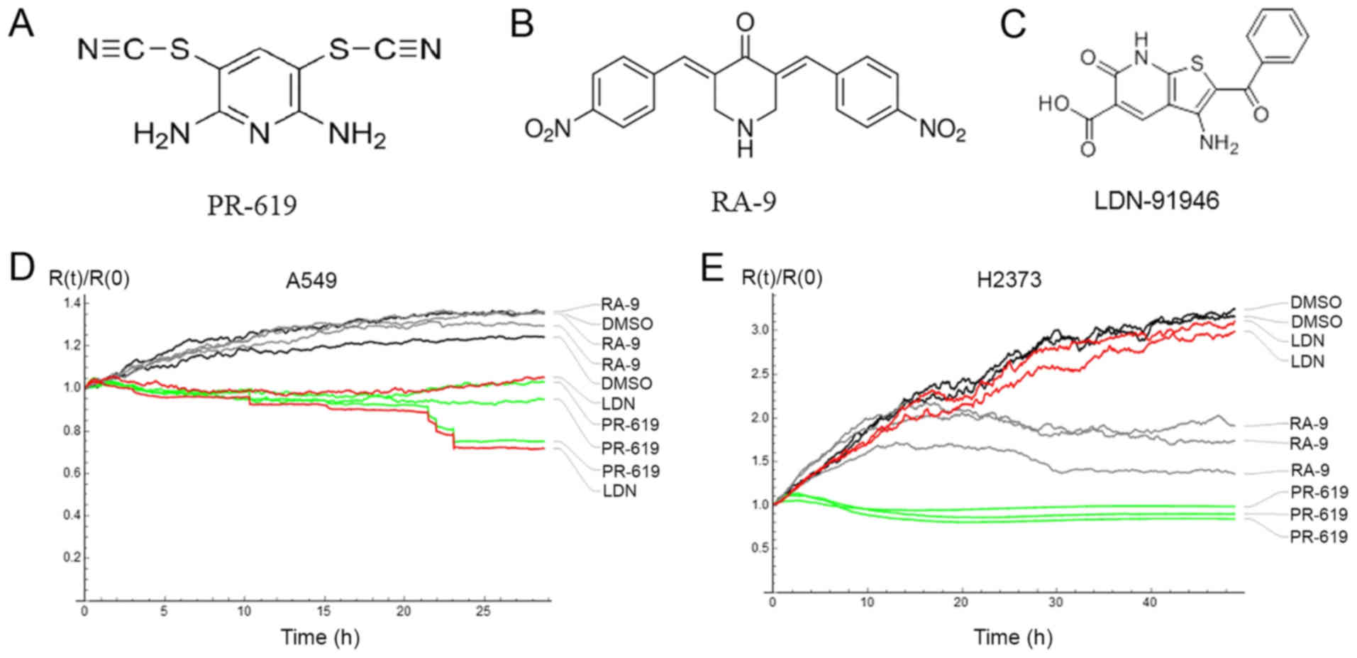
The effect of PR-619, RA-9 and LDN-91946 (Fig. 1A-C) on cell adhesion and
proliferation was evaluated by electrical cell-based impedance
sensing (ECIS) (16). The
resistance time course for control (DMSO) and drug-treated cells
was measured by ECIS as a monotonic function of the fraction of the
covered electrode surface (18).
Therefore, stabilized resistance values for A549 and H2373 cells
were compared as a measure of the drug effect. While LDN-91946 and
PR-619 attenuated the proliferation of the lung cancer cell line
(A549), RA-9 failed to inhibit the proliferation of this cell line.
On the other hand, while PR-619 and RA-9 impeded the proliferation
of the mesothelioma cells (H2373), LDN-91946 did not. For both cell
lines, a statistically significant difference of the drug effect
was ascertained by ANOVA with values of P<0.05 (Fig. 1D and E). Furthermore, Tukey's post
hoc test indicated significant differences in adhesion and
proliferation, as compared with the DMSO control, when treating the
A549 cells with LDN-91946 or PR-619, and when treating the H2373
cells with PR-169 or RA-9 (Table
I).
 | Table IAveraged relative resistances at the
end point evaluation of the cell adhesion and proliferation
assay. |
Table I
Averaged relative resistances at the
end point evaluation of the cell adhesion and proliferation
assay.
| A549 cells
mean R(t)/R(0) at 20-21 h | H2373 cells
mean R(t)/R(0) at 47-48 h |
|---|
| DMSO | 1.284 | 3.166 |
| LDN-91946 | 0.939a | 2.993 |
| PR-619 | 0.944a | 0.909a |
| RA-9 | 1.314 | 1.705a |
Consistent with the observations of the differential
effects of these DUBs inhibitors on lung cancer and mesothelioma,
RA-9 has been observed to inhibit the proliferation of breast,
ovarian and cervical cancer cell lines at concentrations comparable
to those used in the present study (11). The cytotoxic effect of RA-9 has
been attributed to 'proteotoxic stress', where non-degraded
ubiquitinated protein accumulates in the cell (19). These experiments establish potent
single agent activity of both LDN91946 and PR-619 in the models in
the present study. The differential effects of these compounds on
the two cancer cell lines is of interest, as it suggests
specificity and the involvement of distinct DUBs in this
process.
Effect of DUB inhibitors on cell
motility
Cell motility was assessed with a wound healing
assay. It is important to note the cell proliferation was
suppressed (Materials and methods), as is typical in such assays
(20), so that the wound closure
by cell growth is not a confounding factor. The
IncuCyte® Live Cell Analysis System used in the present
study measures relative wound density Y(t)
where W(t) and C(t) are the image pixel densities of
the wound and non-wounded areas respectively. The assay was
replicated 8 times resulting in 8 curves corresponding to wound
density vs. time. Representative data of the replicated
measurements of the A549 and H2373 cells treated with DMSO, PR-619,
or LDN-91946 are shown on
Fig. 2.
In order to compare the curves and to ascertain
statistically-significant differences, it was desirable to describe
each curve with one parameter, i.e., an average migration rate. To
this end, the wound healing process was modelled as follows: The
cell migration was a first order process, i.e., the number of
migrated cells between the wound and the non-wounded area per unit
of time is estimated to be proportional to the number of cells in
the non-wounded area. Mathematically, the pixel densities of the
wound and the non-wounded areas may be modelled by the following
differential equations, which depend on k, the average migration
rate:
Solving the above equations and substituting for
Y(t) yields:
First order series expansion yields
Y(t)=kt+O(t2). As expected, the data follow a
straight line for the first 10 h (Fig.
2). It is also important to note that measuring the initial
slope of the curve provides an additional benefit to the analysis.
Specifically, given the short time necessary to measure the initial
slope, the approach eliminates biases from any residual cell
proliferation (if it was not fully suppressed in the
experiment).
The slope of the initial parts of the curves was
estimated by fitting a straight line with an intercept set to zero.
Each experiment was repeated 8 times, and the resulting slopes were
further analyzed using a one-way ANOVA. The data are presented in
Table II and representative
movies of cell migration for the A549 and H2373 cells with and
without drug treatments are available as supplementary material
(Videos S1-S8). Notably, while
LDN-91946 and RA-9 had no detectable effect on THE migration in
either cell line, PR-619 significantly attenuated the migration of
the mesothelioma cell line (H2373), but increased the migration
rate of the lung cancer cell line (A549). To the best of our
knowledge, the effects of LDN-91946, PR-619 and RA-9 on the
motility of cell lines have not yet been reported. It is believed
that the modeling of the wound healing assay evaluated and
quantified the cell motility in a statistically meaningful
manner.
| Table IIAverage migration rate measured by the
wound healing assay. |
Table II
Average migration rate measured by the
wound healing assay.
| A549 cells | H2373 cells |
|---|
| DMSO | 0.037 | 0.039 |
| LDN-91946 | 0.039 | 0.041 |
| PR-619 | 0.052a | 0.022a |
| RA-9 | 0.035 | 0.040 |
Protein phosphorylation
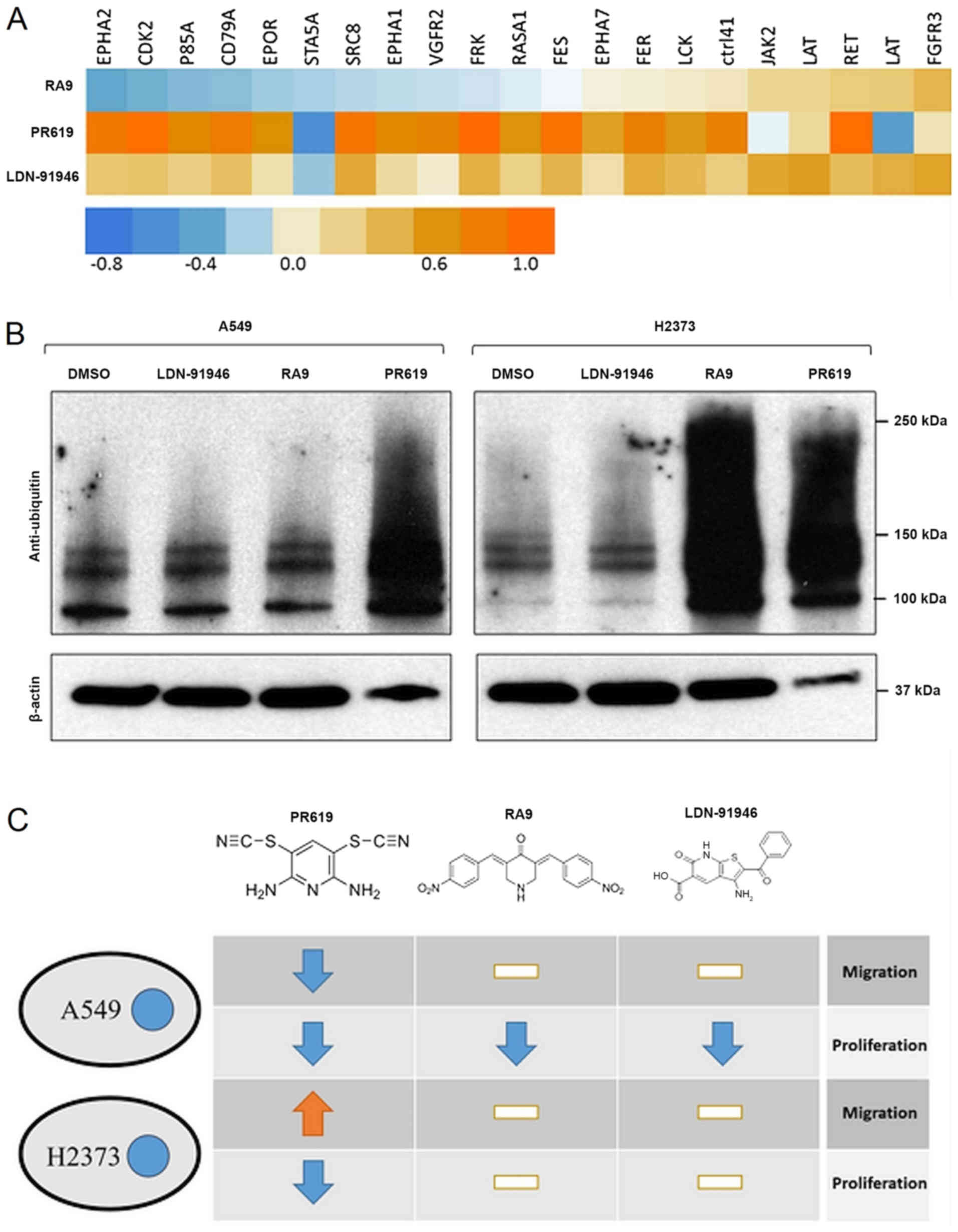
To shed light onto the mechanisms through which
these inhibitors may act, protein phosphorylation kinetics were
measured using an endpoint PamGene assay. The assay provides
surface-immobilized peptides, which are exposed to a cell lysate.
Peptide-specific phosphorylation activity of the lysate is measured
by immunostaining. For this assay, the cells were grown to 75%
confluence, treated with either DMSO (control) or drugs at 10
µM for 5 h. Cell lysates were analyzed by a PamGene chip in
triplicate. The extent of phosphorylation of each peptide was
measured and compared between the treatments. One-way ANOVA was
used to test for significant differences among the treatments. Post
hoc tests revealed which treatments significantly affected
phosphorylation in pairwise comparisons. The present study focused
on those peptides that, amongst the responses to the different
drugs, had at least one statistically significant difference in
comparison with the control.
In the case of the mesothelioma cell line (H2373), a
single peptide was significantly affected by the treatments. The
peptide corresponded to the protein P85A with the phosphorylation
site between amino acid positions 600 and 612. Compared to the DMSO
control, a statistically significant difference was found in the
presence of LDN-91946 (increase), RA-9 (increase) and PR-619
(decrease). P85A is involved in numerous pathways, including the
mTOR and PI3K-Akt signaling pathways as found in the KEGG pathway
database (KEGG, 21). Similarly, for the A549 cells, >20 peptides
were affected, which were involved in 'Pathways in cancer'
(hsa05200), including the PI3K-Akt signaling pathway (hsa04151) and
the Ras signaling pathway (hsa04014), among others. A visual
representation of the data is presented in Fig. 3A.
Verification of DUB inhibition
Given the diverse effects of the DUB inhibitors on
the proliferation and motility of the two cell lines, western blot
analysis was performed to verify that the studied compounds had an
inhibitory effect on deubiquitination (increase in ubiquitin
levels) and whether it was cell line-specific. As shown in Fig. 3B, PR-619, one of the broad spectrum
inhibitors, wsa the only inhibitor that resulted in a robust, cell
line-independent activity, and its effect on both cell lines was
statistically significant (Tables
I and II). LDN-91946, being a
specific DUB inhibitor, unsurprisingly did not result an easily
measurable change in the context of the whole cell ubiquitin
levels, making it difficult to determine whether the impact on
proliferation in A549 was an on-target effect using this method.
RA-9, the other non-specific DUB inhibitor, exhibited a potent
inhibitory effect of DUBs on the H2373 cell line, while having very
little impact on ubiquitin levels in A549 cells. Of note, this
result is in contrast to the results of the proliferation
experiment that revealed the inhibition of A549 proliferation,
while no inhibition of DUB activity.
Variety of responses
It is interesting to note a seemingly unpredictable
response of the cell lines to the drug treatment. One could have
studied additional cell lines, which may or may not behave
respectively the same as the ones we already studied. Moreover, the
current knowledge of the cell lines is not fine grained enough to
be able to tease apart the mechanistic effects resulting from the
interaction of the cell line peculiarities and the drug. In fact,
preliminary studies were conducted with BEAS-2B and Met 5A cell
lines and the same drugs to discover equally unexpected outcomes
(data not shown). The purpose of the present study, however, is to
open up a conversation about the unpredictability of the drug
effects on different types of cells.
In conclusion, the findings of the present study
demonstrate that while PR-619 acts as a proliferation-inhibitor for
both cell lines and exerts a significant effect on overall
ubiquitin levels, it affects cell motility quite differently, which
points to a need for detailed investigations into the role of
ubiquitination in these two important processes (Fig. 3C). Both RA-9 and LDN-91946
exhibited differential effects on proliferation between cell lines,
though in the case of RA-9, the proliferation change was in the
A549 cells, the cell line that did not exhibit any change in
ubiquitin levels. This result is interesting as it not only
indicates that a strong decrease in ubiquitin levels can
potentially have no effect on cell motility and proliferation, but
also indicates that the A549 cell line may have a method for
processing or otherwise negating the effects of the RA-9 inhibitor
that resulted in reduced proliferation. If RA-9 is being rapidly
processed, it could be that the downstream products are toxic. The
mechanism responsible for the escape of the A549 cells from the
effects RA-9 may prove an interesting avenue of research going
forward.
The only noted impact of the change in the
phosphorylation state of the H2373 cellss was the increase in
migration and a decrease in proliferation found when PR-619 was
applied. Increases in the phosphorylation of P85A during exposure
to RA-9 and LDN-91946 had little impact on proliferation or
migration. Moreover, the larger number of phosphorylation changes
observed in the A549 cell line uniformly lead to a reduction in
proliferation, though only PR-619, which had the greatest number of
increased phosphorylation sites, showed a significant reduction in
migration rates. The highly selective effect of altered
phosphorylation on P85A in the mesothelioma cell line, versus a
much larger number of proteins in the same (PI3K) pathway in the
lung cancer cell line, after the application of the same drugs,
emphasizes the subtleties with which proliferation and migration
are coordinated. The effects of the PI3K-AKT signaling pathway
range from feeding into the p53 and NF-κB pathways to cell cycle
control and apoptosis while the mTOR pathway impacts regulation of
the actin cytoskeleton. Insights regarding how these drugs
influence these pathways, and possible off target effects may prove
to be both crucial in developing small molecule DUBs inhibitors as
targeted cancer therapeutics and critical to avoiding
misapplication of these treatments.
Supplementary Data
Funding
The present study was supported by the National
Cancer Institute of the National Institutes of Health under Grants
nos. P30CA033572, U54CA209978 and R01CA218545.
Availability of data and materials
All data generated or analyzed during this study are
included in this published article or are available from the
corresponding author on reasonable request.
Authors' contributions
AP and MSN conducted the analysis. TM, YCT, SS, and
AN designed and performed the experiments. IM, YHCT, KZ, DR and SS
provided reagents and reviewed the manuscript. MWN provided
technical guidance. PK, AP, TM and MSN wrote the manuscript. MWN,
SKB, MS, IM, YHCT, KZ, DR, SS, PK, MSN and RS conceptualized and
designed the experiments and reviewed the manuscript. All authors
have read and approved the final manuscript.
Ethics approval and consent to
participate
Not applicable.
Patient consent for publication
Not applicable.
Competing interests
The authors declare that they have no competing
interests.
Acknowledgments
Not applicable.
References
|
1
|
Kerscher O, Felberbaum R and Hochstrasser
M: Modification of proteins by ubiquitin and ubiquitin-like
proteins. Annu Rev Cell Dev Biol. 22:159–180. 2006. View Article : Google Scholar : PubMed/NCBI
|
|
2
|
Mansour MA: Ubiquitination: Friend and foe
in cancer. Int J Biochem Cell Biol. 101:80–93. 2018. View Article : Google Scholar : PubMed/NCBI
|
|
3
|
Nijman SMB, Luna-Vargas MPA, Velds A,
Brummelkamp TR, Dirac AMG, Sixma TK and Bernards R: A genomic and
functional inventory of deubiquitinating enzymes. Cell.
123:773–786. 2005. View Article : Google Scholar : PubMed/NCBI
|
|
4
|
Amerik AY and Hochstrasser M: Mechanism
and function of deubiquitinating enzymes. Biochim Biophys Acta.
1695:189–207. 2004. View Article : Google Scholar : PubMed/NCBI
|
|
5
|
Poondla N, Chandrasekaran AP, Kim KS and
Ramakrishna S: Deubiquitinating enzymes as cancer biomarkers: New
therapeutic opportunities? BMB Rep. 52:181–189. 2019. View Article : Google Scholar : PubMed/NCBI
|
|
6
|
Patel K, Ahmed ZS, Huang X, Yang Q, Ekinci
E, Neslund-Dudas CM, Mitra B, Elnady FA, Ahn YH, Yang H, et al:
Discovering proteasomal deubiquitinating enzyme inhibitors for
cancer therapy: Lessons from rational design, nature and old drug
reposition. Future Med Chem. 10:2087–2108. 2018. View Article : Google Scholar : PubMed/NCBI
|
|
7
|
Kaushal K, Antao AM, Kim KS and
Ramakrishna S: Deubiquitinating enzymes in cancer stem cells:
Functions and targeted inhibition for cancer therapy. Drug Discov
Today. 23:1974–1982. 2018. View Article : Google Scholar : PubMed/NCBI
|
|
8
|
Yuan T, Yan F, Ying M, Cao J, He Q, Zhu H
and Yang B: Inhibition of Ubiquitin-Specific Proteases as a Novel
Anticancer Therapeutic Strategy. Front Pharmacol. 9:10802018.
View Article : Google Scholar : PubMed/NCBI
|
|
9
|
Harrigan JA, Jacq X, Martin NM and Jackson
SP: Deubiquitylating enzymes and drug discovery: Emerging
opportunities. Nat Rev Drug Discov. 17:57–78. 2018. View Article : Google Scholar
|
|
10
|
Altun M, Kramer HB, Willems LI, McDermott
JL, Leach CA, Goldenberg SJ, Kumar KG, Konietzny R, Fischer R,
Kogan E, et al: Activity-based chemical proteomics accelerates
inhibitor development for deubiquitylating enzymes. Chem Biol.
18:1401–1412. 2011. View Article : Google Scholar : PubMed/NCBI
|
|
11
|
Issaenko OA and Amerik AY: Chalcone-based
small-molecule inhibitors attenuate malignant phenotype via
targeting deubiquitinating enzymes. Cell Cycle. 11:1804–1817. 2012.
View Article : Google Scholar : PubMed/NCBI
|
|
12
|
Mermerian AH, Case A, Stein RL and Cuny
GD: Structure-activity relationship, kinetic mechanism, and
selectivity for a new class of ubiquitin C-terminal hydrolase-L1
(UCH-L1) inhibitors. Bioorg Med Chem Lett. 17:3729–3732. 2007.
View Article : Google Scholar : PubMed/NCBI
|
|
13
|
D'Arcy P, Wang X and Linder S:
Deubiquitinase inhibition as a cancer therapeutic strategy.
Pharmacol Ther. 147:32–54. 2015. View Article : Google Scholar
|
|
14
|
Buus R, Faronato M, Hammond DE, Urbé S and
Clague MJ: Deubiquitinase activities required for hepatocyte growth
factor-induced scattering of epithelial cells. Curr Biol.
19:1463–1466. 2009. View Article : Google Scholar : PubMed/NCBI
|
|
15
|
Lennon FE, Cianci GC, Kanteti R, Riehm JJ,
Arif Q, Poroyko VA, Lupovitch E, Vigneswaran W, Husain A, Chen P,
et al: Unique fractal evaluation and therapeutic implications of
mitochondrial morphology in malignant mesothelioma. Sci Rep.
6:245782016. View Article : Google Scholar : PubMed/NCBI
|
|
16
|
Giaever I and Keese CR: Micromotion of
mammalian cells measured electrically. Proc Natl Acad Sci U S A.
88:7896–7900. 1991. View Article : Google Scholar : PubMed/NCBI
Erratum. Proc Natl Acad Sci U S A.
90:16341993.
|
|
17
|
Szulcek R, Bogaard HJ and van Nieuw
Amerongen GP: Electric cell-substrate impedance sensing for the
quantification of endothelial proliferation, barrier function, and
motility. J Vis Exp. 85:e513002014.
|
|
18
|
Wegener J, Keese CR and Giaever I:
Electric cell-substrate impedance sensing (ECIS) as a noninvasive
means to monitor the kinetics of cell spreading to artificial
surfaces. Exp Cell Res. 259:158–166. 2000. View Article : Google Scholar : PubMed/NCBI
|
|
19
|
Coughlin K, Anchoori R, Iizuka Y, Meints
J, MacNeill L, Vogel RI, Orlowski RZ, Lee MK, Roden RB and Bazzaro
M: Small-molecule RA-9 inhibits proteasome-associated DUBs and
ovarian cancer in vitro and in vivo via exacerbating unfolded
protein responses. Clin Cancer Res. 20:3174–3186. 2014. View Article : Google Scholar : PubMed/NCBI
|
|
20
|
Jonkman JE, Cathcart JA, Xu F, Bartolini
ME, Amon JE, Stevens KM and Colarusso P: An introduction to the
wound healing assay using live-cell microscopy. Cell Adhes Migr.
8:440–451. 2014. View Article : Google Scholar
|
|
21
|
Kanehisa M, Furumichi M, Tanabe M, Sato Y
and Morishima K: KEGG: New perspectives on genomes, pathways,
diseases and drugs. Nucleic Acids Res. 45(D1): D353–D361. 2017.
View Article : Google Scholar :
|