Spandidos Publications Logo
  • About
    • About Spandidos
    • Aims and Scopes
    • Abstracting and Indexing
    • Editorial Policies
    • Reprints and Permissions
    • Job Opportunities
    • Terms and Conditions
    • Contact
  • Journals
    • All Journals
    • Oncology Letters
      • Oncology Letters
      • Information for Authors
      • Editorial Policies
      • Editorial Board
      • Aims and Scope
      • Abstracting and Indexing
      • Bibliographic Information
      • Archive
    • International Journal of Oncology
      • International Journal of Oncology
      • Information for Authors
      • Editorial Policies
      • Editorial Board
      • Aims and Scope
      • Abstracting and Indexing
      • Bibliographic Information
      • Archive
    • Molecular and Clinical Oncology
      • Molecular and Clinical Oncology
      • Information for Authors
      • Editorial Policies
      • Editorial Board
      • Aims and Scope
      • Abstracting and Indexing
      • Bibliographic Information
      • Archive
    • Experimental and Therapeutic Medicine
      • Experimental and Therapeutic Medicine
      • Information for Authors
      • Editorial Policies
      • Editorial Board
      • Aims and Scope
      • Abstracting and Indexing
      • Bibliographic Information
      • Archive
    • International Journal of Molecular Medicine
      • International Journal of Molecular Medicine
      • Information for Authors
      • Editorial Policies
      • Editorial Board
      • Aims and Scope
      • Abstracting and Indexing
      • Bibliographic Information
      • Archive
    • Biomedical Reports
      • Biomedical Reports
      • Information for Authors
      • Editorial Policies
      • Editorial Board
      • Aims and Scope
      • Abstracting and Indexing
      • Bibliographic Information
      • Archive
    • Oncology Reports
      • Oncology Reports
      • Information for Authors
      • Editorial Policies
      • Editorial Board
      • Aims and Scope
      • Abstracting and Indexing
      • Bibliographic Information
      • Archive
    • Molecular Medicine Reports
      • Molecular Medicine Reports
      • Information for Authors
      • Editorial Policies
      • Editorial Board
      • Aims and Scope
      • Abstracting and Indexing
      • Bibliographic Information
      • Archive
    • World Academy of Sciences Journal
      • World Academy of Sciences Journal
      • Information for Authors
      • Editorial Policies
      • Editorial Board
      • Aims and Scope
      • Abstracting and Indexing
      • Bibliographic Information
      • Archive
    • International Journal of Functional Nutrition
      • International Journal of Functional Nutrition
      • Information for Authors
      • Editorial Policies
      • Editorial Board
      • Aims and Scope
      • Abstracting and Indexing
      • Bibliographic Information
      • Archive
    • International Journal of Epigenetics
      • International Journal of Epigenetics
      • Information for Authors
      • Editorial Policies
      • Editorial Board
      • Aims and Scope
      • Abstracting and Indexing
      • Bibliographic Information
      • Archive
    • Medicine International
      • Medicine International
      • Information for Authors
      • Editorial Policies
      • Editorial Board
      • Aims and Scope
      • Abstracting and Indexing
      • Bibliographic Information
      • Archive
  • Articles
  • Information
    • Information for Authors
    • Information for Reviewers
    • Information for Librarians
    • Information for Advertisers
    • Conferences
  • Language Editing
Spandidos Publications Logo
  • About
    • About Spandidos
    • Aims and Scopes
    • Abstracting and Indexing
    • Editorial Policies
    • Reprints and Permissions
    • Job Opportunities
    • Terms and Conditions
    • Contact
  • Journals
    • All Journals
    • Biomedical Reports
      • Information for Authors
      • Editorial Policies
      • Editorial Board
      • Aims and Scope
      • Abstracting and Indexing
      • Bibliographic Information
      • Archive
    • Experimental and Therapeutic Medicine
      • Information for Authors
      • Editorial Policies
      • Editorial Board
      • Aims and Scope
      • Abstracting and Indexing
      • Bibliographic Information
      • Archive
    • International Journal of Epigenetics
      • Information for Authors
      • Editorial Policies
      • Editorial Board
      • Aims and Scope
      • Abstracting and Indexing
      • Bibliographic Information
      • Archive
    • International Journal of Functional Nutrition
      • Information for Authors
      • Editorial Policies
      • Editorial Board
      • Aims and Scope
      • Abstracting and Indexing
      • Bibliographic Information
      • Archive
    • International Journal of Molecular Medicine
      • Information for Authors
      • Editorial Policies
      • Editorial Board
      • Aims and Scope
      • Abstracting and Indexing
      • Bibliographic Information
      • Archive
    • International Journal of Oncology
      • Information for Authors
      • Editorial Policies
      • Editorial Board
      • Aims and Scope
      • Abstracting and Indexing
      • Bibliographic Information
      • Archive
    • Medicine International
      • Information for Authors
      • Editorial Policies
      • Editorial Board
      • Aims and Scope
      • Abstracting and Indexing
      • Bibliographic Information
      • Archive
    • Molecular and Clinical Oncology
      • Information for Authors
      • Editorial Policies
      • Editorial Board
      • Aims and Scope
      • Abstracting and Indexing
      • Bibliographic Information
      • Archive
    • Molecular Medicine Reports
      • Information for Authors
      • Editorial Policies
      • Editorial Board
      • Aims and Scope
      • Abstracting and Indexing
      • Bibliographic Information
      • Archive
    • Oncology Letters
      • Information for Authors
      • Editorial Policies
      • Editorial Board
      • Aims and Scope
      • Abstracting and Indexing
      • Bibliographic Information
      • Archive
    • Oncology Reports
      • Information for Authors
      • Editorial Policies
      • Editorial Board
      • Aims and Scope
      • Abstracting and Indexing
      • Bibliographic Information
      • Archive
    • World Academy of Sciences Journal
      • Information for Authors
      • Editorial Policies
      • Editorial Board
      • Aims and Scope
      • Abstracting and Indexing
      • Bibliographic Information
      • Archive
  • Articles
  • Information
    • For Authors
    • For Reviewers
    • For Librarians
    • For Advertisers
    • Conferences
  • Language Editing
Login Register Submit
  • This site uses cookies
  • You can change your cookie settings at any time by following the instructions in our Cookie Policy. To find out more, you may read our Privacy Policy.

    I agree
Search articles by DOI, keyword, author or affiliation
Search
Advanced Search
presentation
International Journal of Oncology
Join Editorial Board Propose a Special Issue
Print ISSN: 1019-6439 Online ISSN: 1791-2423
Journal Cover
February-2026 Volume 68 Issue 2

Full Size Image

Cover Legend PDF

Sign up for eToc alerts
Recommend to Library

Journals

International Journal of Molecular Medicine

International Journal of Molecular Medicine

International Journal of Molecular Medicine is an international journal devoted to molecular mechanisms of human disease.

International Journal of Oncology

International Journal of Oncology

International Journal of Oncology is an international journal devoted to oncology research and cancer treatment.

Molecular Medicine Reports

Molecular Medicine Reports

Covers molecular medicine topics such as pharmacology, pathology, genetics, neuroscience, infectious diseases, molecular cardiology, and molecular surgery.

Oncology Reports

Oncology Reports

Oncology Reports is an international journal devoted to fundamental and applied research in Oncology.

Experimental and Therapeutic Medicine

Experimental and Therapeutic Medicine

Experimental and Therapeutic Medicine is an international journal devoted to laboratory and clinical medicine.

Oncology Letters

Oncology Letters

Oncology Letters is an international journal devoted to Experimental and Clinical Oncology.

Biomedical Reports

Biomedical Reports

Explores a wide range of biological and medical fields, including pharmacology, genetics, microbiology, neuroscience, and molecular cardiology.

Molecular and Clinical Oncology

Molecular and Clinical Oncology

International journal addressing all aspects of oncology research, from tumorigenesis and oncogenes to chemotherapy and metastasis.

World Academy of Sciences Journal

World Academy of Sciences Journal

Multidisciplinary open-access journal spanning biochemistry, genetics, neuroscience, environmental health, and synthetic biology.

International Journal of Functional Nutrition

International Journal of Functional Nutrition

Open-access journal combining biochemistry, pharmacology, immunology, and genetics to advance health through functional nutrition.

International Journal of Epigenetics

International Journal of Epigenetics

Publishes open-access research on using epigenetics to advance understanding and treatment of human disease.

Medicine International

Medicine International

An International Open Access Journal Devoted to General Medicine.

Journal Cover
February-2026 Volume 68 Issue 2

Full Size Image

Cover Legend PDF

Sign up for eToc alerts
Recommend to Library

  • Article
  • Citations
    • Cite This Article
    • Download Citation
    • Create Citation Alert
    • Remove Citation Alert
    • Cited By
  • Similar Articles
    • Related Articles (in Spandidos Publications)
    • Similar Articles (Google Scholar)
    • Similar Articles (PubMed)
  • Download PDF
  • Download XML
  • View XML

  • Supplementary Files
    • Supplementary_Data1.pdf
    • Supplementary_Data2.pdf
Article Open Access

Regulation and reversal of paclitaxel resistance via the STAT1‑mediated apoptotic pathway in ovarian cancer

  • Authors:
    • Fanchen Wang
    • Xiaolin Xu
    • Bin Guan
    • Xin Li
    • Jia Yuan
    • Wencai Guan
    • Junyu Chen
    • Jingyi Fang
    • Qi Lu
    • Guoxiong Xu
  • View Affiliations / Copyright

    Affiliations: Research Center for Clinical Medicine, Jinshan Hospital, Fudan University, Shanghai 201508, P.R. China
    Copyright: © Wang et al. This is an open access article distributed under the terms of Creative Commons Attribution License.
  • Article Number: 19
    |
    Published online on: December 5, 2025
       https://doi.org/10.3892/ijo.2025.5832
  • Expand metrics +
Metrics: Total Views: 0 (Spandidos Publications: | PMC Statistics: )
Metrics: Total PDF Downloads: 0 (Spandidos Publications: | PMC Statistics: )
Cited By (CrossRef): 0 citations Loading Articles...

This article is mentioned in:


Abstract

Ovarian cancer (OC) is the most lethal disease in women. Resistance to paclitaxel (PTX) is the main cause of treatment failure in patients with OC. The STAT1 protein is a transcription factor implicated in a variety of cellular processes. The present study explored the function and regulatory mechanism of STAT1 in the reversal of PTX resistance in vivo and in vitro. The OC cell lines SK‑OV‑3 and OVCAR‑3 and their counterpart PTX‑resistant OC cell lines SK3R‑PTX and OV3R‑PTX were applied. The Tet‑On STAT1‑overexpression plasmids were constructed using the technique of the Tet‑On gene expression system and were packaged by lentivirus. RNA and protein were detected by reverse transcription‑quantitative PCR (RT‑qPCR) and western blot analysis, respectively. OC cell mRNA‑sequencing and subsequent RT‑qPCR verification revealed that STAT1 expression was downregulated in PTX‑resistant cells compared with their sensitive counterparts (P<0.01), except for STAT1β expression in SK3R cells (P>0.05). Cell viability was assessed using a CCK‑8 assay and PTX sensitivity was detected based on their IC50 values. Overexpression of STAT1 sensitized PTX responses and decreased the tumor volume in xenograft mice. Bioinformatics analysis indicated that STAT1 had favorable effects on the overall survival of patients with OC. Apoptotic cells were detected using flow cytometry. STAT1α overexpression increased the percentage of apoptotic cells to 53.20±0.92 and 36.74±0.77% in OV3R‑PTX and A2780‑PTX cells, respectively, after 1 µM PTX treatment for 24 h. Mechanistically, overexpression of STAT1, especially STAT1α, confirmed by western blot and immunofluorescence staining, induced apoptosis by increasing apoptotic molecules such as Fas cell surface death receptor (FAS) and caspase‑8 (CASP8), which was abolished in the presence of a caspase blocker (Z‑VAD‑FMK). Furthermore, the dual‑luciferase assay confirmed that STAT1 directly bound to the promoter regions of the FAS and CASP8 genes. Thus, the present data demonstrated that STAT1 was a key mediator of the PTX chemotherapy response. Low STAT1 expression was a marker of PTX resistance, whereas overexpression of STAT1 sensitized OC cells to PTX and promoted apoptosis via the FAS/CASP8 signaling pathway. These findings may provide a potential therapeutic strategy to reverse PTX resistance in OC patients by targeting STAT1.
View Figures

Figure 1

Detection of STAT1 expression, and
effect of STAT1 on PTX sensitivity and viability in PTX-resistant
OC cells (SK3R-PTX, OV3R-PTX and A2780-PTX cells) and their
sensitive counterpart cells (SK-OV-3, OVCAR-3 and A2780 cells). (A)
Validation of PTX resistance in OC cells. The dots and curves in
the graphs indicate the relative cell viability at the
corresponding PTX concentrations and non-linear regression fitting
curves. Differences between sensitive cells and resistant cells
were analyzed using two-way ANOVA followed by a Tukey's HSD test.
Data are presented as the mean ± SD (n=3). (B) Differentially
expressed TF genes in PTX-sensitive and PTX-resistant OC cells. The
pie chart on the left represents the percentage of TF genes that
were differentially expressed at a significant level (expression FC
>2.5 and P<0.05). The pie chart on the right represents the
percentage of upregulated and downregulated TF genes. (C) Volcano
plot showing the alteration of TF gene expression between A2780 and
A2780-PTX cells based on the mRNA-sequencing results (n=3). The
gray horizontal line represents the P-value of 0.05, and the two
gray vertical lines represent the locations of FC >2 in gene
expression (A2780-PTX vs. A2780 cells). TF genes with significantly
altered expression (defined as FC <0.4 or >2.5 and P<0.05)
were marked with red dots. The red star indicates STAT1. (D)
Detection of total STAT1, STAT1α and STAT1β mRNA expression in
three paired cells [PTX-sensitive cells (SK-OV-3, OVCAR-3 and
A2780) vs. counterpart resistant cells (SK3R-PTX, OV3R-PTX and
A2780-PTX)] by reverse transcription-quantitative PCR. Expression
values for each group were normalized using β-actin as an internal
reference. Differences between PTX-sensitive cells and
PTX-resistant cells were analyzed using unpaired Student's t-test.
Data are presented as the mean ± SD (n=3). (E) Detection of STAT1
protein expression in three paired cells by western blotting.
Detection of PTX sensitivity in (F) SK3R-PTX, (G) OV3R-PTX and (H)
A2780-PTX cells after overexpression of STAT1α or STAT1β in the
presence of 8 μg/ml Dox and different doses of PTX. The dots
and curves in the graphs indicate the relative cell viability based
on the Cell Counting Kit-8 assay. Blue, red and green vertical
dotted lines in each panel indicated the absolute IC50.
Differences among groups were analyzed by two-way ANOVA followed by
Tukey's HSD test. Data are presented as the mean ± SD [n=4 for (F
and G); n=3 for (H)]. Determination of growth inhibitory effect of
PTX in (I) SK3R-PTX, (J) OV3R-PTX and (K) A2780-PTX cells after
induction of STAT1 overexpression by Dox. A cell viability assay
was performed. Differences among multiple groups were analyzed by
one-way ANOVA followed by Tukey's HSD test. Data are presented as
the mean ± SD (n=3). *P<0.05; **P<0.01;
***P<0.001; ****P<0.0001 resistant
cells vs. sensitive cells in each pair in (A) and (D) or
oe-STAT1α/β vs. oe-NC in (F-K). Dox, doxycycline; FC, fold change;
HSD, Honestly Significant Difference; NC, negative control; ns, not
significant; OC, ovarian cancer; OD, optical density; oe,
overexpression vector; PTX, paclitaxel; TF, transcription
factor.

Figure 2

Effect of STAT1 on tumor formation in
xenograft mice and OS in patients with OC. (A) Brief schematic
diagram of the flow of the animal experiments. (B) Measurement of
the body weight of the mice in each group during the experimental
intervention. Differences among the four groups were analyzed by
one-way ANOVA followed by Tukey's HSD test at each timepoint. Data
are presented as the mean ± SEM (n=8 per group). (C) Images of
empty vector or oe-STAT1α-overexpressing mice without or with PTX
injection. The top row shows the empty vector control group and the
second row shows the STAT1α-overexpressing group. (D) Comparison of
the volume of subcutaneous tumors after saline or PTX treatment.
Differences among the four groups were analyzed by one-way ANOVA
followed by Tukey's HSD test. Data are presented as the mean ± SEM
(n=8 per group). (E) Images of the tumors after the euthanasia of
mice with saline injection on the left. Comparison of the weight of
debulked tumors is shown on the right. (F) Images of the tumors
after the euthanasia of mice with PTX injection on the left.
Comparison of the weight of debulked tumors is shown on the right.
The top row shows the empty vector control group and the second row
shows the STAT1α-overexpressing group. Differences between the two
groups were analyzed using Student's t-test. Data are presented as
the mean ± SEM (n=8 per group). (G) Images from small-animal live
imaging after saline injection are shown on the left. The vector
control group is shown in the top row and the STAT1α-overexpressing
group is shown in the second row. Superimposed color patches on the
right indicate the intensity of the green fluorescent protein
signal. (H) Images from small-animal live imaging after PTX
injection are shown on the left. The vector control group is shown
in the top row and the STAT1α-overexpressing group is shown in the
second row. Superimposed color patches on the right indicate the
intensity of the green fluorescent protein signal. Differences
between the two groups were analyzed using Student's t-test. Data
are presented as the mean ± SEM (n=8 per group). (I) Differential
mRNA expression of STAT1 between samples from
chemotherapy-sensitive OC cases (n=16) and chemotherapy-resistant
OC cases (n=12). Data were extracted from Gene Expression Omnibus
(GSE51373). Differences between the two groups were analyzed using
Student's t-test. Data are presented as the mean ± SD. (J)
Difference in OS of patients with OC (data were downloaded from
TCGA; TCGA-OV dataset; https://www.cancer.gov/ccg/research/genome-sequencing/tcga)
between STAT1-high expression (n=92) and STAT1-low expression
(n=93) groups. The survival times in these two groups were compared
using the log-rank test. The HR was calculated using Cox's
proportional hazard model and the significance of the HR was
compared using the log-likelihood ratio test.
*P<0.05; **P<0.01;
***P<0.001. HR, hazard ratio; HSD, Honestly
Significant Difference; OC, ovarian cancer; ns, not significant;
oe, overexpression vector; OS, overall survival; PTX, paclitaxel;
SEM, standard error of the mean; TCGA, The Cancer Genome Atlas.

Figure 3

Association of STAT1 with PTX
resistance. (A) Analysis of the biological functions of STAT1. High
STAT1 expression (expression level >80th percentile) based on
pan-cancer transcriptomics data was associated with enriched
pathways. Horizontal coordinates indicate the different types of
cancer in The Cancer Genome Atlas and vertical coordinates indicate
the names of cell biological functions. The significance of each
enrichment result was determined using the default setting. (B)
Gene Set Enrichment Analysis of 'HALLMARK_APOPTOSIS' between STAT1
high and low expression samples of ovarian cancer (expression level
>80th and <20th percentile, respectively). (C) Effect of
apoptosis inhibitor Z-VAD-FMK in STAT1-overexpressing PTX-resistant
cells. SK3R-PTX, OV3R-PTX and A2780-PTX cells were stably infected
with empty vector, STAT1α or STAT1β plasmids in the presence of
PTX, followed by 20 μM Z-VAD-FMK or solvent DMSO treatment
for 48 h. Cell viability was detected using a Cell Counting Kit-8
assay. Data were normalized using reads from the empty vector
group. Differences among multiple groups were analyzed by one-way
ANOVA followed by Tukey's Honestly Significant Difference test.
Data are presented as the mean ± SD (n=5). **P<0.01;
***P<0.001 (oe-STAT1α/β vs. Vector). (D) Analysis of
genes closely related to apoptotic pathways obtained after TFoTF
prediction and screening of the apoptosis gene set
('HALLMARK_APOPTOSIS'). Information about these genes is shown in
the table on the right. The scatterplot on the left shows the
visualization of the TFoTF prediction outcomes for these genes. The
horizontal coordinate indicates the R score based on TFoTF and the
vertical coordinate indicates the PWM score
(kmax1). The red circles denote genes in the
extrinsic apoptosis pathway, and the blue circles denote genes in
the intrinsic apoptosis pathway. (E) Correlation of CASP8 and FAS
expression with cellular PTX resistance. The significance of each
regression coefficient was determined using the Wald test with a
t-distribution using the SciPy Python package. (F) Analysis and
screening of STAT1-targeted PTX resistance-associated apoptotic
genes. In the bubble plot, the horizontal coordinate indicates the
Pearson correlation coefficient between the target gene of STAT1
and PTX resistance. The vertical coordinate indicates the
statistical significance of the correlation. The red horizontal
dashed line indicates the position of the regression P-value of
0.05. The size of each bubble indicates the R score based on TFoTF.
The color depth of each bubble indicates the PWM score based on
TFoTF (kmax1). CASP8, caspase-8; FAS, Fas cell
surface death receptor; NES, normalized enrichment score;
NOM_p_val, normalized P-value; ns, not significant; OD, optical
density; oe, overexpression vector; PTX, paclitaxel; PWM, position
weight matrix; r, R-value; TFoTF, Target Finder of Transcription
Factor.

Figure 4

Effect of PTX on CASP8, FAS and STAT1
expression in time-course and dose-dependent experiments, and
effect of STAT1 on apoptosis. mRNA expression levels of CASP8, FAS,
t-STAT1, STAT1α and STAT1β were detected by reverse
transcription-quantitative PCR in (A) OVACR-3 and (B) A2780 cells
after treatment with 0.001, 0.01 and 0.1 μM PTX for 6, 12,
18 and 24 h. Expression values for each group were normalized using
β-actin as an internal reference. Differences among multiple groups
were analyzed using one-way ANOVA followed by Tukey's Honestly
Significant Difference test. Data are presented as the mean ± SD
(n=3). (C) Results of linear trend test for changes in (A). The
vertical coordinate indicates the concentration of PTX, and the
horizontal coordinate indicates the genes. The color of the bubble
represents the slope of the change trend, while the size of the
bubble represents the statistical significance.
*P<0.05; **P<0.01;
***P<0.001; ****P<0.0001. (D) Results
of linear trend test for changes in (B). The vertical coordinate
indicates the concentration of PTX, and the horizontal coordinate
indicates the genes. The color of the bubble represents the slope
of the change trend, while the size represents the statistical
significance. *P<0.05; **P<0.01;
***P<0.001; ****P<0.0001. Detection of
apoptosis in (E) OV3R-PTX and (F) A2780-PTX cells using flow
cytometry. OV3R-PTX and A2780-PTX cells were infected with either
oe-STAT1α or oe-STAT1β in the presence or absence of 0.1, 1 and 10
μM PTX. Quantification of apoptotic cells based on flow
cytometry in STAT1α/β-overexpressing (G) OV3R-PTX and (H) A2780-PTX
cells in the presence or absence of PTX. Data were evaluated and
analyzed using ModFit software and are presented as the mean ± SD
(n=3). *P<0.05; **P<0.01;
***P<0.001 vs. 6 h in (A) and (B) and vs. oe-NC in
(G) and (H). CASP8, caspase-8; FAS, Fas cell surface death
receptor; NC, negative control; ns, not significant; oe,
overexpression vector; PTX, paclitaxel; t-STAT1, total STAT1.

Figure 5

Effect of STAT1 on CASP8 and FAS
expression in PTX-resistant cells. Detection of (A) CASP8 and (B)
FAS mRNA expression in oe-STAT1α- and oe-STAT1β-lentivirus-infected
SK3R-PTX, OV3R-PTX and A2780-PTX cells by reverse
transcription-quantitative PCR. Expression values for each group
were normalized using β-actin as an internal reference. Differences
between the two groups were analyzed using Student's t-test. Data
are presented as the mean ± SD (n=3). *P<0.05;
**P<0.01; ***P<0.001;
****P<0.0001. Detection of STAT1, (C) CASP8 and (D)
FAS protein expression in oe-STAT1α- and
oe-STAT1β-lentivirus-infected SK3R-PTX, OV3R-PTX and A2780-PTX
cells by western blotting. The specific antibodies recognized a
specific target. STAT1α, 91 kDa; STAT1β, 84 kDa; pre-CASP8, 57 kDa;
cleaved-CASP8, 43 kDa; FAS, 40-50 kDa; β-actin, 42 kDa. The numbers
under the bands indicate the densitometric value of the protein
(total CASP3 and FAS) after normalization. Detection of STAT1, (E)
CASP8 and (F) FAS protein expression in oe-STAT1α- and
oe-STAT1β-lentivirus-infected SK3R-PTX, OV3R-PTX and A2780-PTX
cells by immunofluorescence staining. The immunofluorescent signals
were detected after the induction of STAT1α and STAT1β using
doxycycline. DAPI was used to stain the nucleus. Red scale bar, 100
μm. CASP8, caspase-8; FAS, Fas cell surface death receptor;
ns, not significant; oe, overexpression vector; pre-CASP8,
caspase-8 precursor; cleaved-CASP8, cleaved caspase-8; PTX,
paclitaxel.

Figure 6

Detection of the interaction of STAT1
with CASP8 and FAS. (A) Prediction of STAT1 binding site sequence.
The PWM of STAT1 was used to predict the binding site in the
promoter region. The visualized sequence logo of the PWM is
illustrated at the top, and the specific values of the PWM are
shown in the table at the bottom. The x-axis represents the
position in the sequence alignment. The y-axis represents the
information content in bits. A higher value means that position is
highly conserved. (B) Prediction of STAT1 binding sites in the
CASP8 and FAS promoter regions using PWM analysis. Blue wireframes
and arrows indicate high-scoring binding sites (four sites on the
CASP8 promoter and two sites on the FAS promoter). (C) The
correlation regression results of CASP8 and FAS with STAT1 in OC.
The significance of each regression was determined using the Wald
test with the t-distribution using the SciPy Python package. (D)
Schematic diagram showing the full-length promoter (top) and three
truncated fragments of CASP8 with colored squares indicating
predicted binding sites and sequences. (E) Detection of STAT1
binding to CASP8 promoter DNA using dual-luciferase reporter gene
assays. (F) Schematic diagram showing the full-length promoter
(top) and one truncated fragment of FAS (bottom) with colored
squares indicating predicted binding sites and sequences. (G)
Detection of STAT1 directly binding to FAS promoter DNA using
dual-luciferase reporter gene assays. 293T cells were
co-transfected with a dual-luciferase reporter gene plasmid and
oe-STAT1α, oe-STAT1β or empty vector. Differences among multiple
groups were analyzed using one-way ANOVA followed by Tukey's
Honestly Significant Difference test. Data are presented as the
mean ± standard error of the mean (n=3). *P<0.05;
**P<0.01; ***P<0.001 vs. vector. (H)
Schematic illustration of the regulatory mechanism of STAT1 on the
reversal of PTX resistance in OC cells. In PTX-resistant cells, the
expression levels of STAT1 and apoptotic factors FAS and CASP3 were
low. Administration of PTX increased STAT1 expression and the
overexpression of STAT1 upregulated FAS and CASP8 at the
transcriptional level, thus promoting PTX-resistant cell apoptosis.
CASP8, caspase-8; FAS, Fas cell surface death receptor; ns, not
significant; oe, overexpression vector; OC, ovarian cancer; PTX,
paclitaxel; PWM, position weight matrix; r, R-value; TSS,
transcription start site.
View References

1 

Siegel RL, Kratzer TB, Giaquinto AN, Sung H and Jemal A: Cancer statistics, 2025. CA Cancer J Clin. 75:10–45. 2025.PubMed/NCBI

2 

Caruso G, Weroha SJ and Cliby W: Ovarian cancer: A review. JAMA. 334:1278–1291. 2025. View Article : Google Scholar : PubMed/NCBI

3 

Rodolakis I, Pergialiotis V, Liontos M, Haidopoulos D, Loutradis D, Rodolakis A, Bamias A and Thomakos N: Chemotherapy response score in ovarian cancer patients: An overview of its clinical utility. J Clin Med. 12:21552023. View Article : Google Scholar : PubMed/NCBI

4 

Kampan NC, Madondo MT, McNally OM, Quinn M and Plebanski M: Paclitaxel and its evolving role in the management of ovarian cancer. Biomed Res Int. 2015:4130762015. View Article : Google Scholar : PubMed/NCBI

5 

Olawaiye AB, Kim JW, Bagameri A, Bishop E, Chudecka-Głaz A, Devaux A, Gladieff L, Gordinier ME, Korach J, McCollum ME, et al: Clinical trial protocol for ROSELLA: A phase 3 study of relacorilant in combination with nab-paclitaxel versus nab-paclitaxel monotherapy in advanced platinum-resistant ovarian cancer. J Gynecol Oncol. 35:e1112024. View Article : Google Scholar : PubMed/NCBI

6 

Vaidyanathan A, Sawers L, Gannon AL, Chakravarty P, Scott AL, Bray SE, Ferguson MJ and Smith G: ABCB1 (MDR1) induction defines a common resistance mechanism in paclitaxel- and olaparib-resistant ovarian cancer cells. Br J Cancer. 115:431–441. 2016. View Article : Google Scholar : PubMed/NCBI

7 

Pei Y, Yang Z, Li B, Chen X, Mao Y and Ding Y: Unraveling the molecular mechanisms of paclitaxel in high-grade serous ovarian cancer through network pharmacology. Sci Rep. 15:164452025. View Article : Google Scholar : PubMed/NCBI

8 

Hanahan D: Hallmarks of cancer: New dimensions. Cancer Discov. 12:31–46. 2022. View Article : Google Scholar : PubMed/NCBI

9 

Zhang Y, Qiu JG, Wang W, Sun FL, Wang X, Liu WJ, Jia XY, Ji H, Wang L and Jiang BH: Suppression of CYLD by HER3 confers ovarian cancer platinum resistance via inhibiting apoptosis and by inducing drug efflux. Exp Hematol Oncol. 14:212025. View Article : Google Scholar : PubMed/NCBI

10 

Mosadegh M, Noori Goodarzi N and Erfani Y: A comprehensive insight into apoptosis: Molecular mechanisms, signaling pathways, and modulating therapeutics. Cancer Invest. 43:33–58. 2025. View Article : Google Scholar : PubMed/NCBI

11 

Fulda S and Debatin KM: Extrinsic versus intrinsic apoptosis pathways in anticancer chemotherapy. Oncogene. 25:4798–4811. 2006. View Article : Google Scholar : PubMed/NCBI

12 

Carneiro BA and El-Deiry WS: Targeting apoptosis in cancer therapy. Nat Rev Clin Oncol. 17:395–417. 2020. View Article : Google Scholar : PubMed/NCBI

13 

Bertheloot D, Latz E and Franklin BS: Necroptosis, pyroptosis and apoptosis: An intricate game of cell death. Cell Mol Immunol. 18:1106–1121. 2021. View Article : Google Scholar : PubMed/NCBI

14 

Cai W, Rong D, Ding J, Zhang X, Wang Y, Fang Y, Xiao J, Yang S and Wang H: Activation of the PERK/eIF2α axis is a pivotal prerequisite of taxanes to cancer cell apoptosis and renders synergism to overcome paclitaxel resistance in breast cancer cells. Cancer Cell Int. 24:2492024. View Article : Google Scholar

15 

McFadden M, Singh SK, Kinnel B, Varambally S and Singh R: The effect of paclitaxel- and fisetin-loaded PBM nanoparticles on apoptosis and reversal of drug resistance gene ABCG2 in ovarian cancer. J Ovarian Res. 16:2202023. View Article : Google Scholar : PubMed/NCBI

16 

Souto EP, Gong P, Landua JD, Rajaram Srinivasan R, Ganesan A, Dobrolecki LE, Purdy SC, Pan X, Zeosky M, Chung A, et al: Lineage tracing and single-cell RNA sequencing reveal a common transcriptional state in breast cancer tumor-initiating cells characterized by IFN/STAT1 activity. Cancer Res. 85:1390–1409. 2025. View Article : Google Scholar : PubMed/NCBI

17 

Han H, Gong C, Zhang Y, Liu C, Wang Y, Zhao D, Huang J and Gong Z: RBM30 recruits DOT1L to activate STAT1 transcription and drive immune evasion in hepatocellular carcinoma. Oncogene. 44:3955–3973. 2025. View Article : Google Scholar : PubMed/NCBI

18 

Li X, Wang F, Xu X, Zhang J and Xu G: The dual role of STAT1 in ovarian cancer: Insight into molecular mechanisms and application potentials. Front Cell Dev Biol. 9:6365952021. View Article : Google Scholar : PubMed/NCBI

19 

Gerhard DS, Wagner L, Feingold EA, Shenmen CM, Grouse LH, Schuler G, Klein SL, Old S, Rasooly R, Good P, et al: The status, quality, and expansion of the NIH full-length cDNA project: The mammalian gene collection (MGC). Genome Res. 14:2121–2127. 2004. View Article : Google Scholar : PubMed/NCBI

20 

Tian X, Guan W, Zhang L, Sun W, Zhou D, Lin Q, Ren W, Nadeem L and Xu G: Physical interaction of STAT1 isoforms with TGF-β receptors leads to functional crosstalk between two signaling pathways in epithelial ovarian cancer. J Exp Clin Cancer Res. 37:1032018. View Article : Google Scholar

21 

Wang F, Xu X, Li X, Yuan J, Gao X, Wang C, Guan W and Xu G: Target finder of transcription factor (TFoTF): A novel tool to predict transcription factor-targeted genes in cancer. Mol Oncol. 17:1246–1262. 2023. View Article : Google Scholar : PubMed/NCBI

22 

Zhang J, Guan W, Xu X, Wang F, Li X and Xu G: A novel homeostatic loop of sorcin drives paclitaxel-resistance and malignant progression via Smad4/ZEB1/miR-142-5p in human ovarian cancer. Oncogene. 40:4906–4918. 2021. View Article : Google Scholar : PubMed/NCBI

23 

Das AT, Tenenbaum L and Berkhout B: Tet-on systems for doxycycline-inducible gene expression. Curr Gene Ther. 16:156–167. 2016. View Article : Google Scholar : PubMed/NCBI

24 

Tönjes M, Barbus S, Park YJ, Wang W, Schlotter M, Lindroth AM, Pleier SV, Bai AHC, Karra D, Piro RM, et al: BCAT1 promotes cell proliferation through amino acid catabolism in gliomas carrying wild-type IDH1. Nat Med. 19:901–908. 2013. View Article : Google Scholar : PubMed/NCBI

25 

Livak KJ and Schmittgen TD: Analysis of relative gene expression data using real-time quantitative PCR and the 2(-delta delta C(T)) method. Methods. 25:402–408. 2001. View Article : Google Scholar

26 

Xu X, Wang C, Guan W, Wang F, Li X, Yuan J and Xu G: Protoporphyrin IX-loaded albumin nanoparticles reverse cancer chemoresistance by enhancing intracellular reactive oxygen species. Nanomedicine. 51:1026882023. View Article : Google Scholar : PubMed/NCBI

27 

Wu T, Hu E, Xu S, Chen M, Guo P, Dai Z, Feng T, Zhou L, Tang W, Zhan L, et al: clusterProfiler 4.0: A universal enrichment tool for interpreting omics data. Innovation (Camb). 2:1001412021.PubMed/NCBI

28 

Walter W, Sánchez-Cabo F and Ricote M: GOplot: An R package for visually combining expression data with functional analysis. Bioinformatics. 31:2912–2914. 2015. View Article : Google Scholar : PubMed/NCBI

29 

Liberzon A, Birger C, Thorvaldsdóttir H, Ghandi M, Mesirov JP and Tamayo P: The molecular signatures database (MSigDB) hallmark gene set collection. Cell Syst. 1:417–425. 2015. View Article : Google Scholar

30 

Davidson-Pilon C: lifelines: survival analysis in Python. J Open Source Softw. 4:13172019. View Article : Google Scholar

31 

Rozowsky J, Euskirchen G, Auerbach RK, Zhang ZD, Gibson T, Bjornson R, Carriero N, Snyder M and Gerstein MB: PeakSeq enables systematic scoring of ChIP-seq experiments relative to controls. Nat Biotechnol. 27:66–75. 2009. View Article : Google Scholar : PubMed/NCBI

32 

Auerbach RK, Euskirchen G, Rozowsky J, Lamarre-Vincent N, Moqtaderi Z, Lefrançois P, Struhl K, Gerstein M and Snyder M: Mapping accessible chromatin regions using Sono-Seq. Proc Natl Acad Sci USA. 106:14926–14931. 2009. View Article : Google Scholar : PubMed/NCBI

33 

Yu G, Wang LG and He QY: ChIPseeker: An R/Bioconductor package for ChIP peak annotation, comparison and visualization. Bioinformatics. 31:2382–2383. 2015. View Article : Google Scholar : PubMed/NCBI

34 

Lawrence M, Huber W, Pagès H, Aboyoun P, Carlson M, Gentleman R, Morgan MT and Carey VJ: Software for computing and annotating genomic ranges. PLoS Comput Biol. 9:e10031182013. View Article : Google Scholar : PubMed/NCBI

35 

Koti M, Gooding RJ, Nuin P, Haslehurst A, Crane C, Weberpals J, Childs T, Bryson P, Dharsee M, Evans K, et al: Identification of the IGF1/PI3K/NF κB/ERK gene signalling networks associated with chemotherapy resistance and treatment response in high-grade serous epithelial ovarian cancer. BMC Cancer. 13:5492013. View Article : Google Scholar

36 

Virtanen P, Gommers R, Oliphant TE, Haberland M, Reddy T, Cournapeau D, Burovski E, Peterson P, Weckesser W, Bright J, et al: SciPy 1.0: Fundamental algorithms for scientific computing in python. Nat Methods. 17:261–272. 2020. View Article : Google Scholar : PubMed/NCBI

37 

Zhang Y and Liu Z: STAT1 in cancer: Friend or foe? Discov Med. 24:19–29. 2017.PubMed/NCBI

38 

Meissl K, Macho-Maschler S, Müller M and Strobl B: The good and the bad faces of STAT1 in solid tumours. Cytokine. 89:12–20. 2017. View Article : Google Scholar

39 

Liu F, Liu J, Zhang J, Shi J, Gui L and Xu G: Expression of STAT1 is positively correlated with PD-L1 in human ovarian cancer. Cancer Biol Ther. 21:963–971. 2020. View Article : Google Scholar : PubMed/NCBI

40 

Chen G, Wang H, Xie S, Ma J and Wang G: STAT1 negatively regulates hepatocellular carcinoma cell proliferation. Oncol Rep. 29:2303–2310. 2013. View Article : Google Scholar : PubMed/NCBI

41 

Gordziel C, Bratsch J, Moriggl R, Knösel T and Friedrich K: Both STAT1 and STAT3 are favourable prognostic determinants in colorectal carcinoma. Br J Cancer. 109:138–146. 2013. View Article : Google Scholar : PubMed/NCBI

42 

Hix LM, Karavitis J, Khan MW, Shi YH, Khazaie K and Zhang M: Tumor STAT1 transcription factor activity enhances breast tumor growth and immune suppression mediated by myeloid-derived suppressor cells. J Biol Chem. 288:11676–11688. 2013. View Article : Google Scholar : PubMed/NCBI

43 

Kovacic B, Stoiber D, Moriggl R, Weisz E, Ott RG, Kreibich R, Levy DE, Beug H, Freissmuth M and Sexl V: STAT1 acts as a tumor promoter for leukemia development. Cancer Cell. 10:77–87. 2006. View Article : Google Scholar : PubMed/NCBI

44 

Zhang J, Wang F, Liu F and Xu G: Predicting STAT1 as a prognostic marker in patients with solid cancer. Ther Adv Med Oncol. 12:17588359209175582020. View Article : Google Scholar : PubMed/NCBI

45 

Darnell JE Jr: Transcription factors as targets for cancer therapy. Nat Rev Cancer. 2:740–749. 2002. View Article : Google Scholar : PubMed/NCBI

46 

Lee TI and Young RA: Transcriptional regulation and its misregulation in disease. Cell. 152:1237–1251. 2013. View Article : Google Scholar : PubMed/NCBI

47 

Lambert SA, Jolma A, Campitelli LF, Das PK, Yin Y, Albu M, Chen X, Taipale J, Hughes TR and Weirauch MT: The human transcription factors. Cell. 175:598–599. 2018. View Article : Google Scholar : PubMed/NCBI

48 

Spitz F and Furlong EE: Transcription factors: From enhancer binding to developmental control. Nat Rev Genet. 13:613–626. 2012. View Article : Google Scholar : PubMed/NCBI

49 

Bushweller JH: Targeting transcription factors in cancer-from undruggable to reality. Nat Rev Cancer. 19:611–624. 2019. View Article : Google Scholar : PubMed/NCBI

50 

Lambert M, Jambon S, Depauw S and David-Cordonnier MH: Targeting transcription factors for cancer treatment. Molecules. 23:14792018. View Article : Google Scholar : PubMed/NCBI

51 

Blagosklonny MV, Robey R, Sheikh MS and Fojo T: Paclitaxel-induced FasL-independent apoptosis and slow (non-apoptotic) cell death. Cancer Biol Ther. 1:113–117. 2002. View Article : Google Scholar : PubMed/NCBI

52 

Wang TH, Wang HS and Soong YK: Paclitaxel-induced cell death: Where the cell cycle and apoptosis come together. Cancer. 88:2619–2628. 2000. View Article : Google Scholar : PubMed/NCBI

53 

Oda K, Matoba Y, Irie T, Kawabata R, Fukushi M, Sugiyama M and Sakaguchi T: Structural basis of the inhibition of STAT1 activity by sendai virus C protein. J Virol. 89:11487–11499. 2015. View Article : Google Scholar : PubMed/NCBI

54 

Parrini M, Meissl K, Ola MJ, Lederer T, Puga A, Wienerroither S, Kovarik P, Decker T, Müller M and Strobl B: The C-terminal transactivation domain of STAT1 has a gene-specific role in transactivation and cofactor recruitment. Front Immunol. 9:28792018. View Article : Google Scholar : PubMed/NCBI

55 

Wojciak JM, Martinez-Yamout MA, Dyson HJ and Wright PE: Structural basis for recruitment of CBP/p300 coactivators by STAT1 and STAT2 transactivation domains. EMBO J. 28:948–958. 2009. View Article : Google Scholar : PubMed/NCBI
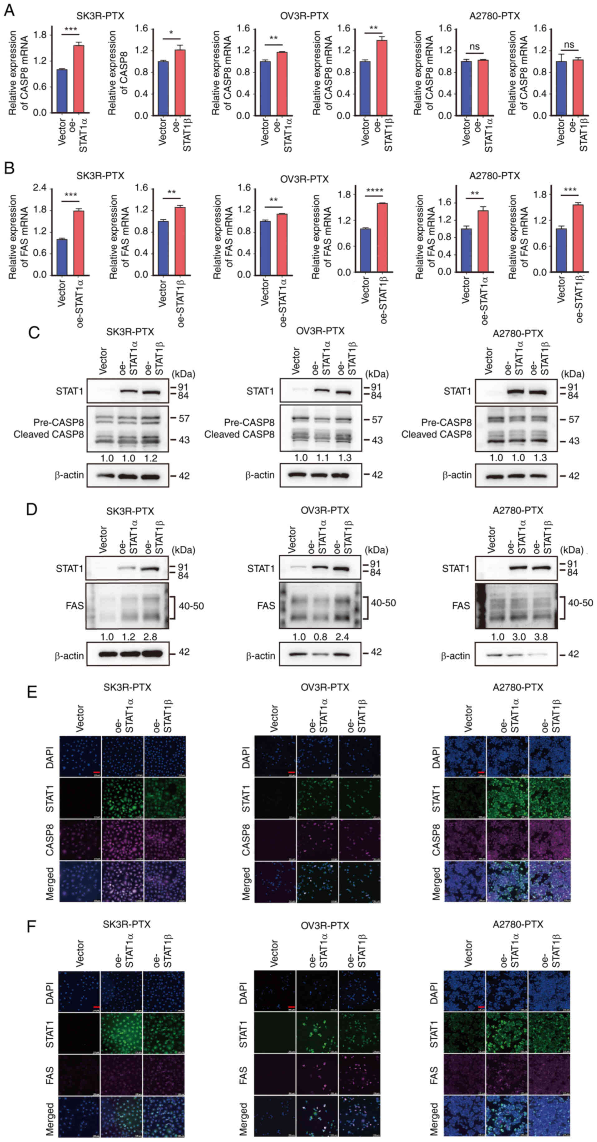
Related Articles

  • Abstract
  • View
  • Download
  • Twitter
Copy and paste a formatted citation
Spandidos Publications style
Wang F, Xu X, Guan B, Li X, Yuan J, Guan W, Chen J, Fang J, Lu Q, Xu G, Xu G, et al: Regulation and reversal of paclitaxel resistance via the STAT1‑mediated apoptotic pathway in ovarian cancer. Int J Oncol 68: 19, 2026.
APA
Wang, F., Xu, X., Guan, B., Li, X., Yuan, J., Guan, W. ... Xu, G. (2026). Regulation and reversal of paclitaxel resistance via the STAT1‑mediated apoptotic pathway in ovarian cancer. International Journal of Oncology, 68, 19. https://doi.org/10.3892/ijo.2025.5832
MLA
Wang, F., Xu, X., Guan, B., Li, X., Yuan, J., Guan, W., Chen, J., Fang, J., Lu, Q., Xu, G."Regulation and reversal of paclitaxel resistance via the STAT1‑mediated apoptotic pathway in ovarian cancer". International Journal of Oncology 68.2 (2026): 19.
Chicago
Wang, F., Xu, X., Guan, B., Li, X., Yuan, J., Guan, W., Chen, J., Fang, J., Lu, Q., Xu, G."Regulation and reversal of paclitaxel resistance via the STAT1‑mediated apoptotic pathway in ovarian cancer". International Journal of Oncology 68, no. 2 (2026): 19. https://doi.org/10.3892/ijo.2025.5832
Copy and paste a formatted citation
x
Spandidos Publications style
Wang F, Xu X, Guan B, Li X, Yuan J, Guan W, Chen J, Fang J, Lu Q, Xu G, Xu G, et al: Regulation and reversal of paclitaxel resistance via the STAT1‑mediated apoptotic pathway in ovarian cancer. Int J Oncol 68: 19, 2026.
APA
Wang, F., Xu, X., Guan, B., Li, X., Yuan, J., Guan, W. ... Xu, G. (2026). Regulation and reversal of paclitaxel resistance via the STAT1‑mediated apoptotic pathway in ovarian cancer. International Journal of Oncology, 68, 19. https://doi.org/10.3892/ijo.2025.5832
MLA
Wang, F., Xu, X., Guan, B., Li, X., Yuan, J., Guan, W., Chen, J., Fang, J., Lu, Q., Xu, G."Regulation and reversal of paclitaxel resistance via the STAT1‑mediated apoptotic pathway in ovarian cancer". International Journal of Oncology 68.2 (2026): 19.
Chicago
Wang, F., Xu, X., Guan, B., Li, X., Yuan, J., Guan, W., Chen, J., Fang, J., Lu, Q., Xu, G."Regulation and reversal of paclitaxel resistance via the STAT1‑mediated apoptotic pathway in ovarian cancer". International Journal of Oncology 68, no. 2 (2026): 19. https://doi.org/10.3892/ijo.2025.5832
Follow us
  • Twitter
  • LinkedIn
  • Facebook
About
  • Spandidos Publications
  • Careers
  • Cookie Policy
  • Privacy Policy
How can we help?
  • Help
  • Live Chat
  • Contact
  • Email to our Support Team