Introduction
SHK is the main active compound of traditional
Chinese herb Zicao (the dried root of Lithospermum
erythrorhizon), which has been shown to exert diverse antitumor
effects. SHK can induce autophagy, apoptosis and necroptosis in
multiple types of cancer cells (1). SHK can also suppress the invasion,
migration and metastasis of cancer cells, inhibit angiogenesis,
affect metabolic reprogramming, and promote cell cycle arrest
(2,3). Indeed, a large number of studies
have investigated the antitumor effects of SHK over the past three
decades, and nearly 1,000 related publications have been published.
However, due to its strong and non-selective toxicity, SHK has not
yet been approved for the clinical treatment of tumors (4). Thus, there is an urgent need for
therapeutic strategies that preserve the potent antitumor activity
of SHK while minimizing its toxicity to normal tissues.
Nanocarriers have emerged as promising platforms for cancer therapy
(5). Encapsulating SHK within
nanocarriers for systemic administration to enhance its localized
accumulation in tumor appears to be a promising direction (6). However, increasing evidence shows
that when nanocarriers are administered through systemic
circulation, a large proportion of nanocarriers still undergo
phagocytosis through the mononuclear phagocyte system or even
phagocytes around the tumor, leading to low drug concentrations at
the tumor site (7). In the
present study, for the first time, SHK was loaded into the
sustained-release hydrogel for intratumoral injection, to ensure
direct and sustained exposure of local tumors to SHK. This strategy
can prevent the high-concentration SHK from entering into the
systemic circulation and reduce the toxic damage to normal
tissues.
Several studies have found that SHK can strongly
induce immunogenic cell death (ICD) in cancer cells. Unlike
apoptosis, ICD not only releases a large number of tumor-specific
antigens, but also damage-associated molecular patterns (DAMPs),
which activates antigen presenting cells (APCs) and triggers the
process of tumor antigen presentation, eventually promoting the
expansion of tumor-specific T cells (8). Furthermore, it has been reported
that SHK reduces the content of lactic acid in the tumor
microenvironment (TME) by inhibiting pyruvate kinase (9), promotes the transformation of M2
subtype macrophages to the M1 subtype (10), and inhibits IDO1 catalytic
activity through interaction with the ferric form of IDO1 (11). All these data suggest that SHK has
favorable potential in reversing the tumor immunosuppressive
microenvironment. Unfortunately, because of SHK's pharmacological
properties in inhibiting the expression of a variety of immune
promoting cytokines and the negative effects on immune activation
(1,12), SHK alone is insufficient to
activate an effective antitumor immune response in immune-cold
tumors.
Photothermal therapy (PTT) has emerged as a novel
paradigm for precise cancer medicine due to its advantages of
controllable irradiation and temperature, non-invasiveness and
localized treatment. Generally, photothermal heating to a high
temperature of >50°C is needed for achieving a harsh environment
for tumor ablation (13). This
excessive hyperthermia increases the risk of injuring the
surrounding normal tissues and may trigger unwanted inflammation
due to the problem of heat transfer. Since PTT compels tumor cells
to death through necrosis at a temperature of >50°C by releasing
intracellular biomolecules and cellular fragments, aggressive local
inflammation may occur, thus damaging healthy tissues and promoting
tumor metastasis (14). Moreover,
it should be noted that overheating at such high temperature may
reduce the production of DAMPs in the TME, leading to insufficient
recruitment and activation of endogenous APCs, as well as
disrupting the immune antigens that evoke antitumor immunity and
immune cells that execute immune response in the TME (15). This, in turn, prevents the immune
system from destroying cancer cells. To surmount these bottleneck
problems, mild PTT (also referred to as mild-temperature or
low-temperature PTT), which exerts multiple effects on local tumors
at relatively low temperatures while minimizing damage to healthy
tissues, has been proposed. In the present study, mild PTT was
operationally defined as temperature-controlled local photothermal
heating maintained at ~43°C for 15 min. Theoretically, mild PTT is
regarded as one of the important variables in constructing a
favorable TME for triggering immune responses (16). Mild PTT can improve the
infiltration of immune cells into tumors, increase the expression
of immune-promoting cytokines in tumor site, and trigger a limited
antitumor immunity (17).
However, a slight increase in temperature alone may not be
sufficient to induce widespread tumor cell death, which results in
an inadequate supply of neoantigens for activating specific
antitumor immunity. Moreover, this increase in temperature may
potentially trigger the upregulation of PD-L1, IDO1, and heat shock
proteins on tumor cells as part of their self-defense mechanisms.
Such a feedback loop could potentially lead to an immunosuppressive
effect, further hindering the desired antitumor immune response
(18). Hence, the antitumor
immunity evoked by mild PTT is generally limited and has a short
lifespan (19). Therefore, it is
important to explore the combination strategies for enhancing and
prolonging the antitumor immunity triggered by mild PTT.
Triple-negative breast cancer (TNBC) is the most
malignant subtype of breast cancer (BC), prone to spread faster and
relapse after treatment (20). As
these cancer cells do not express HER-2, progesterone receptor or
estrogen receptor, there are only a few TNBC-targeted therapies
available (20). A number of
studies have shown that TNBC exhibits improved immunogenicity than
other subtypes, suggesting that immunotherapy is more suitable for
patients with TNBC (21).
However, several recent clinical trials indicated that the current
immunotherapy strategies (for example, anti-PD-1 or anti-PD-L1
antibody) had low efficacy in the whole population of patients with
TNBC as a result of limited or extensive infiltration of immune
cells or immunosuppressive cells, respectively, in the TMEs
(22,23). In other words, most of patients
with TNBC fail to respond to current immunotherapies because of the
'non-inflamed' or 'cold' immune microenvironments and transforming
immune-cold tumor into immune-hot tumor may be a promising solution
to this challenge (22,24). In the present study, 4T1 BC cells,
a poorly immunogenic triple-negative model, were selected as the
primary tumor model (25,26). SHK was wrapped into methoxy
poly(ethylene glycol)-b-poly(ε-caprolactone-co-1,4,8
-trioxa[4.6]spiro-9-undecanone) (mPECT) nanoparticles and loaded
with mPECT-modified gold nanorods (AuNRs) in a supramolecular
hydrogel for intratumoral injection to realize the combination of
mild PTT and SHK local tumor treatment (Fig. 1A). Mild PTT triggered immune
activation rapidly in the immunosuppressive microenvironment, while
SHK strongly induced ICD and persistently inhibited the activation
of IDO1 and expression of PD-L1 induced by mild PTT. The
combination of these two treatments formed a synergistic effect to
turn immune 'cold' tumor into 'hot' tumor, thereby enhancing a
strong and long-lasting antitumor immunity and suppressing
untreated distant tumors (Fig.
1B). The present study demonstrates the potential of low-dose
local SHK administration to induce systemic antitumor immunity and
provides a preclinical framework for the further development of
localized SHK-based combination strategies in immune-cold TNBC.
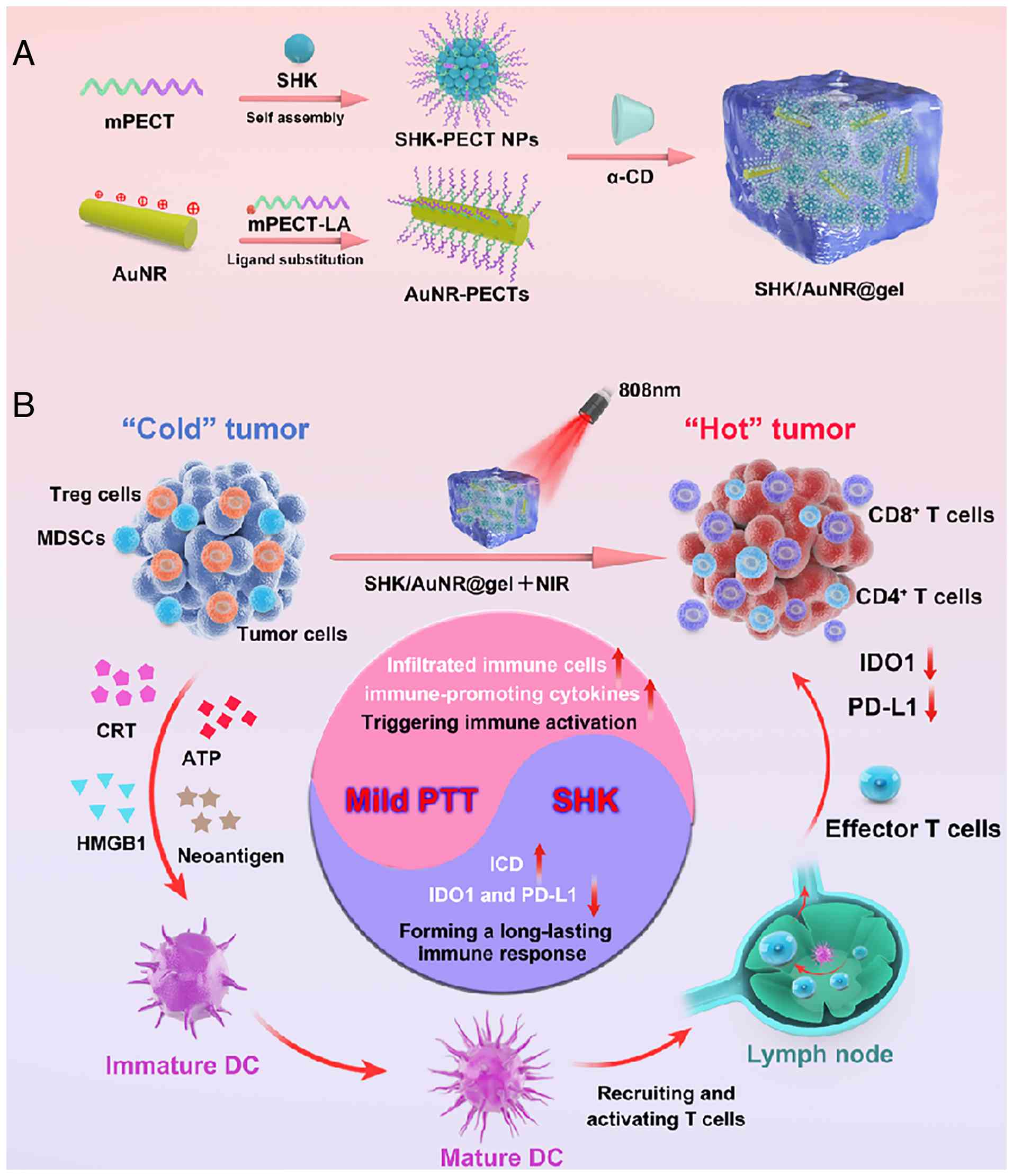 | Figure 1Schematic diagram for the preparation
and function of SHK/AuNR@gel. (A) Preparation of SHK/AuNR@gel. (B)
The mechanism of SHK/AuNR@ gel in tumor tissues. Briefly, mild PTT
increases the infiltration of immune cells into tumors, upregulates
the expression of immune-promoting cytokines, and triggers the
immune activation rapidly in tumor immunosuppressive
microenvironment. SHK strongly induces ICD and persistently
inhibits the activation of IDO1 and expression of PD-L1 induced by
mild PTT. The combination of these two forms a synergistic effect
to turn immune 'cold' tumor into 'hot' tumor, enhance antitumor
immunity and attack the metastatic tumors. SHK, shikonin; AuNRs,
gold nanorods; PTT, photothermal therapy; ICD, immunogenic cell
death; PD-L1, programmed death-ligand 1; IDO1, indoleamine
2,3-dioxygenase 1; HMGB1, high mobility group box 1; CRT,
calreticulin; DC, dendritic cell. |
Materials and methods
Chemicals and reagents
Shikonin (SHK, >98% purity) was obtained from
Yuanye Biotechnology (https://www.shyuanye.com/). Cyclodextrin (α-CD),
cetyltrimethyl ammonium bromide (CTAB) and sodium borohydride
(NaBH4) were supplied by Shanghai Aladdin Biochemical
Technology Co., Ltd. 4-(dimethylamino) pyridine (DMAP),
N,N-dicyclohexyl carbodiimide (DCC), lipoic acid, tetrachloroauric
acid (HAuCl4·4H2O), ascorbic acid and silver nitrate (AgNO3) were
obtained from MilliporeSigma. Diethyl ether, tetrahydrofuran (THF),
dimethyl-sulfoxide (DMSO) and hydrochloric acid were obtained from
Nantong Jiangtian Chemical Co., Ltd.
3-[4,5-dimethylthiazol-2-yl]-2,5-diphenyltetrazolium bromide (MTT)
was supplied by Beyotime Institute of Biotechnology. Cell culture
reagents were purchased from Gibco; Thermo Fisher Scientific,
Inc.).
Cell lines and animals
Two TNBC cell lines (murine TNBC cells 4T1 and human
TNBC cells MDA-MB-231) were supplied by the Chinese Academy of
Sciences and cultured in RPMI-1640 (4T1 cells) or DMEM (MDA-MB-231
cells) containing 10% (vol/vol) FBS (Gibco; Thermo Fisher
Scientific, Inc.) and 1% penicillin/streptomycin (Thermo Fisher
Scientific, Inc.) at 5% CO2 and 37°C as previously
described (27).
BALB/c mice (female, 6-8 weeks old, weighing 18-20
g) were supplied by Vital River Laboratories. All mice were
maintained under specific pathogen-free conditions in an
individually ventilated cage facility with a 12/12-h dark/light
cycle at 23°C and had ad libitum access to water and food.
Animals were monitored at least once daily throughout the study.
Predefined humane endpoints were strictly implemented, and every
effort was made to minimize pain, distress, and suffering. All
animal procedures were conducted in accordance with the Guidelines
for Care and Use of Laboratory Animals of Air Force Medical
University and were approved by the Animal Ethics Committee of Air
Force Medical University (approval no. 20241327; Xi'an China). The
study design and animal welfare measures were conducted in
accordance with internationally recognized principles for animal
research and widely accepted welfare guidelines for oncology
studies involving laboratory animals (28-30). A total of 220 mice were used
across all in vivo experiments. The bilateral tumor
(abscopal) model and the long-term immune memory/rechallenge
studies were performed using independent cohorts of animals.
Animal welfare, tumor monitoring,
anesthesia, blood collection and euthanasia
Animals were assessed at least once daily for
general health and well-being, including activity, posture,
grooming behavior, food and water intake, and signs of pain or
distress. Tumor growth was monitored every 3 days by palpation and
caliper measurements, while body weight was recorded daily. Tumor
volume was calculated using the formula: 0.5 × length ×
width2.
Humane endpoints were defined a priori. Mice
were euthanized if any of the following criteria were met: i) Tumor
ulceration, necrosis, bleeding or infection; ii) impaired mobility
or inability to access food or water; iii) body weight loss of ≥20%
from baseline; or iv) excessive tumor burden, defined as any single
tumor exceeding 2,000 mm3 in volume.
For near-infrared irradiation (NIR) and surgical
procedures, including tumor resection and tissue collection, mice
were anesthetized with inhaled isoflurane (3-4% for induction and
1-2% for maintenance in oxygen) and placed on a warming pad. Local
tumor temperature during irradiation was continuously monitored
using an infrared thermometer or thermal imaging camera.
For pharmacokinetic analysis, 150 μl of blood
was collected from the tail vein at each sampling time point.
Independent cohorts of mice were used for each time point, and no
animal underwent more than two tail-vein blood collections within a
24-h period. For hematological and serum biochemical analyses,
whole blood (500-800 μl per mouse) was collected once at the
experimental endpoint as a terminal procedure via cardiac puncture
under deep isoflurane anesthesia (4-5% for induction, 2-3% for
maintenance in oxygen until loss of the pedal withdrawal reflex),
immediately followed by euthanasia and tissue collection.
For euthanasia, mice were deeply anesthetized with
inhaled isoflurane (4-5% for induction, 2-3% for maintenance in
oxygen) until complete loss of the pedal withdrawal reflex,
followed by cervical dislocation. Death was confirmed by the
absence of respiration, heartbeat, and reflex responses before
necropsy and tissue harvesting.
Preparation of SHK-loaded mPECT
nanoparticles (SHK-mPECTs) and mPECT-modified gold nanorods
(AuNR-mPECTs)
The mPECT diblock copolymers were prepared via
ring-opening polymerization of 1,4,8-trioxa [4.6]
spiro-9-undecanone (TOSUO) and ε-caprolactone at 130°C using mPEG
as an initiator (31). SHK could
be encapsulated in the mPECT cores via a hydrophobic interaction,
and SHK-mPECT nanoparticles were prepared using the
nanoprecipitation approach (32,33). Briefly, mPECT (200 mg) and SHK (2
mg) were dissolved in 2 ml THF, which was added dropwise to 20 ml
deionized water under constant stirring. After removing THF through
vaporization for 24 h, the SHK-mPECTs were produced by
lyophilization (33).
Gold nanorods (AuNRs) were prepared using a facile,
seedless synthesis method (34).
Briefly, 150 μl cold NaBH4 (0.01 M), 700
μl ascorbic acid (78.8 mM), 80 μl HCl (37%) and 2.5
ml AgNO3 (4 mM) were added to a mixture solution (100
ml) of 0.2 M CTAB and 1 mM HAuCl4 (33). After stirring at 30°C for 6 h, the
obtained AuNR-CTAB solution was kept at room temperature overnight.
The AuNR-CTAB solution was purified via centrifugation (11,100 × g,
15 min) and rinsed twice before reaction to eliminate residual CTAB
coating. Lipoic acid (LA)-modified mPECT (LA-mPECT) was prepared as
previously described by the authors (33). Briefly, 0.137 g LA and 0.413 g DCC
were dissolved in 1 ml DCM and then stirred for 12 h. Next, 1 g
mPECT and 0.12 g DMAP in 10 ml DCM was added. After stirring under
nitrogen at 35°C for another 48 h, the mixture was precipitated in
cold ether and vacuum-dried at 25°C. AuNR-mPECTs were prepared by
dropwise addition of 2 ml LA-mPECT solution (10 mg/ml) in DMSO to a
dispersion of 100 ml AuNRs (0.01 mg/ml) under vigorous stirring,
followed by an overnight incubation. After recovery of AuNR-mPECTs
via centrifugation (16,600 × g, 15 min) and washing twice with
deionized water, the mixture was ultimately dispersed in water to
the final concentration of 100 μg/ml.
Preparation of hydrogels with different
loadings
For the preparation of empty mPECT hydrogel, 60 mg
α-CD was added to 1 ml of empty mPECT NPs [200 mg/ml, prepared via
nanoprecipitation (35)], and
then stirred vigorously until the formation of a hydrogel.
For the preparation of SHK@gel (CSHK=2
mg/ml, CmPECT=200 mg/ml, Cα-CD=60 mg/ml), 60
mg α-CD was added to 1 ml of SHK-mPECTs solution (200 mg/ml) and
then stirred vigorously until the formation of a hydrogel.
For the preparation of AuNR@gel (CAuNR=50
μg/ml, CmPECT=200 mg/ml, Cα-CD=60
mg/ml), 200 mg lyophilized powder of empty mPECT NPs was dispersed
in 1 ml AuNR-mPECTs solution (50 μg/ml) and then added with
60 mg α-CD under stirring.
To prepare SHK/AuNR@gel (CSH K= 2 mg/ml,
CAuNR=50 μg/ml, CmPECT=200 mg/ml,
Cα-CD=60 mg/ml), 60 mg α-CD was added to an AuNR-mPECTs
solution (1 ml, 50 μg/ml) containing 200 mg lyophilized
powder of SHK-mPECTs. Finally, the mixture was stirred vigorously
until the formation of a nano-composite hydrogel.
Characterization
TU-1900 spectrophotometer (Purkinje General
Instrument) was used to collect UV-Vis spectra. The fluorescence
spectral data were recorded using a fluorescence spectrophotometer
(Agilent) at 520 nm. The zeta potential and hydrodynamic size of
the nanoparticles were determined by dynamic light scattering (DLS;
Nano ZS90; Malvern Instruments, Ltd.) at 25°C. The concentration of
AuNRs was measured by ICP-MS (7700x; Agilent, Technologies, Inc.).
JEOL 100CXII transmission microscope (accelerating voltage=100 kV)
was utilized to obtain transmission electron microscopy (TEM)
images. The inner morphology of the obtained hydrogel was examined
by S-4800 field-emission scanning electron microscopy (SEM;
Hitachi, Ltd.). Rheological measurement of the hydrogel was
conducted using a fluid rheometer (MCR 302; Anton Paar, Austria).
An infrared thermal camera (I5; FLIR, USA) was used to monitor the
temperatures of the hydrogel and AuNR solution. To investigate the
degradation and SHK content in SHK/AuNR@ gel, 500 μl
hydrogel was placed in a 10-ml test tube and then added with 5 ml
PBS (pH 7.4). At regular intervals, the supernatant (3 ml) was
collected, lyophilized and dissolved in DMF. The SHK levels were
examined using the UV-Vis spectrophotometer.
Investigation of the inhibitory effect of
IDO1
As described previously (11), the 4T1 cells or MDA-MB-231 cells
(5×10⁵ cells/well) were cultured in a 6-well plate. On
the following day, the cells were transfected with pcDNA3.1-mIDO1
or pcDNA3.1-hIDO1 (Synbio Technologies) using Lipofectamine 2000
(Invitrogen; Thermo Fisher Scientific, Inc.). After transfection
for 24 h, the cells (2.5×10⁴ cells/well) were cultured
in a 96-well plate and treated with the indicated agents for 24 h.
Then, the culture medium (200 μl) was mixed with 30%
trichloroacetic acid (100 μl; MilliporeSigma). After
incubation at 65°C for 15 min and centrifugation (12,000 r/min, 10
min), the supernatant (100 μl) was mixed with 2% (w/v)
p-dimethyl-amino-benzaldehyde (100 μl; D2004;
MilliporeSigma) in acetic acid. SpectraMax Plus 384 microplate
reader (Molecular Devices, LLC) was used to measure the
yellow-colored kynurenine at 492 nm.
Luciferase assay
The cells were seeded in 48-well plates at a density
of 3×10⁴ cells/well and cultured under normal conditions
for 24 h. Subsequently, the cells were transfected with 0.2
μg pNFκB-luc (Beyotime Institute of Biotechnology) and 0.01
μg pRL-TK Renilla (Beyotime Institute of Biotechnology)
using Lipofectamine 2000 for 24 h and then treated with different
concentrations of SHK for another 24 h. After treatment, the cells
were lysed using passive lysis buffer, and the luciferase activity
was detected using a Dual Luciferase Reporter kit (Beyotime
Institute of Biotechnology) (36). The experiment was carried out in
triplicate and repeated three times independently.
Western blot analysis
As described previously (37), the total protein or nuclear
protein was isolated using RIPA buffer (Thermo Fisher Scientific,
Inc.) or nuclear and cytoplasmic protein extraction kit (cat. no.
P0027; Beyotime Institute of Biotechnology), respectively. The
protein concentrations were evaluated using a BCA protein assay
kit. After separation through 10% SDS-PAGE, the protein samples (30
μg per lane) were transferred onto PVDF membranes (Wuhan
Servicebio Technology Co., Ltd.). The membranes were blocked in 5%
non-fat milk for 60 min, and then exposed to primary antibodies
overnight at 4°C. The following antibodies were employed:
Anti-PD-L1 (1:1,000; cat. no. GB115704; Wuhan Servicebio Technology
Co., Ltd.), anti-NF-κB p65 (1:1,000; cat. no. GB12142; Wuhan
Servicebio Technology Co., Ltd.), anti-Histone H3 antibody
(1:1,000; cat. no. GB11102; Wuhan Servicebio Technology Co., Ltd.)
and anti-β-actin antibody (1:1,000; cat. no. AF7018; Affinity
Biosciences). After rinsing, the membranes were exposed to
anti-mouse or anti-rabbit HRP-labeled secondary antibodies
(1:5,000; cat. nos. SA00001-1 and SA00001-2, respectively;
Proteintech Group, Inc.) for 1 h. The protein blots were visualized
by ECL reagent (MilliporeSigma). Semi-quantitative densitometry
analysis of protein expression was performed using ImageJ v1.46r
(National Institutes of Health). Band intensities of target
proteins were normalized to the corresponding loading controls and
expressed relative to the corresponding control group.
Assessment of ICD
To assess treatment-induced ICD in vitro, 4T1
or MDA-MB-231 cells were seeded in flat-bottom 96-well plates at a
density of 1×10⁴ cells per well and allowed to adhere
overnight. Cells were then treated for 24 h with various
concentrations of free SHK or the indicated nano-formulations, with
or without NIR irradiation, at equivalent SHK concentrations.
Following treatment, cells were gently washed three times with PBS
and fixed in 4% paraformaldehyde (PFA) for 10 min at room
temperature without permeabilization to restrict antibody binding
to cell-surface calreticulin (CRT). The cells were then blocked
with 5% bovine serum albumin (BSA; cat. no. ST023; Beyotime
Institute of Biotechnology) in PBS for 1 h and incubated with
rabbit anti-CRT primary antibody (cat. no. ab227444; Abcam; 1:200
dilution in 1% BSA/PBS) for 2 h. After three washes with PBS, cells
were incubated with HRP-conjugated goat anti-rabbit IgG secondary
antibody (Proteintech Group, Inc.; 1:5,000 dilution in 1% BSA/PBS)
for 1 h at room temperature. After extensive washing,
3,3',5,5'-tetramethylbenzidine substrate was added, and color
development was allowed to proceed in the dark for 10-15 min before
termination with 2 M H2SO4. Absorbance was
measured at 450 nm using a microplate reader. Meanwhile, cell
viability in replicate wells subjected to identical treatments was
assessed by the MTT assay. CRT signals were normalized to the
corresponding MTT absorbance values at 570 nm to account for
differences in viable cell numbers and expressed as fold-change
relative to untreated controls.
In addition, culture supernatants were collected
after treatment and centrifuged to remove cellular debris prior to
further analysis. The concentrations of ATP and HMGB1 were
determined using a commercial ATP Determination Kit (cat. no.
S0027) and HMGB1 ELISA Kit (cat. no. PH406; both from Beyotime
Institute of Biotechnology), respectively, according to the
manufacturers' instructions.
Evaluation of cytotoxicity
The viability of cells was assessed by MTT assay as
described previously (37,38).
Briefly, the cells were cultured in a 96-well plate at 37°C for 24
h. Then, MTT (5 mg/ml) was added (20 μl/well) and incubated
for another 4 h. After eliminating the supernatant, the formazan
crystals were dissolved in DMSO (150 μl/well). The
absorbance was measured using an ELX 800 microplate reader (BioTek;
Agilent Technologies, Inc.) at 570 nm.
Caspase-3/7 activity assay
Caspase-3/7 activity was measured using the
Caspase-Glo® 3/7 Assay kit (cat. no. G8091; Promega
Corporation) according to the manufacturer's instructions. Briefly,
4T1 cells were seeded into white 96-well plates and allowed to
adhere overnight before treatment with free SHK at the indicated
concentrations for 24 h. Following treatment, an equal volume of
Caspase-Glo 3/7 reagent was added directly to each well containing
cells and culture medium. Plates were gently mixed on an orbital
shaker for 30 sec and incubated at room temperature in the dark for
30-60 min to allow cell lysis and substrate cleavage. Luminescence
was measured using a microplate reader, and background signal from
medium-only wells was subtracted. Caspase-3/7 activity for each
treatment condition was normalized to that of untreated control
cells and expressed as fold-change relative to control. All
treatment conditions were assayed in triplicate wells, and each
experiment was independently repeated at least three times.
Antitumor study of SHK/AuNR@gel on the
local tumors
The left mammary fat pad of each female BALB/c mouse
was orthotopically inoculated with 4T1 cells (5×10⁵
cells suspended in 50 μl PBS). Once tumors reached ~150
mm3, the mice were randomly assigned to 5 groups, and
administered with 50 μl empty mPECT@gel, free SHK solution,
SHK@gel, AuNR@ gel, and SHK/AuNR@gel via intratumoral injection.
Mice were anesthetized with inhaled isoflurane prior to irradiation
as described above. After injection, mice in the NIR-treated groups
were irradiated with an 808-nm laser under real-time temperature
monitoring. The laser power was adjusted as needed to rapidly reach
and maintain the local tumor temperature at ~43°C for 15 min. This
temperature-controlled photothermal stimulation was performed every
other day for a total of four sessions, ending on day 6 after
intratumoral injection.
Body weight (BW) was recorded daily, and tumor
dimensions were measured every 3 days using calipers. Tumor volume
was calculated as 0.5 × length × width2. On day 8 after
intratumoral administration, a portion of tumor tissue was
collected under anesthesia for immune-response analysis. Mice were
monitored until a single tumor exceeded 2,000 mm3 or
predefined humane endpoints were reached, and survival time was
recorded.
Kynurenine (Kyn) and tryptophan (Trp)
measurement in vivo
Detection of Trp and Kyn content in tumor tissues
was conducted as previously described (39). Briefly, the tumor tissues were
harvested from tumor-bearing mice (TBM) treated with different
therapeutics for homogenization using homogenizer. After dissolving
the homogenate in trichloroacetic acid (10%), the contents of Trp
and Kyn in the supernatant were detected using the LC-MS system
(Agilent 1290 Series LC System). Data collection and analysis were
conducted with Analyst software (version 1.6.3; SCIEX). The Kyn to
Trp ratio (Kyn/Trp) was employed as an indicator of IDO
activity.
Immunofluorescence staining
Immunofluorescence staining was conducted as
described previously (40).
Briefly, freshly dissected tumor tissue was fixed in 4% PFA at 25°C
for 48 h, dehydrated through a graded ethanol series (75, 85, 90,
95 and 100% ethanol; 1 h each), embedded in paraffin wax, and
sliced into 4-μm-thick sections. After permeabilization
(0.1% Triton X-100; Beyotime Institute of Biotechnology), the
sections were blocked for 1 h at room temperature with 10% normal
donkey serum (cat. no. ANT051; Wuhan Antgene Biotech Co., Ltd.),
and exposed to primary antibodies, anti-PD-L1 (aPD-L1; 1:100; cat.
no. ab213480; Abcam) overnight at 4°C. Subsequently, the sections
were exposed to fluorescent secondary antibody (1:200; cat. no.
sc-362281; Santa Cruz Biotechnology, Inc.) and DAPI (0.001 mg/ml;
cat. no. KGA215-10; Nanjing KeyGen Biotech Co., Ltd.), the
immunofluorescence image was obtained using a fluorescence
microscope (EVOS FL Auto; Thermo Fisher Scientific, Inc.). Data
analysis was performed with ImageJ software.
In vivo ICD analysis
Immunohistochemistry was performed to observe the
markers of ICD, CRT and HMGB1, using an indirect peroxidase method
(41). The paraffin-embedded
tissue section was dewaxed in xylene and rehydrated in a graded
series of decreasing concentrations of ethanol. After quenching
endogenous peroxidase with hydrogen peroxide (3%), the section was
blocked with goat serum (10%; ZSGB-Bio) for 30 min at room
temperature to inhibit non-specific antibody binding. The tissue
sections were incubated overnight with primary antibody at 4°C.
Antibodies against CRT (1:100; cat. no. ab227444) and HMGB1 (1:350;
cat. no. ab79823) were supplied by Abcam. Subsequently, the
sections were exposed to HRP-conjugated goat anti-rabbit IgG
(1:200; cat. no. SA00001-2; Proteintech Group, Inc.). The
chromogenic substrate used in the present study was
diaminobenzidine (DAB; ZSGB-Bio). After counterstaining with
hematoxylin and mounting in resin, images were obtained using an
Aperio pathology scanner. The IOD values were determined with
ImageJ software.
Immune-response sampling from primary
tumors and tumor-draining lymph nodes
Primary tumors and ipsilateral inguinal/axillary
tumor-draining lymph nodes (TDLNs) were collected 24 h after the
final treatment. TDLNs were used for CD11c+ dendritic
cell (DC) enrichment, qPCR-based analysis of DC maturation markers,
and IL-12p70 ELISA. Primary tumor tissues were used for qPCR-based
assessment of T-cell infiltration- and cytolytic activity-related
genes, as well as CD4/CD25-enriched Foxp3 analysis.
Magnetic enrichment of immune cell
subsets
CD11c+ cells were isolated from TDLNs
using positive magnetic selection with CD11c MicroBeads UltraPure
(mouse; cat. no. 130-125-835; Miltenyi Biotec, Inc.) and LS Columns
(cat. no. 130-042-401; Miltenyi Biotec, Inc.). CD4+
tumor-infiltrating lymphocytes (TILs) were enriched from tumor
tissues using the Tumor Dissociation Kit (mouse; cat. no.
130-096-730; Miltenyi Biotec, Inc.), CD4 (TIL) MicroBeads (mouse;
cat. no. 130-116-475; Miltenyi Biotec, Inc.), and LS Columns. Where
indicated, CD25 surface labeling-based magnetic enrichment was
further performed using mouse CD25 MicroBeads (cat. no.
130-091-072; Miltenyi Biotec, Inc.), and RNA was extracted from the
CD25-enriched CD4+ fraction for Foxp3 analysis. For
ex vivo memory T-cell assays, splenic CD8+ T
cells were purified by negative selection using the MojoSort™ Mouse
CD8+ T Cell Isolation Kit (cat. no. 480008; BioLegend,
Inc.), whereas CD11c+ DCs were enriched using CD11c
MicroBeads (Miltenyi Biotec, 130-108-338).
RT-qPCR analysis
Total RNA was extracted from cells, tumor tissues,
or enriched immune cell fractions using TRIzol™ reagent (cat. no.
15596026; Thermo Fisher Scientific, Inc.), the RNeasy Micro Kit
(cat. no. 74004; Qiagen China Co., Ltd.), or the RNeasy Mini Kit
(cat. no. 74104; Qiagen), as appropriate. RNA was
reverse-transcribed into cDNA using PrimeScript™ RT Master Mix
(cat. no. RR036A; Takara) or PrimeScript™ RT Reagent Kit (cat. no.
RR037A; Takara Biotechnology Co., Ltd.). RT-qPCR was performed on a
CFX96 Real-Time PCR System using TB Green® Premix Ex
Taq™ II (cat. no. RR820A; Takara) under the following thermocycling
conditions: Initial denaturation at 95°C for 30 sec, followed by 40
cycles of denaturation at 95°C for 5 sec and annealing/extension at
60°C for 30 sec. GAPDH was used as the reference gene, and relative
expression was calculated using the 2−ΔΔCq method
(42). For DC maturation
analysis, H2-Ab1, Cd80, Cd86, Il12a and Il12b were quantified in
CD11c+ cells isolated from TDLNs, and the DC maturation
index was calculated as the mean z-score of the normalized
expression values of these five genes (43). The primer sequences for RT-qPCR
were as follows: GAPDH forward, 5'-CATCACTGCCACCCAGAAGACTG-3' and
reverse, 5'-ATGCCAGTGAGCTTCCCGTTCAG-3'; H2-Ab1 forward,
5'-GTGTGCAGACACAACTACGAGG-3' and reverse,
5'-CTGTCACTGAGCAGACCAGAGT-3'; Cd80 forward,
5'-CCTCAAGTTTCCATGTCCAAGGC-3' and reverse,
5'-GAGGAGAGTTGTAACGGCAAGG-3'; Cd86 forward,
5'-ACGTATTGGAAGGAGATTACAGCT-3' and reverse,
5'-TCTGTCAGCGTTACTATCCCGC-3'; Il12a forward,
5'-CTGTGCCTTGGTAGCATCTATG-3' and reverse,
5'-GCAGAGTCTCGCCATTATGATTC-3'; Il12b forward,
5'-TGGTTTGCCATCGTTTTGCTG-3' and reverse,
5'-ACAGGTGAGGTTCACTGTTTCT-3'; Cd3e forward,
5'-GCTCCAGGATTTCTCGGAAGTC-3' and reverse,
5'-ATGGCTACTGCTGTCAGGTCCA-3'; Cd4 forward,
5'-GTTCAGGACAGCGACTTCTGGA-3' and reverse,
5'-GAAGGAGAACTCCGCTGACTCT-3'; Cd8a forward,
5'-ACTACCAAGCCAGTGCTGCGAA-3' and reverse,
5'-ATCACAGGCGAAGTCCAATCCG-3'; Gzmb forward,
5'-CAGGAGAAGACCCAGCAAGTCA-3' and reverse,
5'-CTCACAGCTCTAGTCCTCTTGG-3'; Prf1 forward,
5'-TTTCGCCTGGTACAAAAACC-3' and reverse, 5'-CGTTCAGGCAGTCTCCTACC-3';
Foxp3 forward, 5'-CCCAGGAAAGACAGCAACCTT-3' and reverse,
5'-TTCTCACAACCAGGCCACTTG-3'; Tnf forward,
5'-CCCTCACACTCAGATCATCTTCT-3' and reverse,
5'-GCTACGACGTGGGCTACAG-3'; Ifng forward, 5'-GCCACGGCACAGTCATTGA-3'
and reverse, 5'-TGCTGATGGCCTGATTGTCTT-3'; and Cd274 forward,
5'-TGCGGACTACAAGCGAATCACG-3' and reverse,
5'-CTCAGCTTCTGGATAACCCTCG-3.
ELISA and ELISpot assays
IL-12p70 levels in TDLN homogenates were quantified
using a Mouse IL-12p70 ELISA Kit (cat. no. 88-7121-22; Invitrogen;
Thermo Fisher Scientific, Inc.) and normalized to total protein
content (44). For ex vivo
memory immune-response analysis, granzyme B and perforin
concentrations were quantified using Mouse Granzyme B ELISA (cat.
no. MBS2507805; MyBioSource, Inc.) and Mouse Perforin ELISA (cat.
no. MBS2021832; MyBioSource, Inc.) kits, respectively. IFN-γ,
TNF-α, IL-2, CXCL9 and CXCL10 levels in culture supernatants were
measured using the corresponding commercial ELISA kits according to
the manufacturers' instructions. IFN-γ-producing T cells were
quantified using a Mouse IFN-γ ELISpot Kit (cat. no. 552569; BD
Biosciences), and results were expressed as spot-forming units
(SFU) per 105 T cells. Polyfunctional T cells
simultaneously producing IFN-γ and TNF-α were quantified using a
Dual-Color Mouse ELISpot Kit (cat. no. SPOT-09-2; Mabtech), and
results were expressed as double-positive SFU per 105 T
cells.
Calculation of intratumoral immune
indices
For T-cell infiltration analysis, Cd3e, Cd8a and Cd4
were quantified in tumor tissues, and the T-cell infiltration index
was calculated as the mean z-score of normalized Cd3e, Cd8a and Cd4
expression values (45).
Cytolytic activity (CYT) was quantified by the average of the
log-transformed expression values of Gzmb and Prf1, corresponding
to the geometric mean of their expression on a linear scale
(46). Foxp3 expression was
quantified in CD25-enriched CD4+ TILs and normalized to
the corresponding control group (47).
Detection of SHK concentration in
vivo
To measure SHK levels in plasma and tumor tissues,
4T1 TBM were intratumorally injected with free SHK, SHK@gel, or
SHK/AuNR@ gel. The treatment schedule was identical to that used in
the local tumor therapy study described above. Peripheral blood
(150 μl per mouse per time point) was collected from the
tail vein at predetermined time points into heparinized tubes.
Independent cohorts of mice were used for each sampling time point.
Plasma was obtained by centrifugation at 850 × g for 10 min,
transferred to clean polypropylene tubes and stored at −80°C until
analysis. To determine SHK content in plasma, 50 μl plasma
samples were mixed with 200 μl ethyl acetate, followed by
vortexing (10 min) and centrifugation (15,000 rpm, 10 min, 4°C).
The obtained SHK solution was dried through evaporation and
reconstituted in 100 μl 50% methanol/50% acetonitrile
containing 1% formic acid (48).
To detect SHK content in tumors, the tumor tissues were homogenized
in saline (ratio of tumor weight-to-volume=1:1), and the protein
was precipitated with 5 volume of 50% methanol/50% acetonitrile
containing 1% formic acid. After centrifugation (21,100 × g, 5 min,
4°C), the supernatant was filtered using a 0.22-μm filter,
and subsequently analyzed with the HPLC system (Shimadzu
LC-20AT).
Safety evaluation of the therapies
At the terminal time point, whole blood (500-800
μl per mouse) was collected by cardiac puncture under deep
anesthesia for hematological and serum biochemical analyses,
followed immediately by euthanasia as aforementioned. Hematology
and serum biochemistry were analyzed by the Clinical Laboratory of
Xijing Hospital, including hepatorenal function markers [blood urea
nitrogen (BUN), creatinine (CREAT), alkaline phosphatase (ALP),
alanine aminotransferase (ALT) and aspartate aminotransferase
(AST)] and routine blood parameters [monocyte (Mono), lymphocyte
(LYMPH), hematocrit (HCT), hemoglobin (HGB), platelet (PLT), red
blood cell (RBC) and white blood cell (WBC) counts]. Major organs
were collected, fixed in 4% neutral-buffered PFA, and assessed by
H&E staining. BW was recorded throughout the experiment.
Bilateral tumor model and T-cell
depletion
For the bilateral tumor model, 5×10⁵ 4T1
cells suspended in 50 μl PBS were injected into the right
mammary fat pad to establish the primary tumor. Three days later,
an equal number of 4T1 cells was injected into the contralateral
mammary fat pad to establish the distant tumor, following a
previously reported short-interval bilateral orthotopic tumor model
(49). When the primary tumor
volume reached ~150 mm3, mice were randomly assigned to
five treatment groups and received intratumoral injections (50
μl) of empty mPECT@gel, free SHK solution, SHK@gel, AuNR@gel
+ NIR, or SHK/AuNR@ gel + NIR. Mice were anesthetized with inhaled
isoflurane prior to NIR irradiation and surgical procedures, as
described above. NIR irradiation was performed using the same
temperature-controlled protocol described above. After completion
of the treatment schedule, the primary tumor was surgically
resected. BW was monitored daily, and the volume of untreated
contralateral tumors was measured every 3 days. Immune-related
molecular endpoints in distant tumors, including T-cell
infiltration-related genes, cytolytic effector genes and Foxp3
expression in CD25-enriched CD4+ TILs, were analyzed
using the same magnetic enrichment, RT-qPCR and calculation
procedures described above.
For depletion of CD4+ and CD8+
T cells, bilateral tumor-bearing mice were intraperitoneally
injected with anti-CD8 antibody (10 mg/kg; BioXcell, clone 53-6.7)
or anti-CD4 antibody (10 mg/kg; BioXcell, clone GK1.5), with the
corresponding IgG isotype controls (BioXcell, BE0089 or BE0090),
once every 3 days starting from the implantation of the distant
tumor (50). Intratumoral
administration and NIR irradiation were performed as described
above. BW was monitored daily, and distant tumor growth was
assessed every 3 days. The bilateral tumor model was conducted in
accordance with the same animal monitoring and humane endpoint
criteria described above (28-30,51).
Tumor rechallenge and experimental lung
metastasis models
Female BALB/c mice were orthotopically inoculated
with 4T1 cells (5×10⁵ cells per mouse in 100 μl
PBS) into the mammary fat pad. When tumors reached ~150
mm3, mice were randomly assigned into five treatment
groups and received intratumoral injections (50 μl) of blank
mPECT@gel, free SHK solution, SHK@gel, AuNR@gel + NIR, or
SHK/AuNR@gel + NIR. For the NIR-treated groups, tumors were
irradiated using the same temperature-controlled 808-nm laser
protocol described above. Two days after the final treatment,
primary tumors were surgically resected under anesthesia to
simulate complete local tumor eradication.
On day 30 after treatment, mice were rechallenged
subcutaneously with 4T1 cells (5×10⁵ cells per mouse) in
the contralateral flank to evaluate systemic antitumor immune
memory (52,53). Rechallenge tumor growth was
monitored every 3 days by caliper measurements, and tumor volume
was calculated as 0.5 × length × width2. In a separate
cohort, mice received intravenous injection of 1×10⁵ 4T1
cells on day 30 after treatment to establish an experimental lung
metastasis model. Fourteen days later, mice were euthanized, and
lungs were harvested, rinsed with PBS and fixed in 4% PFA. Surface
metastatic nodules were counted under a dissecting microscope (S9D;
Leica Microsystems GmbH), and lung tissues were processed for
H&E staining to assess pulmonary micrometastasis. Animal
monitoring and humane endpoints were applied as aforementioned.
Ex vivo functional analysis of memory T
cells
To characterize memory immune responses, spleens
were harvested on day 31 after treatment. Single-cell suspensions
were prepared by mechanical dissociation, filtration through
70-μm cell strainers and erythrocyte lysis. Purified
CD8+ T cells and enriched CD11c+ DCs were
prepared as described in the magnetic enrichment of immune cell
subsets section. DCs were pulsed with 4T1 tumor lysates (100
μg/ml protein) for 3 h at 37°C and washed three times to
remove free antigen. Purified CD8+ T cells were then
co-cultured with antigen-loaded DCs at an E:T ratio of 10:1 for
antigen-specific recall assays.
The frequency of IFN-γ-producing T cells and
IFN-γ/TNF-α double-positive T cells was measured by ELISpot as
described in the ELISA and ELISpot assays section. Cytotoxic
activity of memory CD8+ T cells was evaluated using a
colorimetric lactate dehydrogenase (LDH) cytotoxicity assay kit
(cat. no. 88953; Thermo Fisher Scientific, Inc.). Effector
CD8+ T cells were co-incubated with 4T1 target cells at
E:T ratios of 5:1, 10:1, 20:1 and 40:1 for 6 h, followed by
absorbance measurement at 490 nm. Specific lysis was calculated as
[(sample-spontaneous)/(maximum-spontaneous)] ×100%, and overall
cytotoxic activity was summarized as the area under the
dose-response curve using GraphPad Prism v9.0.
For molecular profiling, total RNA was extracted
from purified CD8+ T cells and analyzed by RT-qPCR as
aforementioned. For degranulation analysis, CD8+ T cells
were co-cultured with antigen-pulsed DCs for 4 h, after which
culture supernatants and cell lysates were collected separately.
Degranulation capacity was calculated as [supernatant/(supernatant
+ lysate)] ×100% based on granzyme B and perforin levels, and the
mean release rate of these two markers was used as an overall
measure of cytotoxic granule exocytosis. Recall cytokine responses
were assessed by co-culturing T cells with antigen-pulsed DCs for
48 h, followed by ELISA-based measurement of IFN-γ, TNF-α, IL-2,
CXCL9 and CXCL10 as aforementioned.
Statistical analysis
All data are presented as the mean ± SD. Statistical
tests were performed using SPSS Statistics version 31.0 (IBM
Corp.). The differences among the groups were compared by one-way
ANOVA and Bonferroni's post-hoc test. The intergroup survival rates
were determined using the log-rank test of Kaplan-Meier survival
curves. P<0.05 was considered to indicate a statistically
significant difference.
Results
Free SHK inhibits IDO1 catalytic activity
and PD-L1 expression and induces ICD-like responses in TNBC
cells
Increasing evidence indicates that indoleamine
2,3-dioxygenase 1 (IDO1) plays a vital role in modulating tumor
immune evasion, with IDO1-associated depletion of tryptophan and
accumulation of toxic metabolites frequently observed in
immunosuppressive TMEs (54).
SHK, a naphthoquinone compound (Fig.
2A), has been reported to exert a notable inhibitory effect on
the catalytic activity of IDO1 (11). Consistent with these observations,
the concentration-dependent effects of free SHK were first examined
in 4T1 cells. Exposure to SHK for 24 h resulted in a gradual
reduction in cell viability, with only minimal effects observed at
0.5-1 μM and moderate cytotoxicity at 2-4 μM, where
more than 50% of cells remained viable (Fig. 2B). Within this concentration
range, SHK inhibited IDO1 enzymatic activity in a dose-dependent
manner, yielding an IC50 value of ~1.94 μM
(Fig. 2C), consistent with a
previous study (11). Notably, 1
μM SHK produced only low-to-moderate inhibition of IDO1
activity, whereas inhibition approaching 50% was mainly observed at
~2 μM. Programmed death-ligand 1 (PD-L1), another critical
mediator of tumor-associated immunosuppression (55), was likewise downregulated by SHK
in a concentration-dependent manner (Fig. 2D). Previous studies, including one
by the authors, have demonstrated that SHK suppresses nuclear
factor-κB (NF-κB) signaling in multiple experimental settings
(38,56,57). In agreement with these findings,
SHK significantly reduced NF-κB transcriptional activity, as
assessed using an NF-κB-responsive luciferase reporter assay, and
inhibited nuclear translocation of NF-κB p65 in 4T1 cells (Fig. S1A and B). Given the established
role of NF-κB in promoting PD-L1 transcription (58), these results suggest that
inhibition of the NF-κB pathway may contribute, at least in part,
to the SHK-mediated downregulation of PD-L1. ICD is characterized
by the release or exposure of DAMPs that facilitate antigen uptake
and promote DC maturation (59).
To determine whether SHK induces ICD-related signals in TNBC cells
within the aforementioned concentration range, several canonical
DAMPs and cell-death-associated parameters were quantified in 4T1
cells. Under non-permeabilizing conditions, SHK induced a
concentration-dependent increase in normalized cell-surface CRT
exposure, as determined by cell-based ELISA after normalization to
viable cell number (Fig. 2E).
Consistent with the induction of ICD, extracellular HMGB1 and ATP
levels in culture supernatants were significantly elevated
following treatment with 2 and 4 μM SHK (Fig. 2F and G). Moreover, caspase-3/7
activity increased in a concentration-dependent manner, with
pronounced elevations observed at 1-4 μM (Fig. 2H), indicating engagement of
apoptotic signaling pathways within this moderately cytotoxic
concentration range. To determine whether these effects extended
beyond murine TNBC, parallel studies were performed in human
MDA-MB-231 cells. As revealed in Fig. S2A-F, SHK similarly reduced cell
viability, suppressed IDO1 activity and PD-L1 expression, and
enhanced cell-surface CRT exposure as well as HMGB1 and ATP release
in a concentration-dependent manner. Collectively, these findings
demonstrate that, at concentrations causing only partial loss of
viability, SHK simultaneously inhibits IDO1 catalytic activity and
PD-L1 expression, while promoting the release of hallmark
ICD-associated DAMPs in both murine and human TNBC cells,
consistent with the induction of an immunostimulatory ICD-like
cell-death program.
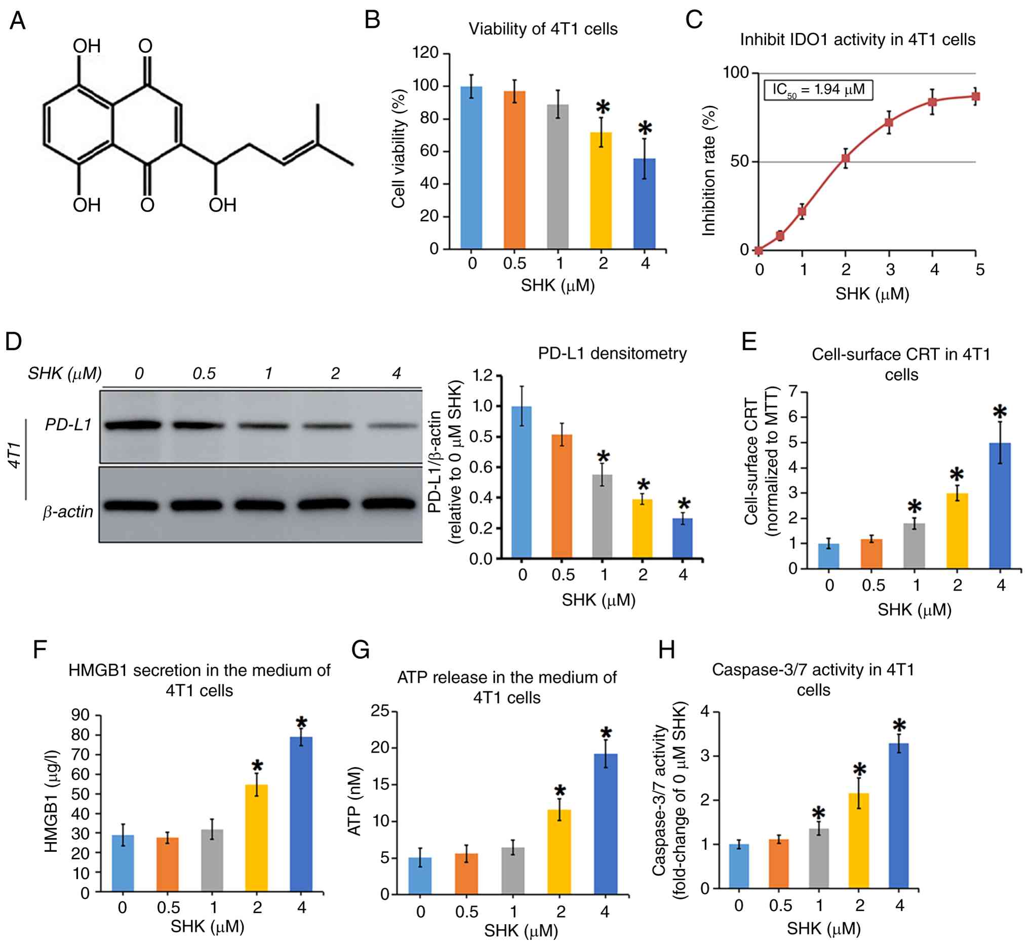 | Figure 2In vitro effects of different
concentrations of free SHK on cell viability, IDO1 activity, PD-L1
expression and ICD-associated damage signals in 4T1 cells. (A) The
chemical structure of SHK. (B) Cell viability of 4T1 cells treated
with increasing concentrations of free SHK for 24 h, as determined
by the MTT assay and expressed as percentage of the 0 μM SHK
group. (C and D) Inhibitory effects of SHK at different
concentrations on IDO1 catalytic activity (C) and PD-L1 expression
(D) in 4T1 cells. (E) CRT levels in 4T1 cells after 24 h treatment
with SHK, quantified by a cell-based ELISA under non-permeabilizing
conditions and normalized to cell viability. (F and G)
Extracellular HMGB1 (F) and ATP (G) levels in culture supernatants
of SHK-treated 4T1 cells. (H) Caspase-3/7 activity in 4T1 cells
after 24 h exposure to SHK, expressed as fold-change relative to
the 0 μM SHK group. Data are presented as the mean ± SD (n=3
independent experiments). *P<0.05 vs. 0 μM
SHK. SHK, shikonin; IDO1, indoleamine 2,3-dioxygenase 1; PD-L1,
programmed death-ligand 1; ICD, immunogenic cell death; CRT,
calreticulin; HMGB1, high mobility group box. |
Preparation and characterization of
AuNR-mPECTs and SHK-mPECTs
In the present study, AuNRs were prepared using the
seedless synthesis method (34),
and AuNR-mPECTs were synthesized through the 'ligand substitution
method' based on the authors' previous procedures (33). Compared with AuNRs, AuNR-mPECTs
exhibited a similar characteristic UV-Vis absorption, as revealed
by longitudinal and transverse plasmonic peaks at 785 and 520 nm,
respectively (Fig. 3A). The
longitudinal plasmonic peak was slightly red-shifted, which might
correspond to the surface modification of mPECTs. Dynamic light
scattering showed a dominant hydrodynamic size peak at 50 nm
(PDI=0.642) for AuNRs and 58 nm (PDI= 0.449) for AuNR-mPECTs
(Fig. 3B). The decreased zeta
potential and increased particle size of AuNR-mPECTs confirmed the
successful substitution of positively charged CTAB on the surface
of AuNR-mPECTs (Fig. 3B and C).
The micro-morphology of AuNR-mPECTs was examined by TEM. Notably,
AuNR-mPECTs showed a typical rod structure, with the long and short
diameters of 48±8 and 11±2 nm, respectively (Fig. 3D). The stability of AuNR-mPECTs is
critical for their biomedical applications. To validate their
dispersion stability under physiological conditions (osmotic
pressure, protein and the microenvironment of acid-base
abnormalities in tumors), AuNR-mPECTs were dispersed in 10%
FBS-containing culture medium, NaCl solution (0.1 M) and PBS
solution (pH 7.4 or 6.8), respectively. After 24 h of incubation,
the UV-Vis spectral data of all samples were obtained, and similar
longitudinal and transverse absorption peaks were observed in each
spectrogram (Fig. S3A),
indicating its optical stability in different media. Furthermore,
the stability of AuNR-mPECTs was evaluated by measuring
hydrodynamic diameter after exposure to various media mimicking the
in vivo microenvironment. It was found that the hydrodynamic
diameter of AuNR-mPECTs showed no obvious changes after incubation
with different media (Fig. S3B).
Furthermore, the UV-Vis spectral data or particle size of
AuNR-mPECTs had no significant changes after laser irradiation,
suggsting the excellent photothermal stability of AuNR-mPECTs
(Fig. S3C and D). Additionally,
no significant changes were observed in UV-Vis spectral data and
particle size after 10 days of storage, indicating AuNR-mPECTs have
favorable storage stability (Fig.
S3E and F). In addition to the aforementioned results, the
surface coating with mPECTs could greatly reduce the toxicity of
AuNRs, and no significant cytotoxicity of AuNR-mPECTs was observed,
even at 50 μg/ml (Fig. S4A
and B).
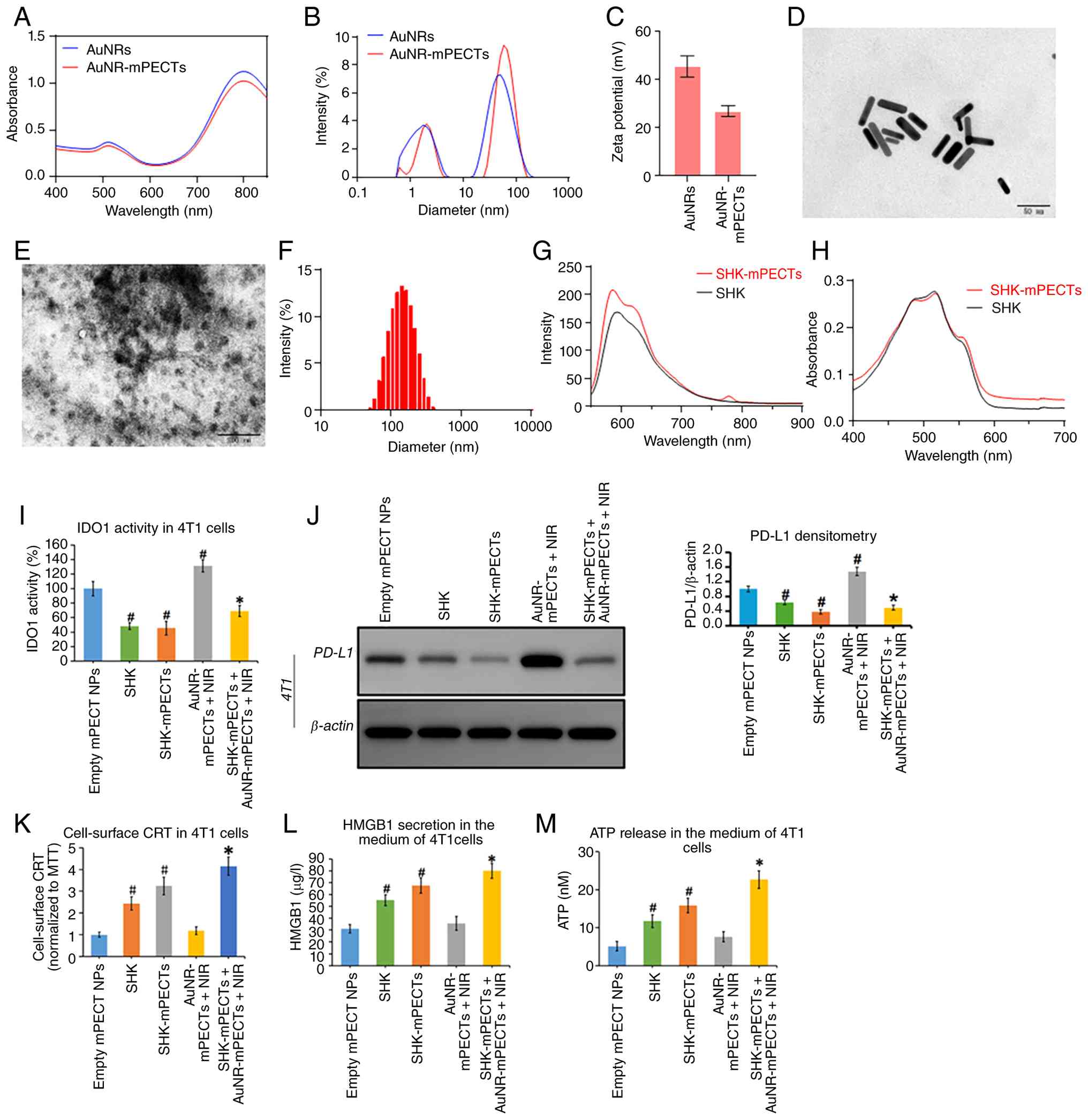 | Figure 3(A-D) UV-Vis absorption spectra (A),
intensity-weighted hydrodynamic size distributions (dynamic light
scattering) (B), zeta potentials (C) and TEM images (D) of
AuNR-mPECTs. (E-H) TEM image (E), size and distributions (F),
fluorescence spectrum (G) and UV-Vis absorption spectrum (H) of
SHK-mPECTs. (I-M) The 4T1 cells were treated with free SHK (2
μM), SHK-mPECTs (the concentration of loaded SHK is equal to
2 μM), mild PTT (43°C, 15 min) generated by AuNR-mPECTs (50
μg/ml) under NIR irradiation (808 nm, heating power: 1.5
W/cm2, maintaining power: 0.5 W/cm2), or
SHK-mPECTs combined with AuNR-mPECTs + NIR-induced mild PTT (43°C,
15 min). After 24 h of continuous culture, IDO1 activity (I), PD-L1
expression (J), cell-surface CRT levels (K), HMGB1 secretion (L),
and ATP release into the supernatant (M) were determined in each
treatment group. Mean ± SD. #P<0.05 vs. empty
mPECT-NPs CON group, *P<0.05 vs. AuNR-mPECTs + NIR
group. TEM, transmission electron microscopy; AuNRs, gold nanorods;
PTT, photothermal therapy; CRT, calreticulin; HMGB1, high mobility
group box. |
SHK, which is a hydrophobic drug, can be
encapsulated in the hydrophobic core of mPECT nanoparticles through
hydrophobic interaction to form drug-loaded SHK-mPECT nanoparticles
(SHK-mPECTs). TEM images showed that SHK-mPECTs had a uniform
spherical structure (Fig. 3E).
The hydrodynamic diameter of SHK-mPECTs was ~141 nm with a narrow
distribution (PDI=0.262) (Fig.
3F). Fluorescence and UV-Vis absorption characterization
revealed that loading SHK into nanoparticles did not affect the
optical properties of SHK compared with bare SHK at the same
concentration, suggesting that the loading of SHK into
nanoparticles did not affect the chemical properties of SHK
(Fig. 3G and H). Moreover, the
effects of different nano-components on IDO1 activity, PD-L1
expression and ICD induction were further assessed. As demonstrated
in Fig. 3I-M, compared with free
SHK, SHK-mPECTs showed the same or even slightly stronger effects
on inhibiting IDO1 activity (Fig.
3I), downregulating PD-L1 expression (Fig. 3J) and inducing ICD (Fig. 3K-M). Mild PTT stimulation
significantly increased IDO1 activity and PD-L1 expression
(Fig. 3I and J), but the ability
of mild PTT to induce ICD was weak (Fig. 3K-M). However, the combination of
mild PTT with SHK-mPECTs significantly reversed the increases in
IDO1 activity and PD-L1 expression induced by mild PTT and strongly
induced ICD in 4T1 cells (Fig.
3I-M). Similar trends were also detected in human TNBC
MDA-MB-231 cells (Fig. S5A-E).
To further exclude the potential effects of the blank nanocarrier
and NIR irradiation itself, a focused two-group control experiment
was performed in 4T1 cells. Empty mPECT-NPs + NIR caused only a
slight temperature increase and did not generate controllable mild
photothermal heating. In addition, compared with empty mPECT-NPs,
empty mPECT-NPs + NIR did not significantly alter IDO1 activity,
PD-L1 mRNA expression, or ICD-associated markers, including CRT,
HMGB1 and ATP (Fig. S6A-F).
Preparation and characterization of the
nano-composite hydrogel co-constructed with AuNR@mPECTs and SHK@
mPECTs (SHK/AuNR@gel)
After adding α-CD into AuNR-mPECTs and SHK-mPECTs,
the mixture solution was converted into a claret-red homogeneous
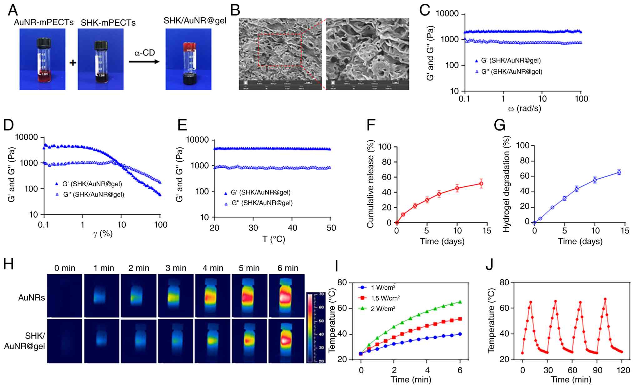
hydrogel at the macroscopic level (Fig. 4A). A homogeneous network structure
was obviously observed in the SEM images of SHK/AuNR@gel (Fig. 4B). The rheological properties of
SHK/AuNR@gel were evaluated subsequently. As shown in Fig. 4C, although the angular frequency
was increased, the storage modulus G' of SHK/AuNR@gel was always
higher than the loss modulus G', suggesting that an elastic
solid-like hydrogel might be formed. Shear thinning is an essential
property for realizing the injectability of a hydrogel. As revealed
in Fig. 4D, with the increase of
the shear strain, SHK/AuNR@ gel exhibited a non-Newtonian shear
thinning (pseudoplastic) behavior, which indicated that
SHK/AuNR@gel was injectable. During the photothermal process, the
stability is crucial for avoiding the unnecessary burst release of
drugs loaded in the hydrogel at the higher treatment temperatures.
As demonstrated in Fig. 4E, with
the rising temperatures, the storage modulus G' of SHK/AuNR@gel was
always higher than the loss modulus G', suggesting that
SHK/AuNR@gel could maintain a stable gelling state at a relatively
higher treatment temperature (≤50°C). In addition, the particle
size distribution of SHK-mPECTs was similar within the temperature
range of 25-60°C, suggesting that SHK-mPECT NPs could remain stable
without disassembly or aggregation during hyperthermia treatment
(Fig. S7). Subsequently, the
release behaviors of SHK from SHK/AuNR@gel were investigated.
Notably, the release of SHK in SHK/AuNR@ gel showed a zero-order
release curve with no obvious burst release phenomenon (Fig. 4F), indicating that SHK/AuNR@ gel
could maintain a controlled, sustainable release behavior.
Furthermore, the degradation curve of SHK/AuNR@gel was similar to
the release curve of SHK, suggesting that the release of SHK mainly
depended on the dissolution and degradation of SHK/AuNR@gel
(Fig. 4F and G).
Photothermal conversion efficiency (PCE) is
important for the application of hydrogels in PTT. As demonstrated
in Fig. 4H, the temperature of
AuNRs and SHK/AuNR@gel both rose rapidly to ~60°C within only 6 min
under the irradiation of 808 nm NIR, indicating that loading AuNRs
into SHK/AuNR@gel did not obviously affect the PCE of AuNRs. The
photothermal conversion properties of SHK/AuNR@gel were highly
dependent on the irradiated power of the laser (Fig. 4I). In other words, the higher the
power irradiated on SHK/AuNR@gel, the faster the temperature rose.
To explore the stability of photothermal conversion, SHK/AuNR@gel
was irradiated with an 808 nm laser for 10 min at 2.0
W/cm2 and then cooled to room temperature; this laser
on/off process involved a total of 4 cycles. As demonstrated in
Fig. 4J, the peak temperatures
did not drop significantly during the whole process, suggesting
that SHK/AuNR@gel had favorable stability of photothermal
conversion even after the long-term and repeated laser irradiation.
Altogether, these results indicate that SHK/AuNR@gel has excellent
efficiency and stability for photothermal conversion.
Antitumor effect of SHK/AuNR@gel + NIR
treatment on local tumors
Based on previous studies (25,26), BALB/c mice bearing 4T1 tumor cells
were chosen as the immune 'cold' TNBC model for in vivo
study. To further verify the previous in vitro findings, the
effects of hydrogel with different compositions on IDO1 activity,
PD-L1 expression and ICD were first detected in local tumors.
Because SHK@ gel does not contain AuNRs, NIR irradiation of SHK@gel
alone was not expected to generate controllable mild photothermal
heating under the present experimental conditions. Therefore,
SHK@gel mainly represented the effect of local sustained SHK
release, whereas SHK/AuNR@gel + NIR represented the combination of
hydrogel-mediated sustained SHK release and AuNR-mediated mild PTT.
The detailed experimental process is shown in Fig. 5A. The activity of IDO1 could be
indirectly reflected by the ratio of Kyn/Trp in tumor tissues
(60). As shown in Fig. 5B-D, intratumoral injection of free
SHK failed to reduce the Kyn/Trp ratio or PD-L1 expression, while
the hydrogel-loaded SHK (SHK@ gel) significantly reduced the ratio
of Kyn/Trp (Fig. 5B) and
expression of PD-L1 (Fig. 5C and
D) in local tumors. It can be inferred that free SHK does not
have a retention capacity after intratumoral injection, and it may
quickly diffuse to the surrounding tissues or enter the systemic
circulation, thus losing its direct and lasting effects on local
tumors. Consistent with previous studies (18,61), the repeated stimulation of mild
PTT (AuNR@gel + NIR) obviously increased the Kyn/Trp ratio and
PD-L1 expression. However, the combination of mild PTT with SHK
(SHK/AuNR@gel + NIR) almost completely reversed these phenomena
(Fig. 5B-D). In Fig. 5E, it is demonstrated that the
effect of AuNR@gel + NIR-mediated mild PTT on ICD induction was not
obvious, while SHK@ gel could significantly induce ICD, and the
effect on ICD induction was further enhanced when combined with
mild PTT (SHK/AuNR@gel + NIR). Furthermore, SHK/AuNR@ gel + NIR
almost completely inhibited the growth of the tumor (Fig. 5F and G) and significantly
prolonged the survival time of tumor-bearing mice (Fig. 5H). All these results indicate that
SHK/AuNR@gel + NIR not only attenuates the increases in Kyn/Trp and
PD-L1 expression induced by mild PTT but also has strong abilities
to induce ICD and inhibit tumor growth.
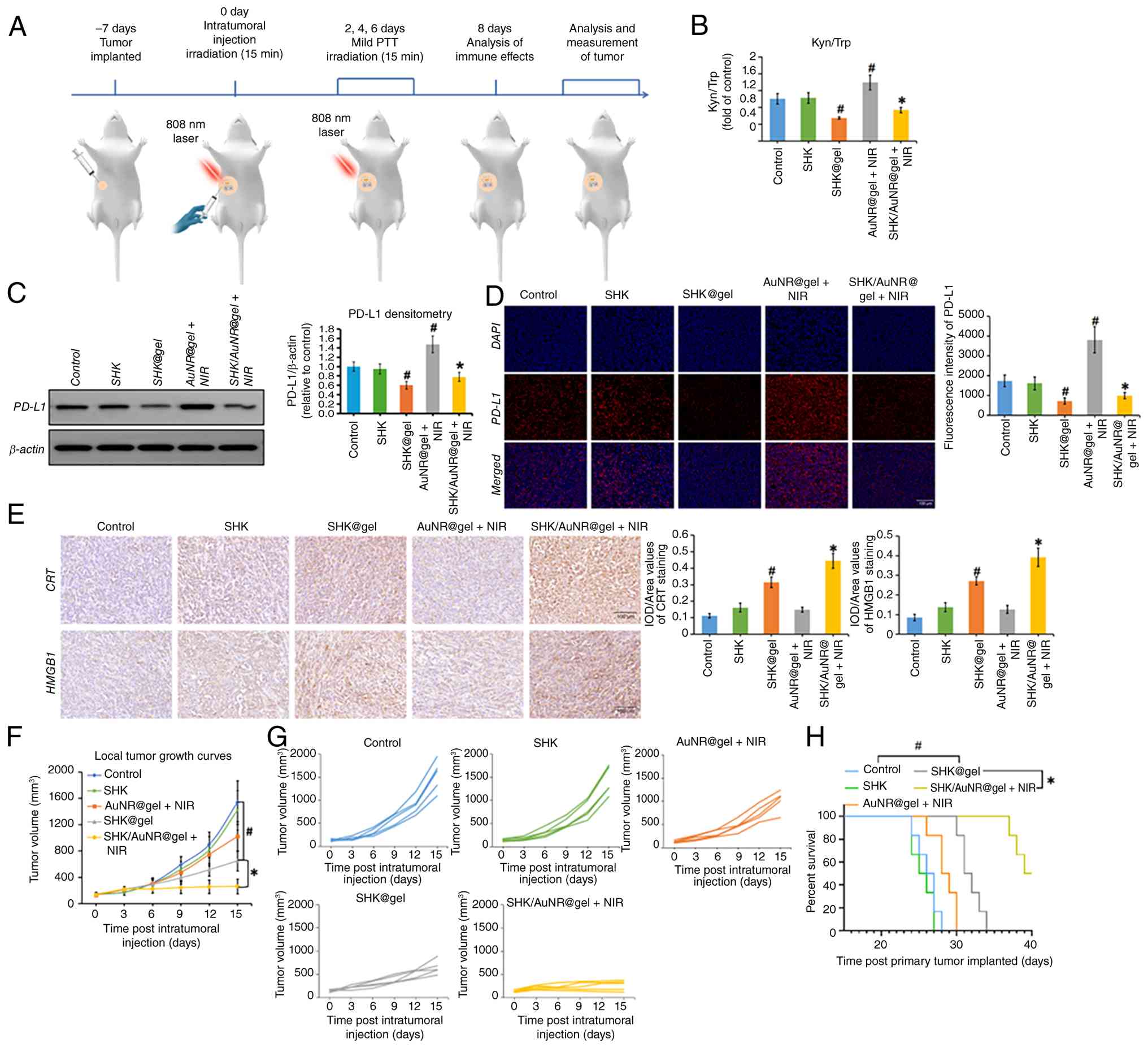 | Figure 5Antitumor effects of SHK/AuNR@gel +
NIR on the local tumors. (A) Schematic illustration of the study
design. (B) Kyn/Trp ratio in the local tumor tissue after different
treatments (n=4). (C and D) The protein expression of PD-L1 was
determined by western blotting (C, n=3) and immunofluorescence (D,
n=3), respectively. (E) Representative Immunohistochemical images
and quantification of CRT and HMGB1 positive areas in tumors
(IOD/area value, n=3). Mean ± SD, #P<0.05 vs. Control
group, *P<0.05 vs. AuNR@gel + NIR group. (F) Local
TGCs with the average tumor volume in tumor-bearing mice after the
indicated treatments (n=5). (G) The TGCs of each mouse in different
treatment groups. (H) Survival curves of mice after the indicated
treatments (n=5). Mean ± SD. #P<0.05 vs. Control
group, *P<0.05 vs. SHK@gel group. SHK, shikonin;
AuNRs, gold nanorods; NIR, near-infrared irradiation; PD-L1,
programmed death-ligand; CRT, calreticulin; HMGB1, high mobility
group box; TGCs, tumor growth curves. |
SHK/AuNR@gel + NIR treatment induces
potent adaptive antitumor immunity and converts immune-cold tumors
into immune-hot tumors
To characterize the antitumor immune response
elicited by the hydrogel-mediated local SHK delivery combined with
mild PTT, a comprehensive analysis of immune-cell activation and
cytokine production was performed in TBM (Fig. 6). Within TDLNs, treatment with
SHK/AuNR@gel + NIR significantly increased the DC maturation score,
defined as the mean z-score of *H2-Ab1*, Cd80, Cd86,
Il12a and Il12b expression levels (Fig. 6A and B). This finding indicates
enhanced antigen-presenting capacity and improved priming of
Th1-oriented immune responses (43,44). Consistent with the pivotal role of
IL-12 in promoting Th1 differentiation and IFN-γ production
(44), IL-12p70 levels in TDLN
homogenates were also significantly elevated following SHK/AuNR@gel
+ NIR treatment (Fig. 6C).
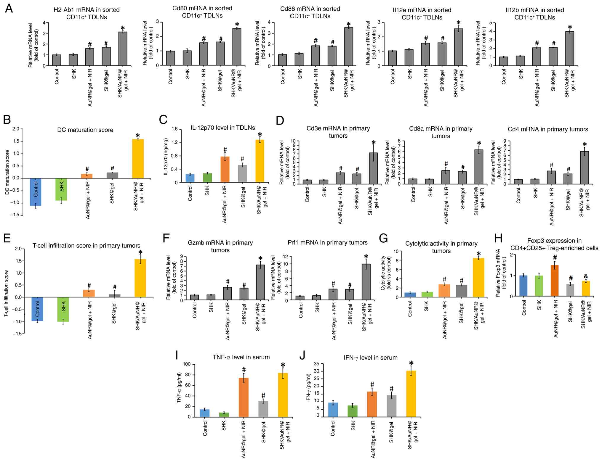 | Figure 6SHK/AuNR@gel + NIR treatment induced
a strong immune response. The treatment details were given in
Fig. 5A. Ipsilateral inguinal and
axillary TDLNs and primary tumors were collected to quantify the
immune cascade from upstream priming to intratumoral effector
dominance. (A-C) DC maturation. (A) qPCR for H2-Ab1, Cd80, Cd86,
Il12a, Il12b in sorted CD11c+ TDLN cells; (B) DC
maturation score (z-mean of the five markers from-ΔCt); (C)
Tissue-level IL-12p70 in TDLN homogenates (ELISA; ng per mg total
protein). (D-G) T-cell infiltration and CYT in primary tumors. (D)
qPCR for Cd3e, Cd8a and Cd4; (E) T-cell infiltration
score (z-mean of the three genes). (F) qPCR for Gzmb and
Prf1; (G) CYT score (geometric mean of Gzmb/Prf1 on the log
scale). (H) Foxp3 mRNA expression in
CD4+CD25+ Treg-enriched cells magnetically
enriched from primary tumor-infiltrating lymphocytes;
#P<0.05 vs. Control; &P<0.05 vs.
AuNR@gel + NIR. (I and J) Serum levels of TNF-α and IFN-γ after
treatment for 8 days. Unless otherwise specified, data are
presented as the mean ± SD (n=4 mice/group). #P<0.05
vs. Control group, *P<0.05 vs. SHK@gel group. SHK,
shikonin; AuNRs, gold nanorods; NIR, near-infrared irradiation;
TDLNs, tumor-draining lymph nodes; DC, dendritic cell; CYT,
cytolytic activity. |
Analysis of the primary tumors revealed a
pronounced increase in T-cell infiltration in the SHK/AuNR@gel +
NIR group, as reflected by a significantly higher T-cell
infiltration index based on the combined expression of Cd3e,
Cd8a and Cd4 (Fig. 6D
and E) (45). Moreover, the
CYT score, calculated as the geometric mean of Gzmb and
Prf1 expression on a logarithmic scale, was substantially
increased (Fig. 6F and G)
(46), indicating enhanced
effector T-cell functionality within the TME. To further assess
Treg-associated immunosuppressive features, CD4+ TILs
were first enriched from primary tumor tissues, followed by CD25
surface labeling-based magnetic enrichment and Foxp3 qPCR analysis
(62). AuNR@gel + NIR treatment
increased Foxp3 expression in the CD4+CD25+
Treg-enriched fraction, whereas SHK@gel and SHK/AuNR@ gel + NIR
reduced this Foxp3 signal (Fig.
6H). Together with the increased T-cell infiltration and CYT
score, these findings suggest that SHK/AuNR@gel + NIR treatment
promoted effector T-cell activity while attenuating CD25-enriched
Foxp3-associated immunosuppressive features in the primary TME.
Finally, levels of the Th1-associated cytokines TNF-α and IFN-γ
were significantly elevated in both tumor tissues and serum from
mice treated with SHK/AuNR@gel + NIR (Fig. S8A and B; Fig. 6I and J). These findings are
indicative of a robust systemic Th1-polarized immune response
(44). Taken together, these data
demonstrate that SHK/AuNR@gel + NIR effectively remodels the
immunosuppressive TME and instigates a potent, systemic adaptive
immune response.
Evaluation of the therapy safety
Previous studies showed that SHK was injected
intraperitoneally every other day for 3 weeks, and no apparent
toxic effect was found at the dose of 5.0 mg/kg BW (36,63). In the present study, 5.0 mg/kg
were also used as the dosage, but only once (through intratumoral
injection). Free SHK, or an equivalent dose of SHK@gel
(CSHK=2 mg/ml, 50 μl) and SHK/AuNR@gel
(CSHK=2 mg/ml, CAuNR=50 μg/ml, 50
μl) were intratumorally injected into the tumors. The
concentrations of SHK in tumor tissues and plasma were quantified
by HPLC analysis (Fig. 7A and B).
As shown in Fig. 7A, after
intratumoral injection, the concentration of free SHK in the tumor
rapidly dropped to the trace level in a short time. On the
contrary, the concentrations of SHK in the tumor of the two
hydrogel groups (SHK@gel or SHK/AuNR@ gel) retained at high levels
for a longer time, indicating that the hydrogel could realize the
sustained release of SHK and maintain a high concentration in local
tumors for a long-lasting time. It should be noted that the local
temperature rise mediated by NIR light has no obvious effect on the
release profile of SHK from the hydrogel (SHK/AuNR@gel + NIR).
After a single intratumoral injection, free SHK rapidly entered the
blood circulation, resulting in a sudden increase in SHK in the
blood and then a rapid decrease (Fig.
7B). On the contrary, the concentrations of SHK in the blood of
the two hydrogel groups did not show this trend and constantly
remained at a relatively low level. These findings indicated that
the mPECT hydrogel used in the present study can achieve the
sustained release of SHK, maintain a relatively high concentration
of SHK in the local tumor for a long time, while preventing SHK
from rushing into the blood circulation to cause potential damage
to normal tissues. In addition, no abnormal changes in blood
routine and hepatorenal functions were found among different
treatment groups (Fig. 7C and D),
and the BW of mice was also not affected during these therapies
(Fig. 7E). Furthermore, H&E
analysis revealed that no obvious histological damage was observed
in the main organs, such as heart, liver, spleen, lung and kidney,
after various treatments (Fig.
7F).
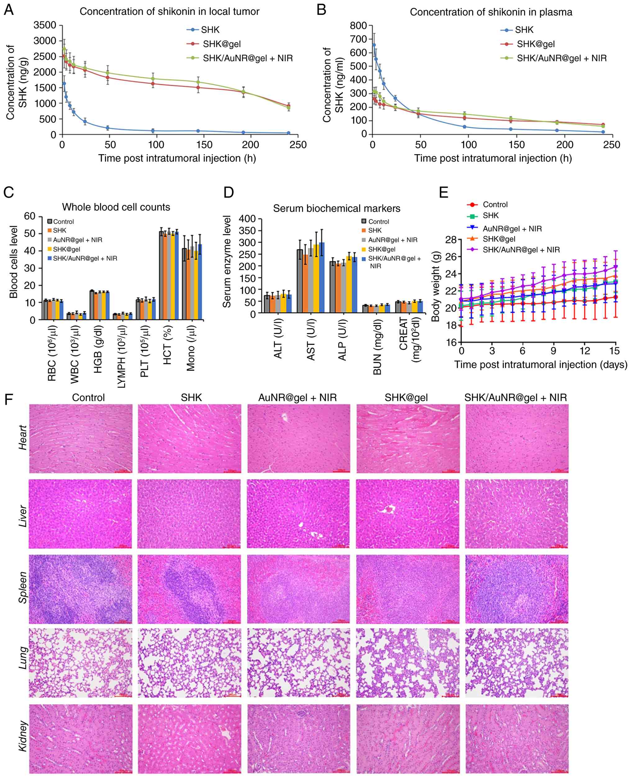 | Figure 7Biosafety evaluation in vivo.
The treatment details are given in Fig. 5A. (A and B) HPLC analysis of SHK
concentrations in tumors (A, n=3) and plasma (B, n=3) after
injection with free SHK, SHK@gel, or SHK/AuNR@gel (dose of SHK is
unified as 5 mg/kg BW). (C and D) Statistical analysis of whole
blood cell counts (C, n=5) and serum biochemical markers (D, n=5)
of TBM after various treatments. All indicators were within normal
biological ranges. (E) The averaged mouse BW profile of each group
during therapies (n=5). (F) Representative images of H&E
morphological analysis of the main organs of TBM after the
indicated treatments (Magnification, 200×; n=3). SHK, shikonin; BW,
body weight; AuNRs, gold nanorods; TBM, tumor-bearing mice. |
SHK/AuNR@gel + NIR induces a systemic
abscopal effect against untreated distal tumors
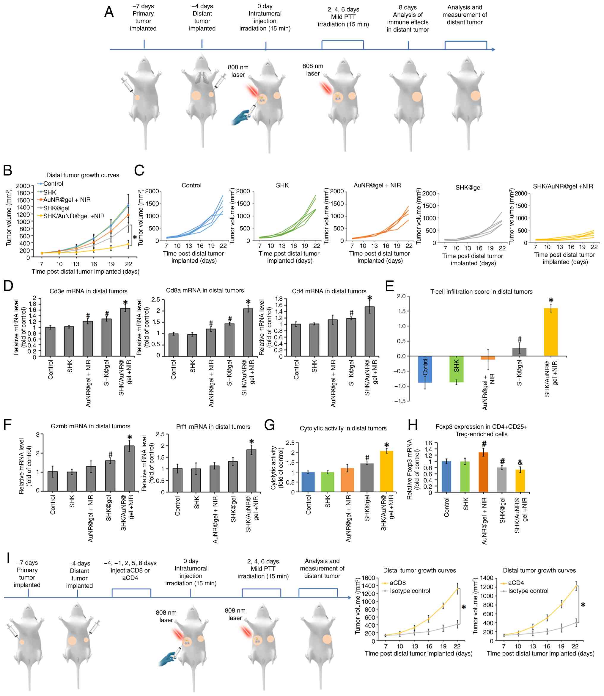
To determine whether localized SHK/AuNR@gel + NIR
therapy could generate systemic antitumor immunity, a bilateral TBM
model was established, in which primary tumors were implanted in
the right mammary fat pad and secondary (distant) tumors were
implanted in the contralateral mammary fat pad three days later
(Fig. 8A). Once the primary
tumors reached ~150 mm3, mice received intratumoral
injections of the indicated formulations or SHK/AuNR@gel + NIR,
followed by NIR irradiation where applicable. Notably, treatment
with SHK/AuNR@gel + NIR resulted in pronounced inhibition of distal
tumor growth compared with all other groups (Fig. 8B and C). To exclude the
possibility that this effect was simply attributable to leakage of
SHK from the treated primary tumor and subsequent systemic drug
distribution, SHK concentrations in distant tumors were quantified
by HPLC following a single intratumoral administration. As revealed
in Fig. S9, SHK levels in
untreated distant tumors remained extremely low and declined
rapidly over time in all treatment groups, remaining well below
concentrations associated with direct cytotoxicity. This abscopal
effect was accompanied by substantial immune activation within the
untreated distal tumors. qPCR analysis revealed significant
upregulation of Cd3e, Cd8a and Cd4 in distal tumors
from the SHK/AuNR@gel + NIR group (Fig. 8D), resulting in a significantly
elevated T-cell infiltration score (Fig. 8E). Similarly, expression levels of
the cytolytic effector molecules Gzmb and Prf1 were
increased (Fig. 8F), leading to a
corresponding increase in the composite CYT score (Fig. 8G). Furthermore, Treg-associated
immunosuppressive features in untreated distant tumors were
assessed using CD25 surface labeling-based magnetic enrichment of
CD4+ TILs. Similar to the primary tumors, AuNR@gel + NIR
treatment increased Foxp3 expression in the
CD4+CD25+ Treg-enriched fraction, whereas
SHK/AuNR@gel + NIR reduced this Foxp3 signal (Fig. 8H). These findings, together with
the increased T-cell infiltration and CYT score in distant tumors,
support a shift toward a more favorable effector-dominant immune
landscape after the combined local treatment. To functionally
validate the contribution of T-cell subsets to the abscopal
response, in vivo depletion studies were performed.
Depletion of either CD8+ or CD4+ T cells
substantially attenuated the ability of SHK/AuNR@gel + NIR
treatment to control distant tumor growth (Fig. 8I), demonstrating that both T-cell
populations are required for the generation of effective systemic
antitumor immunity.
SHK/AuNR@gel + NIR treatment elicits
potent and polyfunctional antitumor immune memory
Having established that SHK/AuNR@gel + NIR
treatment could suppress both tumor rechallenge and experimental
lung metastasis (Fig. 9A-C; and
Fig. S10A-C), it was next sought
to characterize the functional properties of the long-term immune
memory generated by this therapeutic strategy. Splenic T cells were
isolated from treated mice 1 day after intravenous challenge with
4T1 cells, as depicted in Fig.
9A, and subjected to a comprehensive panel of ex vivo
functional analyses. The antigen specificity of the memory T-cell
response was first evaluated. Using an IFN-γ enzyme-linked
immunospot (ELISpot) assay, it was found that T cells isolated from
SHK/AuNR@gel + NIR-treated mice exhibited a significantly stronger
response to 4T1 tumor lysates than those from all other treatment
groups, as reflected by a significantly higher number of SFU per
105 T cells (Fig. 9D).
The cytotoxic effector function of these memory T cells was next
assessed. CD8+ T cells were enriched from the
splenocytes and co-cultured with irradiated 4T1 target cells at
multiple effector-to-target (E:T) ratios in a short-term LDH
release assay. CD8+ T cells derived from the
SHK/AuNR@gel + NIR group displayed potent, dose-dependent tumor
cell killing across all E ratios tested. Consistent with this
observation, the overall cytotoxic capacity, quantified as the area
under the dose-response curve (AUC), was significantly greater than
that observed in all control groups (Fig. 9E).
![SHK/AuNR@gel + NIR treatment induces
a potent and systemic antitumor immune memory. (A) Schematic
diagram of the experimental timeline for assessing lung metastasis.
(B) Representative images (upper) and quantitative analysis (lower)
of metastatic lung nodules in different groups (n=4). (C)
Representative H&E-stained sections of lung tissues. Scale
bars, 500 μm. (D) Antigen-specific T cell response. The
frequency of IFN-γ-producing T cells in response to 4T1 tumor
lysates was measured by ELISpot (antigen-presenting cells:
magnetically isolated CD11c+ DCs pulsed with 4T1 lysates
for 2-4 h and thoroughly washed; APC: T=1:10) and is presented as
spot-forming units per 105 T cells (n=4). (E) Cytotoxic
capacity of memory CD8+ T cells. The tumor-eliminating
activity of enriched CD8+ T cells against 4T1 target
cells was assessed by an LDH release assay at the indicated
effector-to-target (E: T) ratios. The overall cytotoxicity is
summarized by the AUC (n=4). (F) Relative mRNA expression of Gzmb
and Prf1 in enriched CD8+ T cells (n=4). (G)
Degranulation capacity upon antigen-specific stimulation, presented
as the percentage of specific granule release
[supernatant/(supernatant + cell lysate) ×100%] (n=4). (H)
Polyfunctional T cell response. The frequency of T cells
simultaneously producing IFN-γ and TNF-α upon antigen recall was
determined by dual-color ELISpot and is shown as double-positive
spots per 105 T cells (n=4). (I) Comprehensive recall
cytokine signature. A composite recall index was calculated from
the levels of multiple cytokines (IFN-γ, TNF-α, IL-2, CXCL9 and
CXCL10) in the supernatant of re-stimulated untouched T cells
co-cultured with pulsed CD11c+ DCs (n=4). All data are
presented as the mean ± SD. #P<0.05 vs. the Control
group, *P<0.05 vs. the SHK@gel group. SHK, shikonin;
AuNRs, gold nanorods; NIR, near-infrared irradiation; LDH, lactate
dehydrogenase; AUC, area under the dose-response curve; Gzmb,
granzyme B; Prf1, perforin 1; DCs, dendritic cells.](/article_images/ijo/69/4/ijo-69-04-05930-g08.jpg) | Figure 9SHK/AuNR@gel + NIR treatment induces
a potent and systemic antitumor immune memory. (A) Schematic
diagram of the experimental timeline for assessing lung metastasis.
(B) Representative images (upper) and quantitative analysis (lower)
of metastatic lung nodules in different groups (n=4). (C)
Representative H&E-stained sections of lung tissues. Scale
bars, 500 μm. (D) Antigen-specific T cell response. The
frequency of IFN-γ-producing T cells in response to 4T1 tumor
lysates was measured by ELISpot (antigen-presenting cells:
magnetically isolated CD11c+ DCs pulsed with 4T1 lysates
for 2-4 h and thoroughly washed; APC: T=1:10) and is presented as
spot-forming units per 105 T cells (n=4). (E) Cytotoxic
capacity of memory CD8+ T cells. The tumor-eliminating
activity of enriched CD8+ T cells against 4T1 target
cells was assessed by an LDH release assay at the indicated
effector-to-target (E: T) ratios. The overall cytotoxicity is
summarized by the AUC (n=4). (F) Relative mRNA expression of Gzmb
and Prf1 in enriched CD8+ T cells (n=4). (G)
Degranulation capacity upon antigen-specific stimulation, presented
as the percentage of specific granule release
[supernatant/(supernatant + cell lysate) ×100%] (n=4). (H)
Polyfunctional T cell response. The frequency of T cells
simultaneously producing IFN-γ and TNF-α upon antigen recall was
determined by dual-color ELISpot and is shown as double-positive
spots per 105 T cells (n=4). (I) Comprehensive recall
cytokine signature. A composite recall index was calculated from
the levels of multiple cytokines (IFN-γ, TNF-α, IL-2, CXCL9 and
CXCL10) in the supernatant of re-stimulated untouched T cells
co-cultured with pulsed CD11c+ DCs (n=4). All data are
presented as the mean ± SD. #P<0.05 vs. the Control
group, *P<0.05 vs. the SHK@gel group. SHK, shikonin;
AuNRs, gold nanorods; NIR, near-infrared irradiation; LDH, lactate
dehydrogenase; AUC, area under the dose-response curve; Gzmb,
granzyme B; Prf1, perforin 1; DCs, dendritic cells. |
To further elucidate the mechanisms underlying the
enhanced cytotoxic activity of memory T cells, the expression of
key cytolytic effector molecules was analyzed, and degranulation
capacity was assessed. qPCR analysis revealed that the enriched
CD8+ T cells from the SHK/AuNR@gel + NIR group expressed
significantly higher levels of Gzmb and Prf1
(Fig. 9F). Consistent with this
finding, antigen-restimulated CD8+ T cells from the same
group exhibited a significantly greater degree of degranulation, as
reflected by the release of these cytolytic effectors upon
antigen-specific stimulation (Fig.
9G). Beyond cytotoxic potency, the protective efficacy of
memory T cells is strongly influenced by their functional quality,
particularly their capacity for polyfunctional cytokine production.
To evaluate this property, a dual-color ELISpot assay was employed
to quantify T cells capable of simultaneously producing IFN-γ and
TNF-α following antigen recall. The frequency of these
polyfunctional T cells, expressed as double-positive SFU per
105 T cells, was significantly increased in the
SHK/AuNR@gel + NIR group compared with all other treatment groups
(Fig. 9H). To obtain a broader
overview of the recall immune response, a panel of cytokines and
chemokines in the supernatants of antigen-restimulated splenocytes
was further quantified. A composite recall-response index was
calculated as the mean z-score of IFN-γ, TNF-α, IL-2, CXCL9 and
CXCL10 levels. Notably, the SHK/AuNR@gel + NIR group exhibited the
highest recall index among all treatment groups (Fig. 9I). Collectively, these findings
demonstrate that a single intratumoral administration of
SHK/AuNR@gel combined with mild PTT generates a high-quality and
durable antitumor immune memory characterized by strong antigen
specificity, potent cytotoxic activity, enhanced degranulation,
increased T-cell polyfunctionality, and broad cytokine
responsiveness.
Discussion
Conventional systemic chemotherapy for tumors often
causes off-target toxicity in normal tissues. By contrast,
localized drug delivery systems offer several advantages, including
reduced systemic exposure and sustained drug release at the tumor
site, thereby improving therapeutic efficacy (64). Various localized delivery
platforms, such as hydrogels, nanoparticles, liposomes and
micelles, have been investigated. Among these, hydrogels have
attracted considerable attention because of their unique
physicochemical properties. Hydrogels are three-dimensional
networks of crosslinked hydrophilic polymer chains. Injectable
hydrogels, which can form gels in the target area, are widely used
in biomedical applications, including drug and cell delivery and
tissue engineering. In particular, intratumoral drug delivery using
injectable hydrogels has become an area of great interest. These
hydrogels enable sustained and controlled drug release specifically
at the tumor site, thereby minimizing adverse effects caused by
systemic drug exposure. In addition, localized therapy with
injectable hydrogels can help address the poor solubility of
numerous chemotherapeutic agents, reduce the required dose, and
increase the amount of drug reaching the tumor site (64). In the present study, a methoxy
poly(ethylene glycol)-b-poly(ε-caprolactone-co-1,4,
8-trioxa[4.6]spiro-9-undecanone) (mPECT) supramolecular hydrogel
was used, which showed excellent photothermal conversion
performance, stable drug-release characteristics that were not
affected by temperature changes, and favorable biosafety (33). This supramolecular hydrogel
effectively supported the objectives of the present study.
ICD is a form of regulated cell death in which
dying tumor cells release or expose immunostimulatory danger
signals and tumor-associated antigens, thereby promoting
DC-mediated antigen presentation and subsequent T-cell priming
(8). An increasing number of
anticancer therapies have been reported to induce ICD and thereby
enhance antitumor immunity. However, numerous ICD-inducing
treatments, including radiotherapy (65), PTT (18,61), and chemotherapeutic agents such as
paclitaxel (66), doxorubicin
(67) and oxaliplatin (68), can simultaneously activate
compensatory immunosuppressive mechanisms. In particular, these
therapies have been shown to increase PD-L1 expression and/or
enhance IDO1 activity during the process of ICD induction.
Therefore, they often need to be combined with PD-1/PD-L1
antibodies or IDO1 inhibitors to promote and maintain the
long-lasting antitumor immune response. By contrast, in the present
study, SHK enhanced ICD-associated signals while attenuating PD-L1
expression and IDO1 activation, suggesting a therapeutically
favorable profile that may help counteract immunosuppressive
feedback during ICD induction. In line with this functional
profile, SHK reduced NF-κB transcriptional activity and inhibited
the nuclear translocation of NF-κB p65, which may contribute, at
least in part, to PD-L1 downregulation, considering the reported
involvement of NF-κB signaling in PD-L1 regulation. In addition,
the observed inhibition of IDO1 activity is consistent with
previous evidence suggesting that SHK inhibits IDO1 catalytic
activity through interaction with the ferric-IDO1 enzyme. Further
studies are warranted to more precisely define the direct molecular
events linking SHK to PD-L1 regulation and IDO1 inhibition in this
localized treatment system.
Recent advances have expanded the understanding of
SHK in tumor immunology beyond its conventional direct cytotoxic
effects on tumor cells, highlighting its potential role in
regulated cell death-mediated immune activation. A recent study has
shown that SHK can enhance tumor immunogenicity and improve the
response to immune checkpoint blockade, as indicated by increased
calreticulin membrane exposure and Hsp70 upregulation, as well as
synergistic enhancement of DC activation and CD8+ T-cell
responses when combined with PD-1 blockade in tumor models
(69). In addition, SHK has been
reported to induce ferroptosis through GOT1-mediated
ferritinophagy, accompanied by the release of ICD-associated
signals, including ATP and HMGB1 (70). Nanomedicine-based studies have
further demonstrated that Fe(III)-SHK supramolecular nanomedicines
can function as ICD stimulants and multifunctional immunoadjuvants
for tumor vaccination (71).
Moreover, Fe-SHK metal-phenolic networks have been shown to promote
the generation of autologous tumor cell lysates through ferroptosis
and necroptosis, thereby contributing to personalized in
situ nanovaccine-based antitumor immunity (72). More recently, nanosized SHK has
been reported to synergize with manganese to enhance the
cGAS-STING-mediated interferon response and sensitize tumors to
immunotherapy (73).
These findings suggest that the immunomodulatory
effects of SHK may involve multiple interconnected processes,
including ICD, ferroptosis/necroptosis and cGAS-STING-associated
innate immune activation. The present results are consistent with
these recent advances, as local low-dose SHK delivery enhanced
ICD-associated signals and suppressed mild PTT-induced IDO1/PD-L1
immunosuppressive feedback, thereby promoting systemic antitumor
immunity. However, ferroptosis/necroptosis and cGAS-STING signaling
were not directly examined in the present study. Therefore, future
studies are required to determine whether these regulated cell
death pathways and innate immune signaling events contribute to the
long-term systemic antitumor immunity induced by SHK/AuNR@gel plus
mild PTT.
In recent years, PD-1/PD-L1 monoclonal antibodies
have been approved for the clinical treatment of different types of
malignant tumors and are the most common immunotherapeutic strategy
at present. Previous research has shown that PD-1/PD-L1 antibody
can even cure some patients with advanced tumor metastasis, and its
favorable efficacy is encouraging (74). However, PD-1/PD-L1 antibody
therapy has several disadvantages. Firstly, most solid tumors have
an immunosuppressive microenvironment, lacking a sufficient number
of infiltrating immune cells and belonging to the immune 'cold
tumor', which leads to a low clinical response rate of PD-1/PD-L1
antibody therapy (75). Secondly,
the systemic administration of PD-1/PD-L1 antibody is prone to
producing a series of systemic side effects, which trigger the
body's autoimmune response and induce the immune system to attack
normal tissues. Thirdly, PD-1/PD-L1 antibody therapy is costly,
which may increase the global burden of healthcare expenditures.
Compared with the PD-1/PD-L1 antibody, the present study has the
following potential advantages. Firstly, combining the noninvasive
mild PTT with locally administered SHK can trigger immune
activation and induce ICD, while suppressing PD-L1 expression and
IDO1 activation, thereby contributing to strong and long-lasting
antitumor-specific immunity in immune-cold tumors. Secondly, using
a hydrogel as the medium, SHK, which is administered only once, can
target the local part of the tumor for low-dose sustained release.
This may avoid repeated high concentrations of SHK, which is
similar to intravenous administration, entering the blood
circulation and causing toxic damage to systemic organs. Thirdly,
compared with PD-1/PD-L1 antibody therapy, the cost of SHK-based
treatment is considered to be markedly lower.
Despite the potential of SHK/AuNR@gel plus mild PTT
to induce systemic antitumor immunity, an abscopal effect and
immune memory in the present study, several key barriers remain
before further clinical translation can be considered. First,
although the mPECT hydrogel enabled local retention and sustained
release of SHK in the present system, clinically compatible or
medical-grade formulations will need to be further established.
Future studies should systematically evaluate scalable
manufacturing, batch-to-batch consistency, sterilization
procedures, degradation products, quality control and regulatory
approval pathways (76). Second,
AuNRs functioned as the photothermal conversion component for local
mild PTT in this system; however, their long-term in vivo
fate remains an important biosafety concern, including clearance,
reticuloendothelial system uptake, accumulation in the liver,
spleen and other organs, potential chronic toxicity and
dose-related safety (77). Third,
intratumoral injection may not be suitable for all patients with
TNBC. Its clinical feasibility may be limited by tumor location,
lesion size, lesion depth, accessibility, multifocality and the
availability of image-guided injection, suggesting that this
strategy may be more applicable to accessible or image-guided
injectable local lesions (78).
Fourth, although an abscopal antitumor effect and long-term immune
memory were observed in the mouse models used in the present study,
these preclinical findings are not sufficient to suggest that this
local treatment strategy could directly replace current standard
systemic therapies. A more appropriate translational positioning
may be to develop this approach as a local in situ
immune-priming or immune-sensitizing strategy that could be
rationally integrated with current neoadjuvant chemotherapy or
immune checkpoint blockade in selected patients suitable for
intratumoral intervention (79).
Further formulation optimization, long-term safety evaluation and
clinically relevant studies combining this strategy with standard
treatments are still required to support its further translational
development.
In conclusion, the present study demonstrates that
mPECT hydrogel-mediated local administration of SHK/AuNR@gel
combined with mild PTT can remodel the immunosuppressive TME,
enhance ICD-associated immune activation, and suppress
IDO1/PD-L1-mediated immunosuppressive feedback in immune-cold TNBC
models. This localized combination strategy inhibited primary tumor
growth, induced an abscopal antitumor effect against untreated
distant tumors, and generated durable antitumor immune memory. In
addition, the mPECT hydrogel enabled prolonged local retention and
sustained release of SHK, thereby reducing rapid systemic exposure.
Taken together, these findings provide a preclinical basis for the
further development of localized SHK-based combination strategies,
particularly for therapeutic agents with antitumor potential but
limited by systemic toxicity.
Supplementary Data
Availability of data and materials
The data generated in the present study may be
requested from the corresponding author.
Authors' contributions
TP and FZ conceived and designed the study. ZF and
TP synthesized and characterized the hydrogel. TP and TS performed
the experiments. Jun Jiang assisted with the experiments. FZ
analyzed the data and prepared the figures. TP and TS drafted the
manuscript. FZ revised the manuscript. All authors read and
approved the final version of the manuscript. All authors agree to
be accountable for all aspects of the work and to ensure that
questions related to the accuracy or integrity of any part of the
work are appropriately investigated and resolved. TP and FZ confirm
the authenticity of all the raw data.
Ethics approval and consent to
participate
All animal experiments were approved by the Animal
Ethics Committee of Air Force Medical University (approval no.
20241327; Xi'an, China) and were covered by the Laboratory Animal
Use License SYXK (Shaan) 2024-003. All procedures were performed in
accordance with institutional guidelines and the ARRIVE
guidelines.
Patient consent for publication
Not applicable.
Competing interests
The authors declare that they have no competing
interests.
Abbreviations:
|
SHK
|
shikonin
|
|
PTT
|
photothermal therapy
|
|
ICD
|
immunogenic cell death
|
|
IDO1
|
indoleamine 2,3-dioxygenase 1
|
|
PD-L1
|
programmed death-ligand 1
|
|
TNBC
|
triple-negative breast cancer
|
|
mPECT
|
methoxy poly(ethylene
glycol)-b-poly(ε-caprolacto
ne-co-1,4,8-trioxa[4.6]spiro-9-undecanone)
|
|
AuNRs
|
gold nanorods
|
|
NIR
|
near-infrared irradiation
|
|
DAMPs
|
damage-associated molecular
patterns
|
|
APCs
|
antigen-presenting cells
|
|
CRT
|
calreticulin
|
|
HMGB1
|
high mobility group box 1
|
|
Kyn
|
kynurenine
|
|
Trp
|
tryptophan
|
|
DC
|
dendritic cell
|
|
TILs
|
tumor-infiltrating lymphocytes
|
|
TDLNs
|
tumor-draining lymph nodes
|
|
Treg
|
regulatory T cell
|
|
qPCR
|
quantitative polymerase chain
reaction
|
|
ELISA
|
enzyme-linked immunosorbent assay
|
|
CYT
|
cytolytic activity
|
Acknowledgements
Not applicable.
Funding
The present study was supported by grants from the National
Natural Science Foundation of China (grant no. 82100008), the
Natural Science Foundation of Shaanxi (grant no. 2019JQ889), the
State Key Laboratory of Cancer Biology of China (grant no.
CBSKL201710) and the Foundation of Xi'an Medical University (grant
no. 2018DOC02).
References
|
1
|
Guo C, He J, Song X, Tan L, Wang M, Jiang
P, Li Y, Cao Z and Peng C: Pharmacological properties and
derivatives of shikonin-A review in recent years. Pharmacol Res.
149:1044632019. View Article : Google Scholar : PubMed/NCBI
|
|
2
|
Abdulridha MK, Al-Marzoqi AH, Al-Awsi GRL,
Mubarak SMH, Heidarifard M and Ghasemian A: Anticancer effects of
herbal medicine compounds and novel formulations: A literature
review. J Gastrointest Cancer. 51:765–773. 2020. View Article : Google Scholar : PubMed/NCBI
|
|
3
|
Boulos JC, Rahama M, Hegazy MF and Efferth
T: Shikonin derivatives for cancer prevention and therapy. Cancer
Lett. 459:248–267. 2019. View Article : Google Scholar : PubMed/NCBI
|
|
4
|
Wang R, Yin R, Zhou W, Xu D and Li S:
Shikonin and its derivatives: A patent review. Expert Opin Ther
Pat. 22:977–997. 2012. View Article : Google Scholar : PubMed/NCBI
|
|
5
|
Cheng Z, Li M, Dey R and Chen Y:
Nanomaterials for cancer therapy: Current progress and
perspectives. J Hematol Oncol. 14:852021. View Article : Google Scholar : PubMed/NCBI
|
|
6
|
Li S, Zhang T, Xu W, Ding J, Yin F, Xu J,
Sun W, Wang H, Sun M, Cai Z and Hua Y: Sarcoma-targeting
peptide-decorated polypeptide nanogel intracellularly delivers
shikonin for upregulated osteosarcoma necroptosis and diminished
pulmonary metastasis. Theranostics. 8:1361–1375. 2018. View Article : Google Scholar : PubMed/NCBI
|
|
7
|
Korangath P, Barnett JD, Sharma A,
Henderson ET, Stewart J, Yu SH, Kandala SK, Yang CT, Caserto JS,
Hedayati M, et al: Nanoparticle interactions with immune cells
dominate tumor retention and induce T cell-mediated tumor
suppression in models of breast cancer. Sci Adv. 6:eaay16012020.
View Article : Google Scholar : PubMed/NCBI
|
|
8
|
Ahmed A and Tait SWG: Targeting
immunogenic cell death in cancer. Mol Oncol. 14:2994–3006. 2020.
View Article : Google Scholar : PubMed/NCBI
|
|
9
|
Wang H, Tang Y, Fang Y, Zhang M, Wang H,
He Z, Wang B, Xu Q and Huang Y: Reprogramming tumor immune
microenvironment (TIME) and metabolism via biomimetic targeting
codelivery of shikonin/JQ1. Nano Lett. 19:2935–2944. 2019.
View Article : Google Scholar : PubMed/NCBI
|
|
10
|
Li J, Zhao M, Sun M, Wu S, Zhang H, Dai Y
and Wang D: Multifunctional nanoparticles boost cancer
immunotherapy based on modulating the immunosuppressive tumor
microenvironment. ACS Appl Mater Interfaces. 12:50734–50747. 2020.
View Article : Google Scholar : PubMed/NCBI
|
|
11
|
Guo W, Yao S, Sun P, Yang TB, Tang CP,
Zheng MY, Ye Y and Meng LH: Discovery and characterization of
natural products as novel indoleamine 2,3-dioxygenase 1 inhibitors
through high-throughput screening. Acta Pharmacol Sin. 41:423–431.
2020. View Article : Google Scholar :
|
|
12
|
Nasrollahzadeh Sabet M, Biglari S, Khorram
Khorshid HR and Esmaeilzadeh E: Shikonin ameliorates experimental
autoimmune encephalomyelitis (EAE) via immunomodulatory,
anti-apoptotic and antioxidative activity. J Pharm Pharmacol.
72:1970–1976. 2020. View Article : Google Scholar : PubMed/NCBI
|
|
13
|
Zhou F, Wu S, Song S, Chen WR, Resasco DE
and Xing D: Antitumor immunologically modified carbon nanotubes for
photothermal therapy. Biomaterials. 33:3235–3242. 2012. View Article : Google Scholar : PubMed/NCBI
|
|
14
|
Orosz P, Echtenacher B, Falk W, Rüschoff
J, Weber D and Männel DN: Enhancement of experimental metastasis by
tumor necrosis factor. J Exp Med. 177:1391–1398. 1993. View Article : Google Scholar : PubMed/NCBI
|
|
15
|
Frey B, Weiss EM, Rubner Y, Wunderlich R,
Ott OJ, Sauer R, Fietkau R and Gaipl US: Old and new facts about
hyperthermia-induced modulations of the immune system. Int J
Hyperthermia. 28:528–542. 2012. View Article : Google Scholar : PubMed/NCBI
|
|
16
|
Li Y, He L, Dong H, Liu Y, Wang K, Li A,
Ren T, Shi D and Li Y: Fever-inspired immunotherapy based on
photothermal CpG nanotherapeutics: The critical role of mild heat
in regulating tumor microenvironment. Adv Sci (Weinh).
5:17008052018. View Article : Google Scholar : PubMed/NCBI
|
|
17
|
Payne M, Bossmann SH and Basel MT: Direct
treatment versus indirect: Thermo-ablative and mild hyperthermia
effects. Wiley Interdiscip Rev Nanomed Nanobiotechnol.
12:e16382020. View Article : Google Scholar : PubMed/NCBI
|
|
18
|
Peng J, Xiao Y, Li W, Yang Q, Tan L, Jia
Y, Qu Y and Qian Z: Combined photothermal therapy and
immunotherapy: Photosensitizer micelles together with IDO inhibitor
enhance cancer photothermal therapy and immunotherapy (Adv. Sci.
5/2018). Adv Sci (Weinh). 5:18700312018. View Article : Google Scholar :
|
|
19
|
Liu X, Su Q, Song H, Shi X, Zhang Y, Zhang
C, Huang P, Dong A, Kong D and Wang W: PolyTLR7/8a-conjugated,
antigen-trapping gold nanorods elicit anticancer immunity against
abscopal tumors by photothermal therapy-induced in situ
vaccination. Biomaterials. 275:1209212021. View Article : Google Scholar : PubMed/NCBI
|
|
20
|
Bai X, Ni J, Beretov J, Graham P and Li Y:
Immunotherapy for triple-negative breast cancer: A molecular
insight into the microenvironment, treatment, and resistance. J
Natl Cancer Cent. 1:75–87. 2021.PubMed/NCBI
|
|
21
|
Kim H, Choi JM and Lee KM: Immune
checkpoint blockades in triple-negative breast cancer: Current
state and molecular mechanisms of resistance. Biomedicines.
10:11302022. View Article : Google Scholar : PubMed/NCBI
|
|
22
|
Li ZL, Zhang HL, Huang Y, Huang JH, Sun P,
Zhou NN, Chen YH, Mai J, Wang Y, Yu Y, et al: Autophagy deficiency
promotes triple-negative breast cancer resistance to T
cell-mediated cytotoxicity by blocking tenascin-C degradation. Nat
Commun. 11:38062020. View Article : Google Scholar : PubMed/NCBI
|
|
23
|
Song X, Zhou Z, Li H, Xue Y, Lu X, Bahar
I, Kepp O, Hung MC, Kroemer G and Wan Y: Pharmacologic Suppression
of B7-H4 glycosylation restores antitumor immunity in immune-cold
breast cancers. Cancer Discov. 10:1872–1893. 2020. View Article : Google Scholar : PubMed/NCBI
|
|
24
|
Keenan TE and Tolaney SM: Role of
immunotherapy in triple-negative breast cancer. J Natl Compr Canc
Netw. 18:479–489. 2020. View Article : Google Scholar : PubMed/NCBI
|
|
25
|
Revuri V, Rajendrakumar SK, Park MS,
Mohapatra A, Uthaman S, Mondal J, Bae WK, Park IK and Lee YK:
Heat-confined tumor-docking reversible thermogel potentiates
systemic antitumor immune response during near-infrared
photothermal ablation in triple-negative breast cancer. Adv Healthc
Mater. 10:e21009072021. View Article : Google Scholar : PubMed/NCBI
|
|
26
|
Wu C, Xu J, Xie Z, Huang H, Li N, Wei X,
Li T, Yang H, Li S, Qin X and Liu Y: Light-responsive hyaluronic
acid nanomicelles co-loaded with an IDO inhibitor focus targeted
photoimmunotherapy against 'immune cold' cancer. Biomater Sci.
9:8019–8031. 2021. View Article : Google Scholar : PubMed/NCBI
|
|
27
|
Wang W, Wu Y, Chen S, Liu X, He J, Wang S,
Lu W, Tang Y and Huang J: Shikonin is a novel and selective IMPDH2
inhibitor that target triple-negative breast cancer. Phytother Res.
35:463–476. 2021. View Article : Google Scholar
|
|
28
|
National Research Council (US) Committee
for the Update of the Guide for the Care Use of Laboratory Animals:
Guide for the care and use of laboratory animals. 8th edition.
Washington (DC): National Academies Press (US); 2011
|
|
29
|
European Parliament Council of the
European Union: Directive 2010/63/EU of the European Parliament and
of the Council of 22 September 2010 on the protection of animals
used for scientific purposes. Official Journal of the European
Union Publications Office of the European Union, Luxembourg. 33–79.
2010.
|
|
30
|
Workman P, Aboagye EO, Balkwill F, Balmain
A, Bruder G, Chaplin DJ, Double JA, Everitt J, Farningham DA,
Glennie MJ, et al: Guidelines for the welfare and use of animals in
cancer research. Br J Cancer. 102:1555–1577. 2010. View Article : Google Scholar : PubMed/NCBI
|
|
31
|
Xiang Y, Xiao M, Han S, Xu S, Cao Y, Lv Z,
Liu J, Liu J, Deng L and Dong A: Preparation and investigation of
high solid content PTX-loaded nanoparticles dispersion via
nanoprecipitation method. J Biomater Sci Polym Ed. 25:1144–1158.
2014. View Article : Google Scholar : PubMed/NCBI
|
|
32
|
Yin L, Xu S, Feng Z, Deng H, Zhang J, Gao
H, Deng L, Tang H and Dong A: Supramolecular hydrogel based on
high-solid-content mPECT nanoparticles and cyclodextrins for local
and sustained drug delivery. Biomater Sci. 5:698–706. 2017.
View Article : Google Scholar : PubMed/NCBI
|
|
33
|
Liu M, Huang P, Wang W, Feng Z, Zhang J,
Deng L and Dong A: An injectable nanocomposite hydrogel
co-constructed with gold nanorods and paclitaxel-loaded
nanoparticles for local chemo-photothermal synergetic cancer
therapy. J Mater Chem B. 7:2667–2677. 2019. View Article : Google Scholar
|
|
34
|
Ali MRK, Snyder B and El-Sayed MA:
Synthesis and optical properties of small Au nanorods using a
seedless growth technique. Langmuir. 28:9807–9815. 2012. View Article : Google Scholar : PubMed/NCBI
|
|
35
|
Xu S, Yin L, Xiang Y, Deng H, Deng L, Fan
H, Tang H, Zhang J and Dong A: Supramolecular hydrogel from
nanoparticles and cyclodextrins for local and sustained
nanoparticle delivery. Macromol Biosci. 16:1188–1199. 2016.
View Article : Google Scholar : PubMed/NCBI
|
|
36
|
Pan T, Zhang F, Li F, Gao X, Li Z, Li X
and Ren X: Shikonin blocks human lung adenocarcinoma cell migration
and invasion in the inflammatory microenvironment via the
IL-6/STAT3 signaling pathway. Oncol Rep. 44:1049–1063. 2020.
View Article : Google Scholar : PubMed/NCBI
|
|
37
|
Pan T, Chang Y, He M, He Z, Jiang J, Ren X
and Zhang F: β-Hydroxyisovalerylshikonin regulates macrophage
polarization via the AMPK/Nrf2 pathway and ameliorates sepsis in
mice. Pharm Biol. 60:729–742. 2022. View Article : Google Scholar : PubMed/NCBI
|
|
38
|
Zhang F, Pan T, Wu X, Gao X, Li Z and Ren
X: Non-cytotoxic doses of shikonin inhibit
lipopolysaccharide-induced TNF-α expression via activation of the
AMP-activated protein kinase signaling pathway. Exp Ther Med.
20:452020.
|
|
39
|
Feng B, Hou B, Xu Z, Saeed M, Yu H and Li
Y: Self-amplified drug delivery with light-inducible nanocargoes to
enhance cancer immunotherapy. Adv Mater. 31:e19029602019.
View Article : Google Scholar : PubMed/NCBI
|
|
40
|
Tu J, Xu H, Ma L, Li C, Qin W, Chen X, Yi
M, Sun L, Liu B and Yuan X: Nintedanib enhances the efficacy of
PD-L1 blockade by upregulating MHC-I and PD-L1 expression in tumor
cells. Theranostics. 12:747–766. 2022. View Article : Google Scholar : PubMed/NCBI
|
|
41
|
Si Y, Yue J, Liu Z, Li M, Du F, Wang X,
Dai Z, Hu N, Ju J, Gao S, et al: Phase-transformation
nanoparticle-mediated sonodynamic therapy: An effective modality to
enhance anti-tumor immune response by inducing immunogenic cell
death in breast cancer. Int J Nanomedicine. 16:1913–1926. 2021.
View Article : Google Scholar : PubMed/NCBI
|
|
42
|
Livak KJ and Schmittgen TD: Analysis of
relative gene expression data using real-time quantitative PCR and
the 2(-Delta Delta C(T)) method. Methods. 25:402–408. 2001.
View Article : Google Scholar
|
|
43
|
Banchereau J and Steinman RM: Dendritic
cells and the control of immunity. Nature. 392:245–252. 1998.
View Article : Google Scholar : PubMed/NCBI
|
|
44
|
Trinchieri G: Interleukin-12 and the
regulation of innate resistance and adaptive immunity. Nat Rev
Immunol. 3:133–146. 2003. View Article : Google Scholar : PubMed/NCBI
|
|
45
|
Danaher P, Warren S, Dennis L, D'Amico L,
White A, Disis ML, Geller MA, Odunsi K, Beechem J and Fling SP:
Gene expression markers of tumor infiltrating leukocytes. J
Immunother Cancer. 5:182017. View Article : Google Scholar : PubMed/NCBI
|
|
46
|
Rooney MS, Shukla SA, Wu CJ, Getz G and
Hacohen N: Molecular and genetic properties of tumors associated
with local immune cytolytic activity. Cell. 160:48–61. 2015.
View Article : Google Scholar : PubMed/NCBI
|
|
47
|
Sakaguchi S, Yamaguchi T, Nomura T and Ono
M: Regulatory T cells and immune tolerance. Cell. 133:775–787.
2008. View Article : Google Scholar : PubMed/NCBI
|
|
48
|
Qiu N, Liu Y, Liu Q, Chen Y, Shen L, Hu M,
Zhou X, Shen Y, Gao J and Huang L: Celastrol nanoemulsion induces
immunogenicity and downregulates PD-L1 to boost abscopal effect in
melanoma therapy. Biomaterials. 269:1206042021. View Article : Google Scholar : PubMed/NCBI
|
|
49
|
Oba T, Long MD, Keler T, Marsh HC,
Minderman H, Abrams SI, Liu S and Ito F: Overcoming primary and
acquired resistance to anti-PD-L1 therapy by induction and
activation of tumor-residing cDC1s. Nat Commun. 11:54152020.
View Article : Google Scholar : PubMed/NCBI
|
|
50
|
Brackett CM, Kojouharov B, Veith J, Greene
KF, Burdelya LG, Gollnick SO, Abrams SI and Gudkov AV: Toll-like
receptor-5 agonist, entolimod, suppresses metastasis and induces
immunity by stimulating an NK-dendritic-CD8+ T-cell axis. Proc Natl
Acad Sci USA. 113:E874–E883. 2016. View Article : Google Scholar : PubMed/NCBI
|
|
51
|
Hartung T: Comparative analysis of the
revised directive 2010/63/EU for the protection of laboratory
animals with its predecessor 86/609/EEC-a t4 report. ALTEX.
27:285–303. 2010. View Article : Google Scholar
|
|
52
|
Jin L, Shen S, Huang Y, Li D and Yang X:
Corn-like Au/Ag nanorod-mediated NIR-II photothermal/photodynamic
therapy potentiates immune checkpoint antibody efficacy by
reprogramming the cold tumor microenvironment. Biomaterials.
268:1205822021. View Article : Google Scholar
|
|
53
|
Draganov D, Han Z, Rana A, Bennett N,
Irvine DJ and Lee PP: Ivermectin converts cold tumors hot and
synergizes with immune checkpoint blockade for treatment of breast
cancer. NPJ Breast Cancer. 7:222021. View Article : Google Scholar : PubMed/NCBI
|
|
54
|
Gajewski TF, Meng Y, Blank C, Brown I,
Kacha A, Kline J and Harlin H: Immune resistance orchestrated by
the tumor microenvironment. Immunol Rev. 213:131–145. 2006.
View Article : Google Scholar : PubMed/NCBI
|
|
55
|
Jiang X, Wang J, Deng X, Xiong F, Ge J,
Xiang B, Wu X, Ma J, Zhou M, Li X, et al: Role of the tumor
microenvironment in PD-L1/PD-1-mediated tumor immune escape. Mol
Cancer. 18:102019. View Article : Google Scholar : PubMed/NCBI
|
|
56
|
Yu Z, Gao J, Zhang X, Peng Y, Wei W, Xu J,
Li Z, Wang C, Zhou M, Tian X, et al: Characterization of a
small-molecule inhibitor targeting NEMO/IKKβ to suppress colorectal
cancer growth. Signal Transduct Target Ther. 7:712022. View Article : Google Scholar
|
|
57
|
Sun Q, Gong T, Liu M, Ren S, Yang H, Zeng
S, Zhao H, Chen L, Ming T, Meng X and Xu H: Shikonin, a naphthalene
ingredient: Therapeutic actions, pharmacokinetics, toxicology,
clinical trials and pharmaceutical researches. Phytomedicine.
94:1538052022. View Article : Google Scholar
|
|
58
|
Antonangeli F, Natalini A, Garassino MC,
Sica A, Santoni A and Di Rosa F: Regulation of PD-L1 expression by
NF-κB in cancer. Front Immunol. 11:5846262020. View Article : Google Scholar
|
|
59
|
Chen HM, Wang PH, Chen SS, Wen CC, Chen
YH, Yang WC and Yang NS: Shikonin induces immunogenic cell death in
tumor cells and enhances dendritic cell-based cancer vaccine.
Cancer Immunol Immunother. 61:1989–2002. 2012. View Article : Google Scholar : PubMed/NCBI
|
|
60
|
Huang Z, Chen Y, Zhang J, Li W, Shi M,
Qiao M, Zhao X, Hu H and Chen D: Laser/GSH-activatable
oxaliplatin/phthalocyanine-based coordination polymer nanoparticles
combining chemophotodynamic therapy to improve cancer
immunotherapy. ACS Appl Mater Interfaces. 13:39934–39948. 2021.
View Article : Google Scholar : PubMed/NCBI
|
|
61
|
Huang L, Li Y, Du Y, Zhang Y, Wang X, Ding
Y, Yang X, Meng F, Tu J, Luo L and Sun C: Mild photothermal therapy
potentiates anti-PD-L1 treatment for immunologically cold tumors
via an all-in-one and all-in-control strategy. Nat Commun.
10:48712019. View Article : Google Scholar
|
|
62
|
Zheng Y and Rudensky AY: Foxp3 in control
of the regulatory T cell lineage. Nat Immunol. 8:457–462. 2007.
View Article : Google Scholar
|
|
63
|
Thakur R, Trivedi R, Rastogi N, Singh M
and Mishra DP: Inhibition of STAT3, FAK and Src mediated signaling
reduces cancer stem cell load, tumorigenic potential and metastasis
in breast cancer. Sci Rep. 5:101942015. View Article : Google Scholar : PubMed/NCBI
|
|
64
|
Norouzi M, Nazari B and Miller DW:
Injectable hydrogel-based drug delivery systems for local cancer
therapy. Drug Discov Today. 21:1835–1849. 2016. View Article : Google Scholar : PubMed/NCBI
|
|
65
|
Sato H, Okonogi N and Nakano T: Rationale
of combination of anti-PD-1/PD-L1 antibody therapy and radiotherapy
for cancer treatment. Int J Clin Oncol. 25:801–809. 2020.
View Article : Google Scholar : PubMed/NCBI
|
|
66
|
Yang Q, Shi G, Chen X, Lin Y, Cheng L,
Jiang Q, Yan X, Jiang M, Li Y, Zhang H, et al: Nanomicelle protects
the immune activation effects of Paclitaxel and sensitizes tumors
to anti-PD-1 Immunotherapy. Theranostics. 10:8382–8399. 2020.
View Article : Google Scholar : PubMed/NCBI
|
|
67
|
Liu Y, Xie J, Zhao X, Zhang Y, Zhong Z and
Deng C: A polymeric IDO inhibitor based on poly(ethylene
glycol)-b-poly(L-tyrosine-co-1-methyl-D-tryptophan) enables facile
trident cancer immunotherapy. Biomater Sci. 10:5731–5743. 2022.
View Article : Google Scholar : PubMed/NCBI
|
|
68
|
Feng B, Zhou F, Hou B, Wang D, Wang T, Fu
Y, Ma Y, Yu H and Li Y: Binary cooperative prodrug nanoparticles
improve immunotherapy by synergistically modulating immune tumor
microenvironment. Adv Mater. 30:18030012018. View Article : Google Scholar
|
|
69
|
Chen J, Liu J, Liu X, Wang J, Wang X, Ye
X, Xie Q, Liang J and Li Y: Shikonin improves the effectiveness of
PD-1 blockade in colorectal cancer by enhancing immunogenicity via
Hsp70 upregulation. Mol Biol Rep. 51:862024. View Article : Google Scholar : PubMed/NCBI
|
|
70
|
Li W, Fu H, Fang L, Chai H, Gao T, Chen Z
and Qian S: Shikonin induces ferroptosis in multiple myeloma via
GOT1-mediated ferritinophagy. Front Oncol. 12:10250672022.
View Article : Google Scholar : PubMed/NCBI
|
|
71
|
Feng W, Shi W, Cui Y, Xu J, Liu S, Gao H,
Zhu S, Liu Y and Zhang H: Fe(III)-Shikonin supramolecular
nanomedicines as immunogenic cell death stimulants and
multifunctional immunoadjuvants for tumor vaccination.
Theranostics. 13:5266–5289. 2023. View Article : Google Scholar : PubMed/NCBI
|
|
72
|
Shi W, Feng W, Li S, Cui Y, Liu S, Jiang
H, Liu Y and Zhang H: Ferroptosis and necroptosis produced
autologous tumor cell lysates co-delivering with combined
Immnoadjuvants as personalized in situ nanovaccines for antitumor
immunity. ACS Nano. 17:14475–14493. 2023. View Article : Google Scholar : PubMed/NCBI
|
|
73
|
Yu S, Li J, Zhang J, Zeng G, Zeng B, Song
S, Lao Z, Chen H, Wen Z, Yang Z, et al: Nanosized shikonin disrupts
tumor-cell mismatch repair and synergizes with manganese to
sensitize squamous carcinoma to immunotherapy. ACS Nano.
19:13889–13905. 2025. View Article : Google Scholar : PubMed/NCBI
|
|
74
|
Pardoll DM: The blockade of immune
checkpoints in cancer immunotherapy. Nat Rev Cancer. 12:252–264.
2012. View Article : Google Scholar : PubMed/NCBI
|
|
75
|
Majidpoor J and Mortezaee K: The efficacy
of PD-1/PD-L1 blockade in cold cancers and future perspectives.
Clin Immunol. 226:1087072021. View Article : Google Scholar : PubMed/NCBI
|
|
76
|
Carballo-Pedrares N, Giménez VMM and
Alonso MJ: Clinical translation of injectable hydrogels: From
bioactive polymers to long-acting drug delivery systems. Drug Deliv
Transl Res. 16:1685–1708. 2026. View Article : Google Scholar : PubMed/NCBI
|
|
77
|
Lankveld DP, Rayavarapu RG, Krystek P,
Oomen AG, Verharen HW, van Leeuwen TG, de Jong WH and Manohar S:
Blood clearance and tissue distribution of PEGylated and
non-PEGylated gold nanorods after intravenous administration in
rats. Nanomedicine (Lond). 6:339–349. 2011. View Article : Google Scholar : PubMed/NCBI
|
|
78
|
Sheth RA, Wehrenberg-Klee E, Patel SP,
Brock KK, Fotiadis N and de Baère T: Intratumoral injection of
immunotherapeutics: State of the art and future directions.
Radiology. 312:e2326542024. View Article : Google Scholar : PubMed/NCBI
|
|
79
|
Schmid P, Cortes J, Dent R, McArthur H,
Pusztai L, Kümmel S, Denkert C, Park YH, Hui R, Harbeck N, et al:
Overall survival with pembrolizumab in early-stage triple-negative
breast cancer. N Engl J Med. 391:1981–1991. 2024. View Article : Google Scholar : PubMed/NCBI
|