Spandidos Publications Logo
  • About
    • About Spandidos
    • Aims and Scopes
    • Abstracting and Indexing
    • Editorial Policies
    • Reprints and Permissions
    • Job Opportunities
    • Terms and Conditions
    • Contact
  • Journals
    • All Journals
    • Oncology Letters
      • Oncology Letters
      • Information for Authors
      • Editorial Policies
      • Editorial Board
      • Aims and Scope
      • Abstracting and Indexing
      • Bibliographic Information
      • Archive
    • International Journal of Oncology
      • International Journal of Oncology
      • Information for Authors
      • Editorial Policies
      • Editorial Board
      • Aims and Scope
      • Abstracting and Indexing
      • Bibliographic Information
      • Archive
    • Molecular and Clinical Oncology
      • Molecular and Clinical Oncology
      • Information for Authors
      • Editorial Policies
      • Editorial Board
      • Aims and Scope
      • Abstracting and Indexing
      • Bibliographic Information
      • Archive
    • Experimental and Therapeutic Medicine
      • Experimental and Therapeutic Medicine
      • Information for Authors
      • Editorial Policies
      • Editorial Board
      • Aims and Scope
      • Abstracting and Indexing
      • Bibliographic Information
      • Archive
    • International Journal of Molecular Medicine
      • International Journal of Molecular Medicine
      • Information for Authors
      • Editorial Policies
      • Editorial Board
      • Aims and Scope
      • Abstracting and Indexing
      • Bibliographic Information
      • Archive
    • Biomedical Reports
      • Biomedical Reports
      • Information for Authors
      • Editorial Policies
      • Editorial Board
      • Aims and Scope
      • Abstracting and Indexing
      • Bibliographic Information
      • Archive
    • Oncology Reports
      • Oncology Reports
      • Information for Authors
      • Editorial Policies
      • Editorial Board
      • Aims and Scope
      • Abstracting and Indexing
      • Bibliographic Information
      • Archive
    • Molecular Medicine Reports
      • Molecular Medicine Reports
      • Information for Authors
      • Editorial Policies
      • Editorial Board
      • Aims and Scope
      • Abstracting and Indexing
      • Bibliographic Information
      • Archive
    • World Academy of Sciences Journal
      • World Academy of Sciences Journal
      • Information for Authors
      • Editorial Policies
      • Editorial Board
      • Aims and Scope
      • Abstracting and Indexing
      • Bibliographic Information
      • Archive
    • International Journal of Functional Nutrition
      • International Journal of Functional Nutrition
      • Information for Authors
      • Editorial Policies
      • Editorial Board
      • Aims and Scope
      • Abstracting and Indexing
      • Bibliographic Information
      • Archive
    • International Journal of Epigenetics
      • International Journal of Epigenetics
      • Information for Authors
      • Editorial Policies
      • Editorial Board
      • Aims and Scope
      • Abstracting and Indexing
      • Bibliographic Information
      • Archive
    • Medicine International
      • Medicine International
      • Information for Authors
      • Editorial Policies
      • Editorial Board
      • Aims and Scope
      • Abstracting and Indexing
      • Bibliographic Information
      • Archive
  • Articles
  • Information
    • Information for Authors
    • Information for Reviewers
    • Information for Librarians
    • Information for Advertisers
    • Conferences
  • Language Editing
Spandidos Publications Logo
  • About
    • About Spandidos
    • Aims and Scopes
    • Abstracting and Indexing
    • Editorial Policies
    • Reprints and Permissions
    • Job Opportunities
    • Terms and Conditions
    • Contact
  • Journals
    • All Journals
    • Biomedical Reports
      • Information for Authors
      • Editorial Policies
      • Editorial Board
      • Aims and Scope
      • Abstracting and Indexing
      • Bibliographic Information
      • Archive
    • Experimental and Therapeutic Medicine
      • Information for Authors
      • Editorial Policies
      • Editorial Board
      • Aims and Scope
      • Abstracting and Indexing
      • Bibliographic Information
      • Archive
    • International Journal of Epigenetics
      • Information for Authors
      • Editorial Policies
      • Editorial Board
      • Aims and Scope
      • Abstracting and Indexing
      • Bibliographic Information
      • Archive
    • International Journal of Functional Nutrition
      • Information for Authors
      • Editorial Policies
      • Editorial Board
      • Aims and Scope
      • Abstracting and Indexing
      • Bibliographic Information
      • Archive
    • International Journal of Molecular Medicine
      • Information for Authors
      • Editorial Policies
      • Editorial Board
      • Aims and Scope
      • Abstracting and Indexing
      • Bibliographic Information
      • Archive
    • International Journal of Oncology
      • Information for Authors
      • Editorial Policies
      • Editorial Board
      • Aims and Scope
      • Abstracting and Indexing
      • Bibliographic Information
      • Archive
    • Medicine International
      • Information for Authors
      • Editorial Policies
      • Editorial Board
      • Aims and Scope
      • Abstracting and Indexing
      • Bibliographic Information
      • Archive
    • Molecular and Clinical Oncology
      • Information for Authors
      • Editorial Policies
      • Editorial Board
      • Aims and Scope
      • Abstracting and Indexing
      • Bibliographic Information
      • Archive
    • Molecular Medicine Reports
      • Information for Authors
      • Editorial Policies
      • Editorial Board
      • Aims and Scope
      • Abstracting and Indexing
      • Bibliographic Information
      • Archive
    • Oncology Letters
      • Information for Authors
      • Editorial Policies
      • Editorial Board
      • Aims and Scope
      • Abstracting and Indexing
      • Bibliographic Information
      • Archive
    • Oncology Reports
      • Information for Authors
      • Editorial Policies
      • Editorial Board
      • Aims and Scope
      • Abstracting and Indexing
      • Bibliographic Information
      • Archive
    • World Academy of Sciences Journal
      • Information for Authors
      • Editorial Policies
      • Editorial Board
      • Aims and Scope
      • Abstracting and Indexing
      • Bibliographic Information
      • Archive
  • Articles
  • Information
    • For Authors
    • For Reviewers
    • For Librarians
    • For Advertisers
    • Conferences
  • Language Editing
Login Register Submit
  • This site uses cookies
  • You can change your cookie settings at any time by following the instructions in our Cookie Policy. To find out more, you may read our Privacy Policy.

    I agree
Search articles by DOI, keyword, author or affiliation
Search
Advanced Search
presentation
Molecular and Clinical Oncology
Join Editorial Board Propose a Special Issue
Print ISSN: 2049-9450 Online ISSN: 2049-9469
Journal Cover
February-2018 Volume 8 Issue 2

Full Size Image

Sign up for eToc alerts
Recommend to Library

Journals

International Journal of Molecular Medicine

International Journal of Molecular Medicine

International Journal of Molecular Medicine is an international journal devoted to molecular mechanisms of human disease.

International Journal of Oncology

International Journal of Oncology

International Journal of Oncology is an international journal devoted to oncology research and cancer treatment.

Molecular Medicine Reports

Molecular Medicine Reports

Covers molecular medicine topics such as pharmacology, pathology, genetics, neuroscience, infectious diseases, molecular cardiology, and molecular surgery.

Oncology Reports

Oncology Reports

Oncology Reports is an international journal devoted to fundamental and applied research in Oncology.

Experimental and Therapeutic Medicine

Experimental and Therapeutic Medicine

Experimental and Therapeutic Medicine is an international journal devoted to laboratory and clinical medicine.

Oncology Letters

Oncology Letters

Oncology Letters is an international journal devoted to Experimental and Clinical Oncology.

Biomedical Reports

Biomedical Reports

Explores a wide range of biological and medical fields, including pharmacology, genetics, microbiology, neuroscience, and molecular cardiology.

Molecular and Clinical Oncology

Molecular and Clinical Oncology

International journal addressing all aspects of oncology research, from tumorigenesis and oncogenes to chemotherapy and metastasis.

World Academy of Sciences Journal

World Academy of Sciences Journal

Multidisciplinary open-access journal spanning biochemistry, genetics, neuroscience, environmental health, and synthetic biology.

International Journal of Functional Nutrition

International Journal of Functional Nutrition

Open-access journal combining biochemistry, pharmacology, immunology, and genetics to advance health through functional nutrition.

International Journal of Epigenetics

International Journal of Epigenetics

Publishes open-access research on using epigenetics to advance understanding and treatment of human disease.

Medicine International

Medicine International

An International Open Access Journal Devoted to General Medicine.

Journal Cover
February-2018 Volume 8 Issue 2

Full Size Image

Sign up for eToc alerts
Recommend to Library

  • Article
  • Citations
    • Cite This Article
    • Download Citation
    • Create Citation Alert
    • Remove Citation Alert
    • Cited By
  • Similar Articles
    • Related Articles (in Spandidos Publications)
    • Similar Articles (Google Scholar)
    • Similar Articles (PubMed)
  • Download PDF
  • Download XML
  • View XML
Case Report Open Access

Radiation‑induced angiosarcoma of the omentum diagnosed by laparoscopy: A case report

  • Authors:
    • Haruko Kunitomi
    • Kouji Banno
    • Hayato Iseki
    • Asako Sera
    • Azumi Miyauchi
    • Yusuke Kobayashi
    • Shigenori Hayashi
    • Eiichiro Tominaga
    • Aya Sasaki
    • Miho Kawaida
    • Kaori Kameyama
    • Daisuke Aoki
  • View Affiliations / Copyright

    Affiliations: Department of Obstetrics and Gynecology, Keio University School of Medicine, Tokyo 160‑8582, Japan, Division of Diagnostic Pathology, Keio University School of Medicine, Tokyo 160‑8582, Japan
    Copyright: © Kunitomi et al. This is an open access article distributed under the terms of Creative Commons Attribution License.
  • Pages: 264-268
    |
    Published online on: November 27, 2017
       https://doi.org/10.3892/mco.2017.1513
  • Expand metrics +
Metrics: Total Views: 0 (Spandidos Publications: | PMC Statistics: )
Metrics: Total PDF Downloads: 0 (Spandidos Publications: | PMC Statistics: )
Cited By (CrossRef): 0 citations Loading Articles...

This article is mentioned in:


Abstract

Angiosarcoma is a rare malignant tumor with an aggressive clinical course and a poor prognosis. Intraperitoneal angiosarcoma, especially originating from the omentum, is extremely rare. We report a case of radiation‑induced angiosarcoma of the omentum that arose in a 38‑year‑old female seven years after concurrent chemoradiotherapy for cervical cancer. The primary tumor was unknown until diagnostic laparoscopy revealed an unresectable omental mass. Pathological examination revealed high‑grade malignant cells positive for endothelial markers. Although the small number of cases limits the consensus on optimal therapy for advanced angiosarcoma, the patient was managed successfully by taxane‑based chemotherapy, leading to complete response and consequent complete cytoreductive surgery. Our report is the fifth case of radiation‑induced angiosarcoma of the omentum, and all have developed after treatment for gynecologic cancer. Although very rare, this complication should be considered after radiation therapy in cancer treatment, particularly given the increasing importance of this therapy.

Introduction

Angiosarcoma is a rare aggressive malignant tumor of vascular endothelial origin (1) that mainly causes cutaneous lesions. Angiosarcoma originating in the omentum is extremely rare, with only few previous reported cases (2–5). Here, we report a case of angiosarcoma of the greater omentum arising in a prior radiation field 7 years after concurrent chemoradiotherapy for cervical cancer.

Case report

A 38-year-old female visited our outpatient clinic complaining of abdominal pain and distension. She had a history of concurrent chemoradiotherapy for squamous cell carcinoma of the cervix 7 years ago, which had led to complete clinical remission. The prior treatment included whole pelvic irradiation (50.4 Gy/28Fr), intracavitary brachytherapy (18 Gy/3Fr), and three courses of systemic cisplatin administration. She had no other relevant medical history and familial history.

On presentation, the patient was generally ill and had a markedly distended abdomen. The pelvic examination was normal 
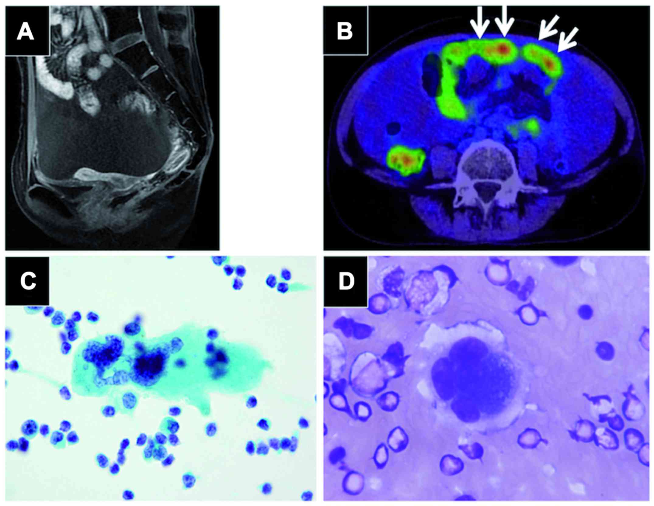
and pelvic MRI scan showed an atrophic but otherwise normal uterus and bilateral adnexa (Fig. 1A). Positron emission tomography CT scans revealed an omental mass with prominent FDG uptake, massive bloody ascites, and signs suspicious of peritonitis carcinomatosis (Fig. 1B). Gastroscopy and colonoscopy findings were unremarkable. Cervical and endometrial cytology were negative for malignancy. Ascites cytology obtained by paracentesis revealed solitary, scattered multinucleated giant cells with prominent nucleoli (Fig. 1C,d); however, the findings did not prove malignancy due to the small number of suspicious cells. CA125 was elevated (237 mIU/ml), while other tumor markers (SCC, CEA and CA19-9) were negative.

Figure 1.

Preoperative evaluation. (A) Pelvic MRI showing a normal uterus. (B) PET/CT revealed massive bloody ascites, an omental mass (arrows), and a diffusely distended small intestine suggestive of peritonitis carcinomatosis. (C) Papanicolaou stain and (D) Giemsa stain of the ascites revealed solitary multinucleated giant tumor cells with prominent nucleoli. Magnification, ×100.

Intra-abdominal cancer of unknown primary origin was 
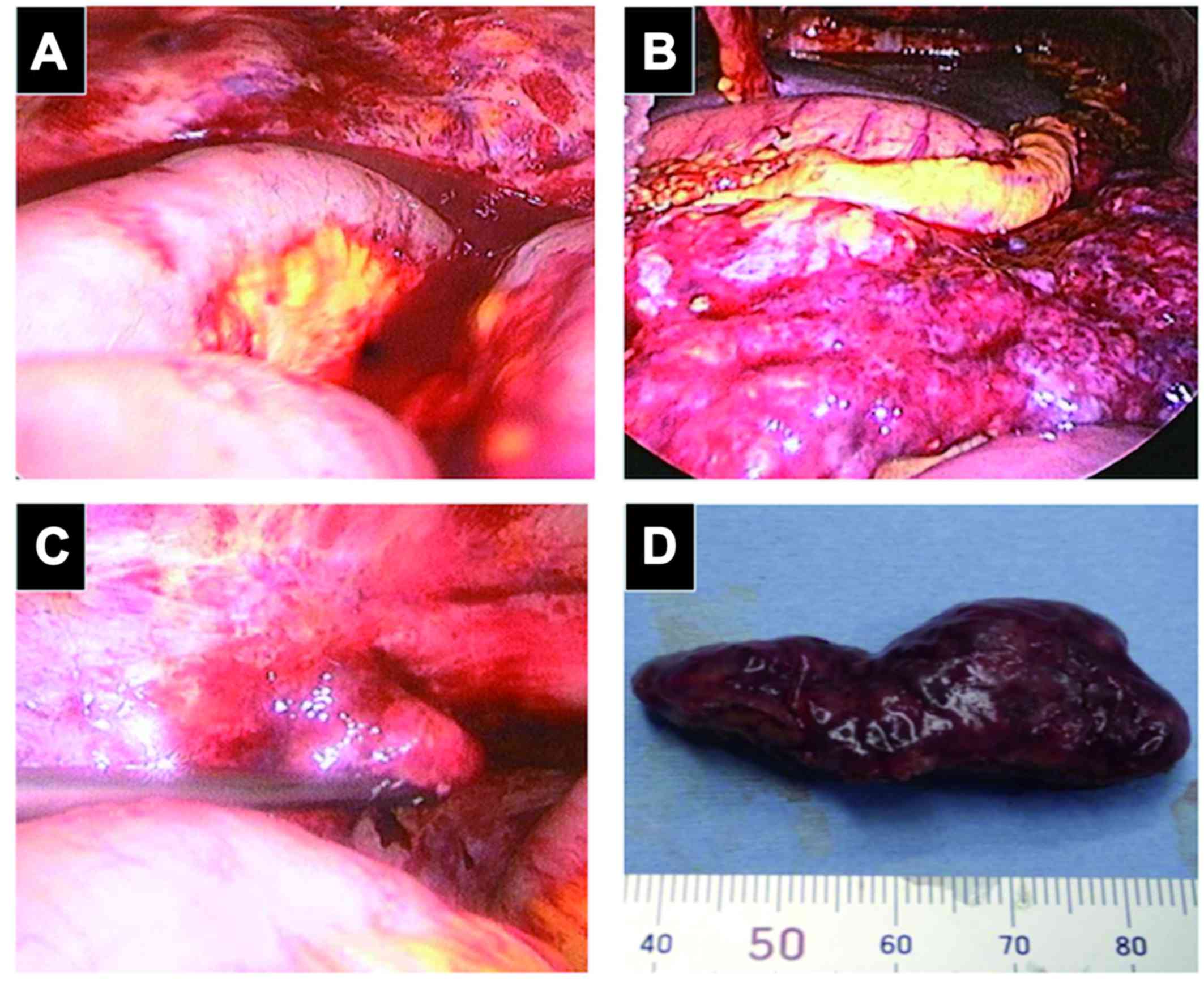
suspected and diagnostic laparoscopy was performed. Laparoscopic examination revealed intensive peritoneal inflammation, massive bloody ascites (Fig. 2A) and a congested greater omental mass of 15 cm in diameter (Fig. 2B). This mass reached the splenic flexure and had invaded close to the splenic hilum. There was a small papillary lesion on the surface of the left ovary, which was suspicious for dissemination (Fig. 2C). The right adnexa (Fig. 2D) and the uterus were atrophic and adhered to the peritoneal wall with no apparent lesion. Biopsy of the omental mass, bilateral partial oophorectomy and right salpingectomy were performed. A frozen section of the omental mass was diagnosed as poorly differentiated malignant tumor. The operation time was 65 min and blood loss was 1,150 ml.

Figure 2.

Laparoscopic findings. (A) Massive bloody ascites and peritoneal inflammation were observed. (B) A congested omental mass invaded close to the splenic hilum. (C) A small papillary lesion (arrow) is seen on the surface of the left Fallopian tube. (D) Macroscopic findings of the biopsied omental mass.

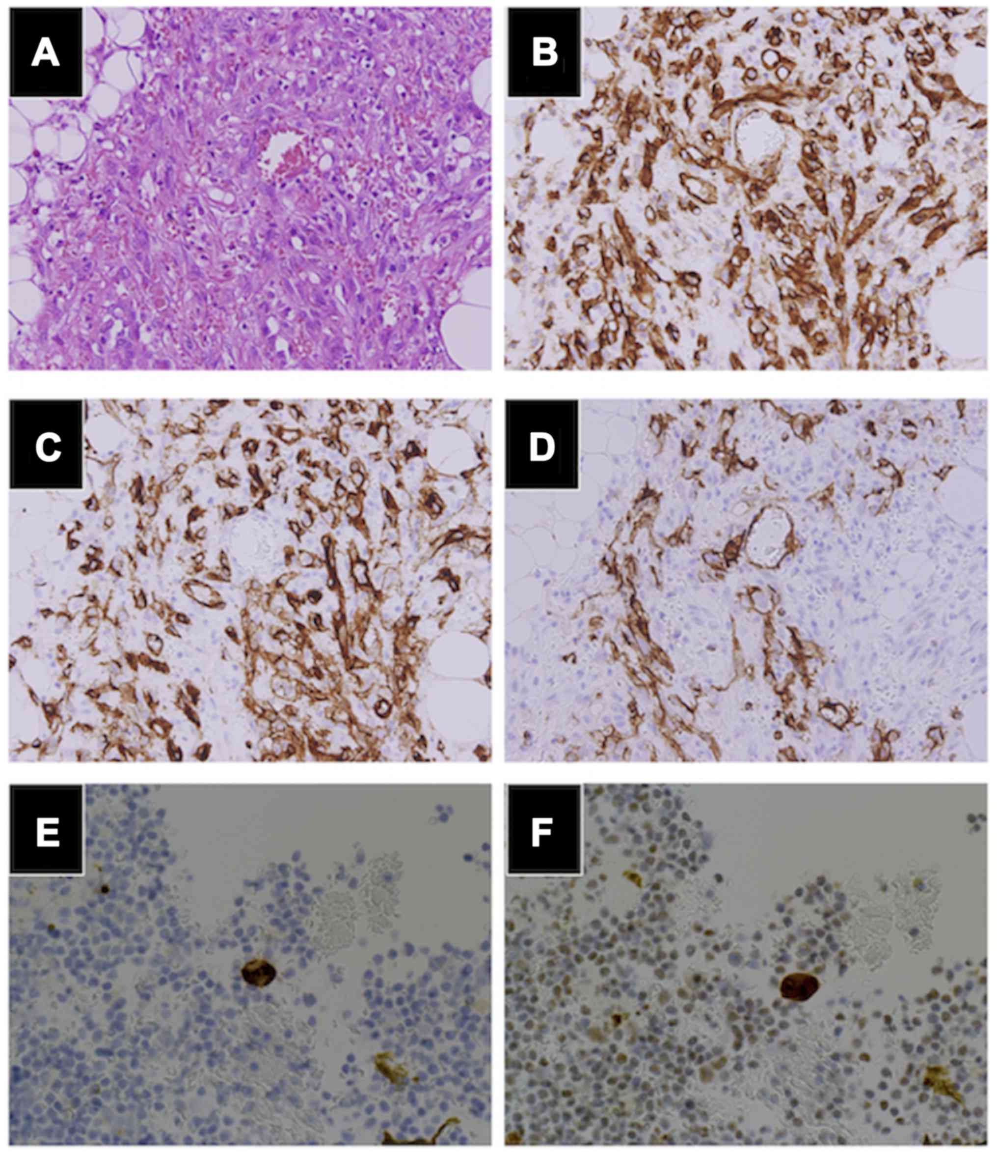
Microscopically, the tumors were composed of high-grade malignant cells with frequent mitosis, forming irregular anastomosing vascular channels containing erythrocytes (Fig. 3A). The same findings were present in the left Fallopian tube, indicating dissemination. Immunohistochemistry revealed positive staining for endothelial markers CD31 (Fig. 3B), CD34 (Fig. 3C) and ERG. The lymphatic endothelial marker D2-40 (Fig. 3D) was positive, whereas cytokeratin (AE1/AE3), estrogen receptor, progesterone receptor and WT-1 were negative. The cell block technique for ascites revealed a few cells positive for CD34 (Fig. 3E) and ERG (Fig. 3F).

Figure 3.

Histopathological findings. (A) H&E stains of the omental mass showed high-grade malignant cells with frequent mitosis, forming irregular anastomosing vascular channels. Immunohistochemistry was positive for (B) CD31, (C) CD34 and partially positive for (D) D2-40, while pan-keratin was grossly negative. The cell block method was used for immunohistochemical investigation of ascites, and showed a small number of cells positive for (E) CD34 and (F) ERG. (A-C) Magnification, ×20; (E-F) Magnification, ×40.

The diagnostic criteria for radiation-induced sarcoma include pathologically proven sarcoma developing in a prior radiation field, normal findings prior to radiotherapy, and at least a three-year period after radiotherapy (6). Fulfilling the above criteria, the clinical and pathological findings led to the diagnosis of radiation-induced angiosarcoma of the greater omentum. The advanced tumor was unresectable; therefore, weekly paclitaxel (100 mg/m2 on days 1, 8, 15, 22, 29, 36 in a 7-week cycle, as approved in Japan) was started 11 days after surgery, which lead to complete response after six courses, and complete cytoreductive surgery was performed consequently. Currently, the patient remains disease free 3 months post surgery.

Discussion

Angiosarcoma is a rare, aggressive malignant tumor of vascular endothelial origin (1) that comprises approximately 2% of soft-tissue sarcomas. The most common site is the head and neck in elderly patients, whereas cases of intraperitoneal origin are extremely rare. The prognosis of angiosarcoma is generally poor, 19-43.9% of cases present with advanced disease (7,8) and the median survival period is 7–12 months (7,8).

Secondary soft tissue sarcoma is an uncommon but important adverse event following radiation therapy. As the importance of radiation therapy in cancer treatment has grown over the years, the number of recognized radiation-induced sarcomas is also growing. In a study of 4,884 adult patients with soft tissue sarcoma, 2.5% were diagnosed as radiation-induced, of which 15% were patents with angiosarcoma (9). Radiation-induced angiosarcoma accounts for 3–12.2% of all angiosarcomas (7), and among patients who received radiotherapy for primary cancer, the standardized incidence ratio of angiosarcoma was 6.0 (95% CI 2.7–11), compared to patients who received other treatment (10). Compared to other subtypes, radiation-induced angiosarcoma has a particularly poor prognosis; even local lesions recur in 87.5% of cases, and the median survival period is 7–8 months (7).

The patient described herein developed angiosarcoma in a prior radiation field 7 years after cervical cancer treatment, and in this respect this is a typical case of radiation-induced angiosarcoma. However, most radiation-induced angiosarcomas occur in the skin of patients after radiation treatment for breast cancer (7). Only four previous cases of angiosarcoma originating in the greater omentum have been reported, all of which were consequences of radiation therapy for gynecologic cancer (2–5) (Table I). All cases presented with advanced disease, which were complicated with massive hemorrhagic ascites and peritoneal dissemination. While the cases receiving palliative care described the rapidly progressing character of the disease (3,5), Chudecha-Glaz et al reported the achievement of partial response by platinum-based chemotherapy, which lead to cytoreductive surgery and control of the disease at least temporarily (4). Swiftly making the diagnosis and enabling chemotherapy administration seems to be the key in controling the disease.

Table I.

Radiation-induced angiosarcoma arising in the omentum.

Table I.

Radiation-induced angiosarcoma arising in the omentum.

Authors (year)AgeSexPrevious malignancyRadiation to onsetIntra-peritoneal hemorrhageTreatmentPrognosis(Refs.)
Westenberg et al (1989)59FCervical cancer8 years+UnknownUnknown(7)
Sakemi et al (2011)74FCervical cancer5 years+Palliative34 days died(8)
Chudecka-Glaz et al (2014)55FOvarian cancer20 years+Chemotherapy, Surgery16 months died(9)
Narayanan et al (2015)77FCervical cancerUnknown+Surgery, palliative careUnknown(10)
Present case38FCervical cancer7 years+Chemotherapy, surgeryCurrently disease Free (8 months)

[i] F, female.

Due to the low prevalence and lack of specific markers, prompt diagnosis of intraperitoneal angiosarcoma is a challenge. Our current experience suggests the clinical usefulness of diagnostic laparoscopy. The importance of diagnostic laparoscopy is emerging for intraperitoneal cancer of unknown primary origin, where minimal invasiveness is required for high-risk patients. This method permits precise evaluation of the cancer origin, determination of the feasibility of complete cytoreduction, and conversion to laparotomy as needed. Further, for an unresectable tumor, diagnostic laparoscopy allows sufficient biopsy for pathological examination with permissive complications (11). With use of diagnostic laparoscopy, we were able to initiate systemic treatment only 11 days postoperatively in the current case.

For advanced stage angiosarcoma, systemic cytotoxic chemotherapy is the treatment of choice. Due to the lack of clinical trials for angiosarcoma patients, anthracycline- and ifosfamide-based regimens have been considered as first line chemotherapy following the results of clinical trials for other soft tissue sarcomas (12,13). However, recently, few studies have shown the efficacy of taxane-based therapy for angiosarcoma. In a retrospective study in 32 patients with angiosarcoma who received triweekly or weekly paclitaxel, the EORTC soft tissue and bone group found a response rate of 62% and a median time to progression of 7.6 months (12). In the ANGIOTAX phase II trial (14), weekly paclitaxel (80 mg/m2 on days 1, 8, and 15 of a 4-week cycle) was administered to 30 patients with unresectable angiosarcoma. The response rate was 19% after 6 courses, but the regimen achieved a relatively high non-progression rate (75% at 3 months and 24% at 6 months), and the median progression free survival (PFS) and overall survival (OS) were 4 and 8 months, respectively. Taking these studies into account, the NCCN guidelines currently recommend weekly paclitaxel as first line chemotherapy for angiosarcoma; however, further clinical trials are needed to optimize standard therapy.

Herein, we have reported a case of radiation-induced angiosarcoma originating in the greater omentum in a 38-year-old female. Radiation-induced angiosarcoma following treatment of gynecologic cancer is clearly a rare complication, but should be considered, especially with the increasing importance of radiation therapy for treatment of invasive cervical cancer.

Acknowledgements

Written informed consent was obtained from the patient for the publication of this case report, and ethics approval was given from the ethical committee of our institute (approval nο. 20070081). We thank the patient for allowing discussion of her case with the academic community.

References

1 

Young RJ, Brown NJ, Reed MW, Hughes D and Woll PJ: Angiosarcoma. Lancet Oncol. 11:983–991. 2010. View Article : Google Scholar : PubMed/NCBI

2 

Westenberg AH, Wiggers T, Henzen-Logmans SC, Verweij J, Meerwaldt JA and van Geel AN: Post-irradiation angiosarcoma of the greater omentum. Eur J Surg Oncol. 15:175–178. 1989.PubMed/NCBI

3 

Sakemi M, Sakemi R, Harada M, So S, Uchiyama D, Morimitsu Y, Kakiuchi S, Ishihara Y, Kubo Y, Matsugaki S, et al: A case of postirradiation angiosarcoma of the greater omentum with hemorrhage. Clin J Gastroenterol. 4:302–306. 2011. View Article : Google Scholar : PubMed/NCBI

4 

Chudecka-Głaz A, Menkiszak J, Kuźniak S, Lewandowska M, Burak M and Walecka A: A rare case of peritoneal disseminated angiosarcoma 20 years after ovarian cancer diagnosis. Gynecol Obstet Invest. 77:68–72. 2014. View Article : Google Scholar : PubMed/NCBI

5 

Narayanan S, Parker M, Shayo J, Zheng M, Matulewicz T and Parker G: The Impact of Radiation on an Unusual Case of Omental Epithelioid Angiosarcoma. Case Rep Surg. 2015:8490592015.PubMed/NCBI

6 

Arlen M, Higinbotham NL, Huvos AG, Marcove RC, Miller T and Shah IC: Radiation-induced sarcoma of bone. Cancer. 28:1087–1099. 1971. View Article : Google Scholar : PubMed/NCBI

7 

Abraham JA, Hornicek FJ, Kaufman AM, Harmon DC, Springfield DS, Raskin KA, Mankin HJ, Kirsch DG, Rosenberg AE, Nielsen GP, et al: Treatment and outcome of 82 patients with angiosarcoma. Ann Surg Oncol. 14:1953–1967. 2007. View Article : Google Scholar : PubMed/NCBI

8 

Fayette J, Martin E, Piperno-Neumann S, Le Cesne A, Robert C, Bonvalot S, Ranchère D, Pouillart P, Coindre JM and Blay JY: Angiosarcomas, a heterogeneous group of sarcomas with specific behavior depending on primary site: A retrospective study of 161 cases. Ann Oncol. 18:2030–2036. 2007. View Article : Google Scholar : PubMed/NCBI

9 

Cha C, Antonescu CR, Quan ML, Maru S and Brennan MF: Long-term results with resection of radiation-induced soft tissue sarcomas. Ann Surg. 239:903–910. 2004. View Article : Google Scholar : PubMed/NCBI

10 

Virtanen A, Pukkala E and Auvinen A: Angiosarcoma after radiotherapy: A cohort study of 332,163 Finnish cancer patients. Br J Cancer. 97:115–117. 2007. View Article : Google Scholar : PubMed/NCBI

11 

Marmor RA, Kelly KJ, Lowy AM and Baumgartner JM: Laparoscopy is safe and accurate to evaluate peritoneal surface metastasis prior to cytoreductive surgery. Ann Surg Oncol. 23:1461–1467. 2016. View Article : Google Scholar : PubMed/NCBI

12 

Schlemmer M, Reichardt P, Verweij J, Hartmann JT, Judson I, Thyss A, Hogendoorn PC, Marreaud S, Van Glabbeke M and Blay JY: Paclitaxel in patients with advanced angiosarcomas of soft tissue: A retrospective study of the EORTC soft tissue and bone sarcoma group. Eur J Cancer. 44:2433–2436. 2008. View Article : Google Scholar : PubMed/NCBI

13 

Agulnik M, Yarber JL, Okuno SH, von Mehren M, Jovanovic BD, Brockstein BE, Evens AM and Benjamin RS: An open-label, multicenter, phase II study of bevacizumab for the treatment of angiosarcoma and epithelioid hemangioendotheliomas. Ann Oncol. 24:257–263. 2013. View Article : Google Scholar : PubMed/NCBI

14 

Penel N, Bui BN, Bay JO, Cupissol D, Ray-Coquard I, Piperno-Neumann S, Kerbrat P, Fournier C, Taieb S, Jimenez M, et al: Phase II trial of weekly paclitaxel for unresectable angiosarcoma: The ANGIOTAX Study. J Clin Oncol. 26:5269–5274. 2008. View Article : Google Scholar : PubMed/NCBI

Related Articles

  • Abstract
  • View
  • Download
  • Twitter
Copy and paste a formatted citation
Spandidos Publications style
Kunitomi H, Banno K, Iseki H, Sera A, Miyauchi A, Kobayashi Y, Hayashi S, Tominaga E, Sasaki A, Kawaida M, Kawaida M, et al: Radiation‑induced angiosarcoma of the omentum diagnosed by laparoscopy: A case report. Mol Clin Oncol 8: 264-268, 2018.
APA
Kunitomi, H., Banno, K., Iseki, H., Sera, A., Miyauchi, A., Kobayashi, Y. ... Aoki, D. (2018). Radiation‑induced angiosarcoma of the omentum diagnosed by laparoscopy: A case report. Molecular and Clinical Oncology, 8, 264-268. https://doi.org/10.3892/mco.2017.1513
MLA
Kunitomi, H., Banno, K., Iseki, H., Sera, A., Miyauchi, A., Kobayashi, Y., Hayashi, S., Tominaga, E., Sasaki, A., Kawaida, M., Kameyama, K., Aoki, D."Radiation‑induced angiosarcoma of the omentum diagnosed by laparoscopy: A case report". Molecular and Clinical Oncology 8.2 (2018): 264-268.
Chicago
Kunitomi, H., Banno, K., Iseki, H., Sera, A., Miyauchi, A., Kobayashi, Y., Hayashi, S., Tominaga, E., Sasaki, A., Kawaida, M., Kameyama, K., Aoki, D."Radiation‑induced angiosarcoma of the omentum diagnosed by laparoscopy: A case report". Molecular and Clinical Oncology 8, no. 2 (2018): 264-268. https://doi.org/10.3892/mco.2017.1513
Copy and paste a formatted citation
x
Spandidos Publications style
Kunitomi H, Banno K, Iseki H, Sera A, Miyauchi A, Kobayashi Y, Hayashi S, Tominaga E, Sasaki A, Kawaida M, Kawaida M, et al: Radiation‑induced angiosarcoma of the omentum diagnosed by laparoscopy: A case report. Mol Clin Oncol 8: 264-268, 2018.
APA
Kunitomi, H., Banno, K., Iseki, H., Sera, A., Miyauchi, A., Kobayashi, Y. ... Aoki, D. (2018). Radiation‑induced angiosarcoma of the omentum diagnosed by laparoscopy: A case report. Molecular and Clinical Oncology, 8, 264-268. https://doi.org/10.3892/mco.2017.1513
MLA
Kunitomi, H., Banno, K., Iseki, H., Sera, A., Miyauchi, A., Kobayashi, Y., Hayashi, S., Tominaga, E., Sasaki, A., Kawaida, M., Kameyama, K., Aoki, D."Radiation‑induced angiosarcoma of the omentum diagnosed by laparoscopy: A case report". Molecular and Clinical Oncology 8.2 (2018): 264-268.
Chicago
Kunitomi, H., Banno, K., Iseki, H., Sera, A., Miyauchi, A., Kobayashi, Y., Hayashi, S., Tominaga, E., Sasaki, A., Kawaida, M., Kameyama, K., Aoki, D."Radiation‑induced angiosarcoma of the omentum diagnosed by laparoscopy: A case report". Molecular and Clinical Oncology 8, no. 2 (2018): 264-268. https://doi.org/10.3892/mco.2017.1513
Follow us
  • Twitter
  • LinkedIn
  • Facebook
About
  • Spandidos Publications
  • Careers
  • Cookie Policy
  • Privacy Policy
How can we help?
  • Help
  • Live Chat
  • Contact
  • Email to our Support Team