Spandidos Publications Logo
  • About
    • About Spandidos
    • Aims and Scopes
    • Abstracting and Indexing
    • Editorial Policies
    • Reprints and Permissions
    • Job Opportunities
    • Terms and Conditions
    • Contact
  • Journals
    • All Journals
    • Oncology Letters
      • Oncology Letters
      • Information for Authors
      • Editorial Policies
      • Editorial Board
      • Aims and Scope
      • Abstracting and Indexing
      • Bibliographic Information
      • Archive
    • International Journal of Oncology
      • International Journal of Oncology
      • Information for Authors
      • Editorial Policies
      • Editorial Board
      • Aims and Scope
      • Abstracting and Indexing
      • Bibliographic Information
      • Archive
    • Molecular and Clinical Oncology
      • Molecular and Clinical Oncology
      • Information for Authors
      • Editorial Policies
      • Editorial Board
      • Aims and Scope
      • Abstracting and Indexing
      • Bibliographic Information
      • Archive
    • Experimental and Therapeutic Medicine
      • Experimental and Therapeutic Medicine
      • Information for Authors
      • Editorial Policies
      • Editorial Board
      • Aims and Scope
      • Abstracting and Indexing
      • Bibliographic Information
      • Archive
    • International Journal of Molecular Medicine
      • International Journal of Molecular Medicine
      • Information for Authors
      • Editorial Policies
      • Editorial Board
      • Aims and Scope
      • Abstracting and Indexing
      • Bibliographic Information
      • Archive
    • Biomedical Reports
      • Biomedical Reports
      • Information for Authors
      • Editorial Policies
      • Editorial Board
      • Aims and Scope
      • Abstracting and Indexing
      • Bibliographic Information
      • Archive
    • Oncology Reports
      • Oncology Reports
      • Information for Authors
      • Editorial Policies
      • Editorial Board
      • Aims and Scope
      • Abstracting and Indexing
      • Bibliographic Information
      • Archive
    • Molecular Medicine Reports
      • Molecular Medicine Reports
      • Information for Authors
      • Editorial Policies
      • Editorial Board
      • Aims and Scope
      • Abstracting and Indexing
      • Bibliographic Information
      • Archive
    • World Academy of Sciences Journal
      • World Academy of Sciences Journal
      • Information for Authors
      • Editorial Policies
      • Editorial Board
      • Aims and Scope
      • Abstracting and Indexing
      • Bibliographic Information
      • Archive
    • International Journal of Functional Nutrition
      • International Journal of Functional Nutrition
      • Information for Authors
      • Editorial Policies
      • Editorial Board
      • Aims and Scope
      • Abstracting and Indexing
      • Bibliographic Information
      • Archive
    • International Journal of Epigenetics
      • International Journal of Epigenetics
      • Information for Authors
      • Editorial Policies
      • Editorial Board
      • Aims and Scope
      • Abstracting and Indexing
      • Bibliographic Information
      • Archive
    • Medicine International
      • Medicine International
      • Information for Authors
      • Editorial Policies
      • Editorial Board
      • Aims and Scope
      • Abstracting and Indexing
      • Bibliographic Information
      • Archive
  • Articles
  • Information
    • Information for Authors
    • Information for Reviewers
    • Information for Librarians
    • Information for Advertisers
    • Conferences
  • Language Editing
Spandidos Publications Logo
  • About
    • About Spandidos
    • Aims and Scopes
    • Abstracting and Indexing
    • Editorial Policies
    • Reprints and Permissions
    • Job Opportunities
    • Terms and Conditions
    • Contact
  • Journals
    • All Journals
    • Biomedical Reports
      • Information for Authors
      • Editorial Policies
      • Editorial Board
      • Aims and Scope
      • Abstracting and Indexing
      • Bibliographic Information
      • Archive
    • Experimental and Therapeutic Medicine
      • Information for Authors
      • Editorial Policies
      • Editorial Board
      • Aims and Scope
      • Abstracting and Indexing
      • Bibliographic Information
      • Archive
    • International Journal of Epigenetics
      • Information for Authors
      • Editorial Policies
      • Editorial Board
      • Aims and Scope
      • Abstracting and Indexing
      • Bibliographic Information
      • Archive
    • International Journal of Functional Nutrition
      • Information for Authors
      • Editorial Policies
      • Editorial Board
      • Aims and Scope
      • Abstracting and Indexing
      • Bibliographic Information
      • Archive
    • International Journal of Molecular Medicine
      • Information for Authors
      • Editorial Policies
      • Editorial Board
      • Aims and Scope
      • Abstracting and Indexing
      • Bibliographic Information
      • Archive
    • International Journal of Oncology
      • Information for Authors
      • Editorial Policies
      • Editorial Board
      • Aims and Scope
      • Abstracting and Indexing
      • Bibliographic Information
      • Archive
    • Medicine International
      • Information for Authors
      • Editorial Policies
      • Editorial Board
      • Aims and Scope
      • Abstracting and Indexing
      • Bibliographic Information
      • Archive
    • Molecular and Clinical Oncology
      • Information for Authors
      • Editorial Policies
      • Editorial Board
      • Aims and Scope
      • Abstracting and Indexing
      • Bibliographic Information
      • Archive
    • Molecular Medicine Reports
      • Information for Authors
      • Editorial Policies
      • Editorial Board
      • Aims and Scope
      • Abstracting and Indexing
      • Bibliographic Information
      • Archive
    • Oncology Letters
      • Information for Authors
      • Editorial Policies
      • Editorial Board
      • Aims and Scope
      • Abstracting and Indexing
      • Bibliographic Information
      • Archive
    • Oncology Reports
      • Information for Authors
      • Editorial Policies
      • Editorial Board
      • Aims and Scope
      • Abstracting and Indexing
      • Bibliographic Information
      • Archive
    • World Academy of Sciences Journal
      • Information for Authors
      • Editorial Policies
      • Editorial Board
      • Aims and Scope
      • Abstracting and Indexing
      • Bibliographic Information
      • Archive
  • Articles
  • Information
    • For Authors
    • For Reviewers
    • For Librarians
    • For Advertisers
    • Conferences
  • Language Editing
Login Register Submit
  • This site uses cookies
  • You can change your cookie settings at any time by following the instructions in our Cookie Policy. To find out more, you may read our Privacy Policy.

    I agree
Search articles by DOI, keyword, author or affiliation
Search
Advanced Search
presentation
Molecular and Clinical Oncology
Join Editorial Board Propose a Special Issue
Print ISSN: 2049-9450 Online ISSN: 2049-9469
Journal Cover
April-2018 Volume 8 Issue 4

Full Size Image

Sign up for eToc alerts
Recommend to Library

Journals

International Journal of Molecular Medicine

International Journal of Molecular Medicine

International Journal of Molecular Medicine is an international journal devoted to molecular mechanisms of human disease.

International Journal of Oncology

International Journal of Oncology

International Journal of Oncology is an international journal devoted to oncology research and cancer treatment.

Molecular Medicine Reports

Molecular Medicine Reports

Covers molecular medicine topics such as pharmacology, pathology, genetics, neuroscience, infectious diseases, molecular cardiology, and molecular surgery.

Oncology Reports

Oncology Reports

Oncology Reports is an international journal devoted to fundamental and applied research in Oncology.

Experimental and Therapeutic Medicine

Experimental and Therapeutic Medicine

Experimental and Therapeutic Medicine is an international journal devoted to laboratory and clinical medicine.

Oncology Letters

Oncology Letters

Oncology Letters is an international journal devoted to Experimental and Clinical Oncology.

Biomedical Reports

Biomedical Reports

Explores a wide range of biological and medical fields, including pharmacology, genetics, microbiology, neuroscience, and molecular cardiology.

Molecular and Clinical Oncology

Molecular and Clinical Oncology

International journal addressing all aspects of oncology research, from tumorigenesis and oncogenes to chemotherapy and metastasis.

World Academy of Sciences Journal

World Academy of Sciences Journal

Multidisciplinary open-access journal spanning biochemistry, genetics, neuroscience, environmental health, and synthetic biology.

International Journal of Functional Nutrition

International Journal of Functional Nutrition

Open-access journal combining biochemistry, pharmacology, immunology, and genetics to advance health through functional nutrition.

International Journal of Epigenetics

International Journal of Epigenetics

Publishes open-access research on using epigenetics to advance understanding and treatment of human disease.

Medicine International

Medicine International

An International Open Access Journal Devoted to General Medicine.

Journal Cover
April-2018 Volume 8 Issue 4

Full Size Image

Sign up for eToc alerts
Recommend to Library

  • Article
  • Citations
    • Cite This Article
    • Download Citation
    • Create Citation Alert
    • Remove Citation Alert
    • Cited By
  • Similar Articles
    • Related Articles (in Spandidos Publications)
    • Similar Articles (Google Scholar)
    • Similar Articles (PubMed)
  • Download PDF
  • Download XML
  • View XML
Article

Diagnostic usefulness of KL-6 concentration of bile in biliary tract cancer

  • Authors:
    • Takumi Onoyama
    • Kazuya Matsumoto
    • Hiroki Koda
    • Taro Yamashita
    • Hiroki Kurumi
    • Soichiro Kawata
    • Yohei Takeda
    • Kenichi Harada
    • Kazuo Yashima
    • Hajime Isomoto
  • View Affiliations / Copyright

    Affiliations: Division of Medicine and Clinical Science, Department of Multidisciplinary Internal Medicine, Faculty of Medicine, Tottori University, Yonago 683‑8504, Japan
  • Pages: 561-566
    |
    Published online on: February 12, 2018
       https://doi.org/10.3892/mco.2018.1571
  • Expand metrics +
Metrics: Total Views: 0 (Spandidos Publications: | PMC Statistics: )
Metrics: Total PDF Downloads: 0 (Spandidos Publications: | PMC Statistics: )
Cited By (CrossRef): 0 citations Loading Articles...

This article is mentioned in:


Abstract

The sensitivity of bile cytology for biliary tract cancer varies from 6‑64%, and hence remains unsatisfactory. Sialylated carbohydrate antigen KL‑6 mucin is positive in biliary tract cancer tissues and serum KL‑6 levels are significantly increased in intrahepatic ductal adenocarcinoma patients compared with healthy individuals. The aim of the present study was to evaluate the usefulness of the KL‑6 concentration of bile for the diagnosis of biliary tract cancer. Bile cytology and measurements of bile KL‑6 concentration were conducted for 43 patients (25 biliary tract cancers and 18 benign biliary disease). The concentration of KL‑6 in the bile of the biliary tract cancer group was compared with the benign biliary disease group. The diagnostic ability was assessed by using receiver operating characteristic curves (ROC). The mean KL‑6 concentration of bile for biliary tract cancer (34.6±51.6 U/ml) was increased compared with benign biliary disease (5.2±3.9 U/ml, P<0.001). The area under the ROC for diagnosis of biliary tract cancer was 0.84 for benign biliary disease. When the cut‑off level of the KL‑6 concentration of bile was 8.6 U/ml, the sensitivity, specificity, and accuracy of the KL‑6 concentration of bile alone for the diagnosis of biliary tract cancer were 72, 89, and 79%, respectively. Adding the bile KL‑6 concentration to bile cytology measurements, the sensitivity for the diagnosis of biliary tract cancer was increased significantly (100%, P=0.0184). The KL‑6 concentration of bile may strengthen the sensitivity of bile cytology for biliary tract cancer.

Introduction

Epstein-Barr virus (EBV) is an established cause of various types of lymphoma including Burkitt lymphoma and Hodgkins lymphoma, and nasopharyngeal carcinoma (1). Similar to Helicobacter pylori, EBV is accepted as an infective agent that plays an important pathogenic role in gastric cancer (2,3). Gastric carcinoma with lymphoid stroma (GCLS) is a rare histological subgroup of gastric cancers, constituting approximately 1–4% of all gastric carcinomas. Approximately 80% of the reported GCLS cases are associated with an EBV infection (1,4). To diagnose GCLS accurately, the immunohistochemical approach of the tumor was important in addition to sophisticated observation of the endoscopic findings, while standardized diagnostic criteria are lacking (2,4). Early gastric cancer has favorable outcomes with curative resection including gastrectomy and endoscopic submucosal resection (4,5). The current prognosis for GCLS is favorable due to its lower lymph node metastatic rate and a higher survival rate despite deep submucosal invasion of tumor cells.

In the present study, we report a case of EBV-associated early GCLS accompanying lymph node metastasis treated by laparoscopic distal gastrectomy.

Case report

A 61-year-old woman was referred to Kochi Medical School Hospital as a result of an evaluation of a gastric mass lesion that was initially diagnosed by a local medical doctor. Laboratory investigations, including serum carcinoembryonic antigen and cancer antigen 19-9 screening showed no significant 
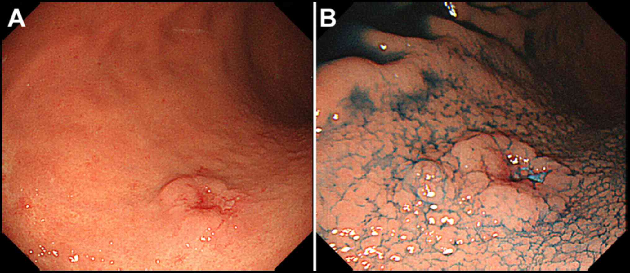
abnormalities. Esophagogastroduodenoscopy (EGD) revealed an elevated lesion with a central irregularly depressed area in the posterior wall of the middle third of the stomach (Fig. 1A). The elevated lesion descended in a gradual slope to the surrounding mucosa when indigo carmine dye was used in chromoendoscopy (Fig. 1B). 
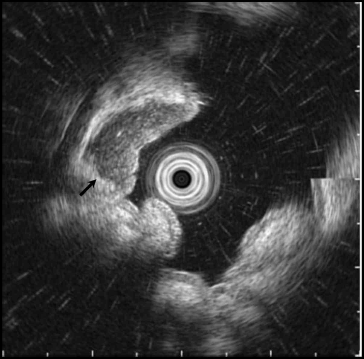
Endoscopic ultrasonography (EUS) revealed a well-circumscribed hypoechoic mass located predominantly within the submucosa and the mucosa (Fig. 2, arrow). Biopsy specimens of the lesion showed prominent lymphocyte infiltration with a lymphoepithelial lesion, suspected of being carcinoma with lymphoid stroma.

Figure 1.

Esophagogastroduodenoscopy shows a slightly elevated lesion with central irregularly depressed area in the middle third of the stomach (A), which descends in a gradual slope to the surrounding mucosa using chromoendoscopy with indigo carmine dye (B).

Figure 2.

Endoscopic ultrasonography revealed a well-circumscribed hypoechoic mass located predominantly within the submucosa and the mucosa.

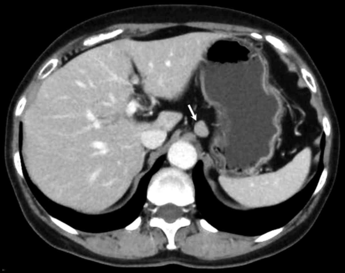
Abdominal contrast-enhanced computed tomography (CT) revealed a well-defined mass with homogeneous enhancement approximately 1.2 cm in diameter in the middle part of the stomach. The CT analysis also revealed lymphadenopathy in the perigastric 
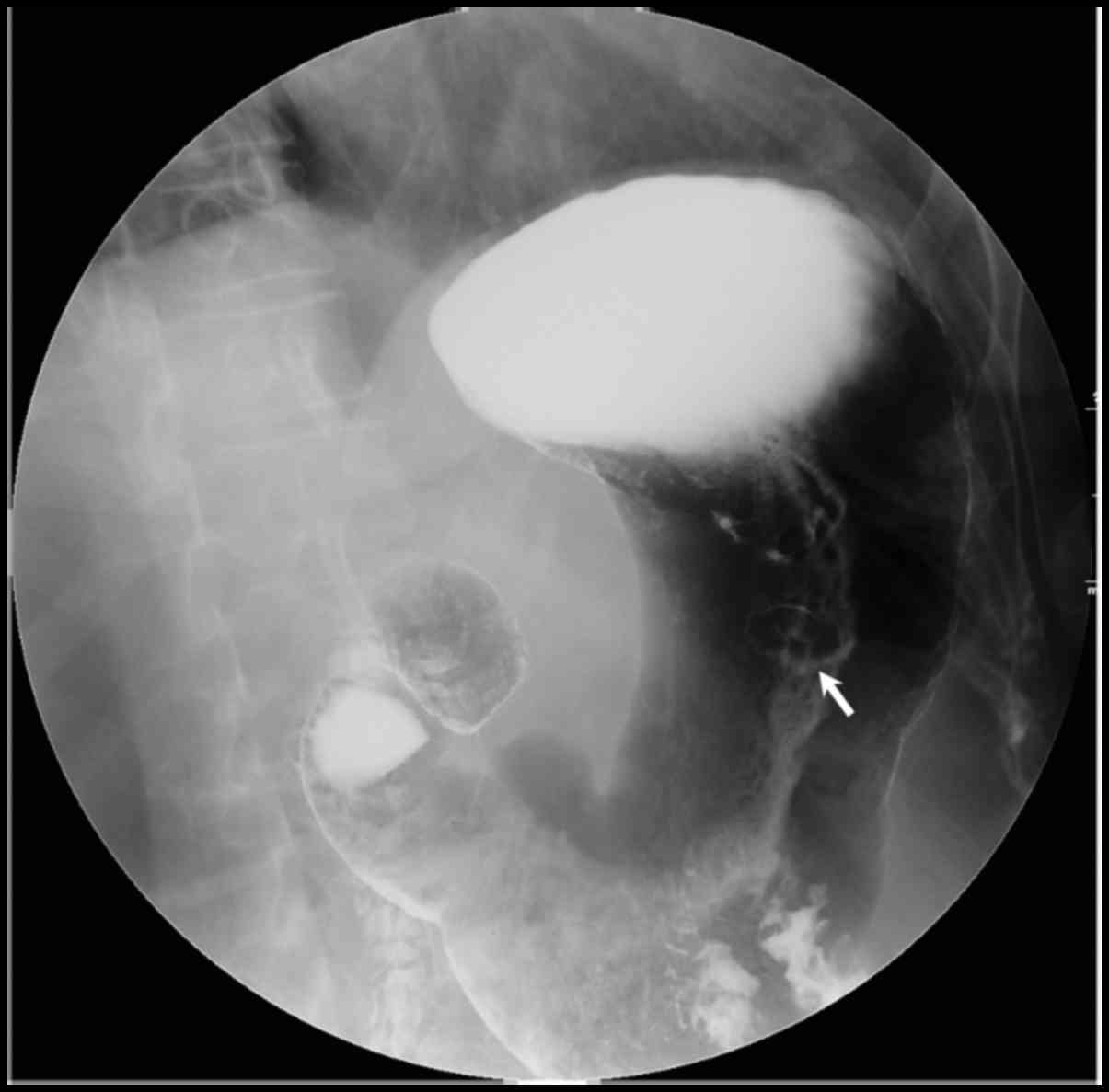
area with a maximum size of 1.4 cm in diameter (Fig. 3, arrow). Double-contrast upper gastrointestinal imaging shows a round filling defect with central collection of Barium measuring 1.2 cm with a clear margin in the middle part of the stomach (Fig. 4, arrow).

Figure 3.

Abdominal computed tomography shows a well-defined ovoid mass with homogeneous enhancement measured in the perigastric area, with a maximum size of 1.4 cm in diameter (arrow).

Figure 4.

Double-contrast upper gastrointestinal imaging shows a round filling defect with central collection of Barium measuring 1.2 cm in diameter with a clear margin in the middle part of the stomach (arrow).

As a result of these investigations we suspected that the patient had a gastric carcinoma with lymphoid stroma accompanying lymph node metastases. The patient underwent laparoscopic distal gastrectomy with reginal lymphadenectomy 
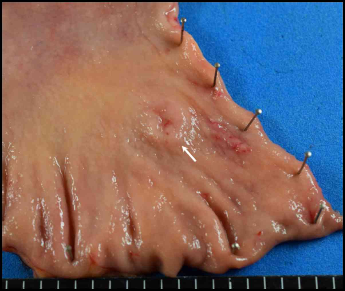
followed by Billroth I reconstruction. Macroscopic examination of the resected specimen showed a slightly elevated lesion-like submucosal tumor with a central depression measuring 1.2×1.2 cm (Fig. 5, arrow).

Figure 5.

Gross appearance of the surgically resected specimen shows a slightly elevated lesion-like submucosal tumor with central depression measuring 1.2×1.2 cm in the posterior wall side of middle third of the stomach (arrow).

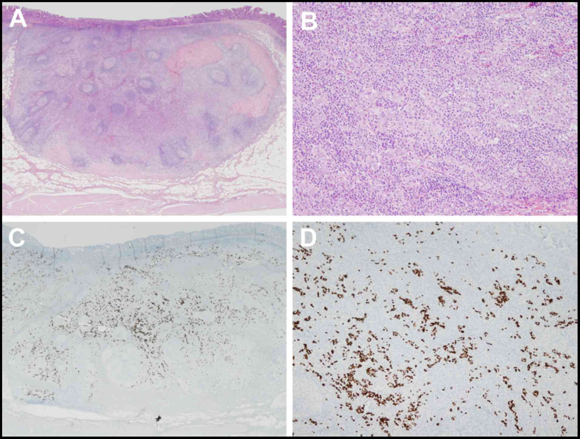
Microscopic examination of the patient specimens showed a bulging mass consists of poorly differentiated adenocarcinoma. There was expansive growth into the submucosa that formed tubular structures and cancer nests with a submucosal depth of invasion of 4,000 µm (Fig. 6A and B). The tumor cells were positive for EBV-encoded RNA by in situ hybridization (EBER-ISH; Fig. 6C and D), and the expression of Ki-67 in cancer cells was 25%. 
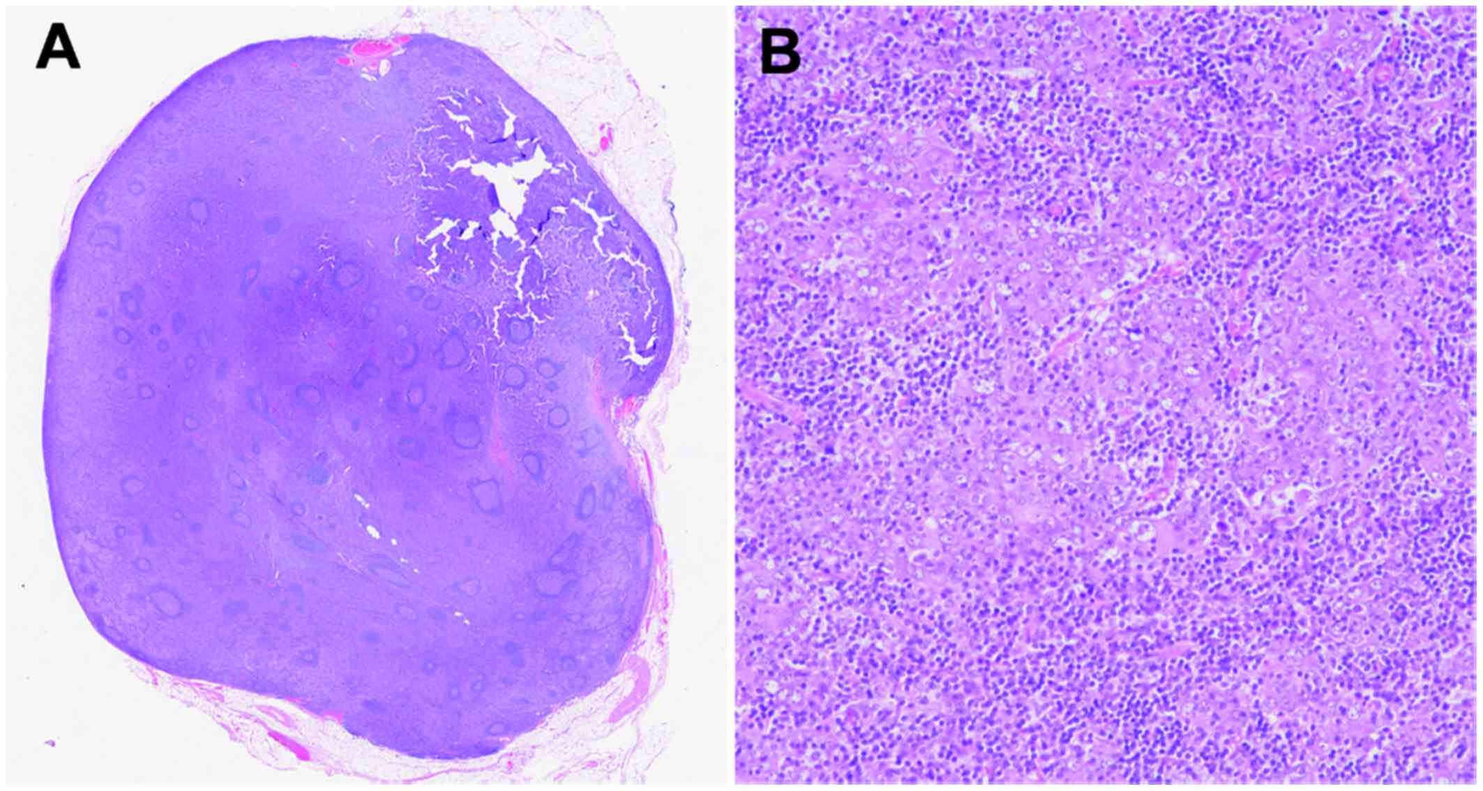
In addition, there was one lymph node metastasis in 13 dissected lymph nodes, which was detected as lymphadenopathy in the CT imaging but there was no lymphovascular infiltration. The lymph node station number with metastasis was 3 according to anatomical definition of Japanese classification of gastric carcinoma (6), which was 2.0 cm in diameter, consisting of diffuse spreading of adenocarcinoma in the lymph node (Fig. 7A and B). The results of the other immunohistochemical investigations of the primary gastric cancer and metastatic lymph node showed positive immunostaining for cytokeratin (AE1/AE3) and negative for CDX-2. Marked infiltration of lymphoid cells was observed in the tumor stroma and these were negative for EBER-ISH. The postoperative course was uneventful and the patient has been well without evidence of recurrence for two months following the operation.

Figure 6.

Histological examination of the resected specimen demonstrates a poorly differentiated adenocarcinoma with expansive growth into the submucosa [(A) low magnification; (B) high magnification]. Immunohistochemical examination shows that the tumor is positive for EBV-encoded RNA by in situ hybridization, whereas no signal can be seen in the lymphocytes. [(C) low magnification; (D) high magnification]. Stained with hematoxylin and eosin.

Figure 7.

Histological examination of the lymph node which was 2.0 cm in diameter shows diffuse spreading of adenocarcinoma [(A) low magnification; (B) high magnification]. Stained with hematoxylin and eosin.

Discussion

GCLS has distinct clinical characteristics that occur in old age and predominantly in males. These tumors arise in the cardia or middle portion of the stomach and have prominent lymphocyte infiltration, particularly in the submucosa (7,8). EBER-ISH is the gold standard for determining EBV infection in histological sections. Carcinoma cells and dysplastic epithelial cells are positive for EBV, but not the normal epithelium or lymphoid stroma in EBV-associated GCLS. The estimate of EBV positivity in gastric cancer is 8.7% overall according to a meta-analysis, and the frequencies are not statistically different among different anatomical locations (1). Although EBV is considered to be the main cause of the lymphocytic response, the mechanism by which EBV contributes to the carcinogenesis of gastric mucosa has not been elucidated (9).

The rate of metastasis to the lymph node in submucosal cancers with GCLS is significantly lower than in conventional early gastric cancer, especially those involving an EBV infection (2,8). The spread of tumors through the gastric wall may be prevented by abundant lymphocytic reactions, which regulate the anti-tumor effect of immunity, resulting in a more favorable prognosis as a result of the immune response to the tumor (8,9). An investigation of 41 GCLS in early gastric cancer reported that there was no lymph node metastasis if the submucosal depth of invasion was ≤2,000 µm from the muscularis mucosa, while the incidence rate of lymph node metastasis was 28.6% if the SM depth of invasion was >2,000 µm (8).

Regarding the association between the sizes of gastric carcinoma and lymph node metastasis, Kim et al reported that there was no lymph node metastasis in the tumors with sizes <1.0 cm, and 2-dimensional tumor size was the only significant risk factor for lymph node metastasis in the analysis for 574 patients with differentiated minute submucosal cancer (10). Shin et al reported that tumor size was smaller in GCLS, 2.1 cm, than in no-GCLS, 3.1 cm, and only two patients with GCSL (3.4%) showed lymph node metastasis in the analysis of 1696 patients with early gastric cancer (11). Therefore, the lymph node metastasis of small size GCLS as the present case seems to be extremely rare.

In the present case, it was difficult to detect the submucosal invasion during EGD without EUS, because the majority of the lesion was a bulging mass with abundant lymphoid stroma that was located in the submucosa by pathological investigation. Standard radical gastrectomy with regional lymphadenectomy is important, recommending in cases with submucosal invasion of tumor cells to a depth of more than 2,000 µm, regardless of tumor size. EUS seems to be an essential modality to estimate the depth of tumor invasion in the diagnostic and therapeutic management of GCLS.

Since GCLS is a rare disease, clinicians should recognize the features of this entity to make an accurate diagnosis and select the appropriate treatment. Further studies and assessments of additional cases are required to establish standardized recommendation criteria and management for this entity.

References

1 

Murphy G, Pfeiffer R, Camargo MC and Rabkin CS: Meta-analysis shows that prevalence of Epstein-Barr virus-positive gastric cancer differs based on sex and anatomic location. Gastroenterology. 137:824–833. 2009. View Article : Google Scholar : PubMed/NCBI

2 

Liang Q, Yao X, Tang S, Zhang J, Yau TO, Li X, Tang CM, Kang W, Lung RW, Li JW, et al: Integrative identification of Epstein-Barr virus-associated mutations and epigenetic alterations in gastric cancer. Gastroenterology. 147:1350–1362.e4. 2014. View Article : Google Scholar : PubMed/NCBI

3 

Lim H, Park YS, Lee JH, Son DH, Ahn JY, Choi KS, Kim DH, Choi KD, Song HJ, Lee GH, et al: Features of gastric carcinoma with lymphoid stroma associated with Epstein-Barr virus. Clin Gastroenterol Hepatol. 13:1738–1744.e2. 2015. View Article : Google Scholar : PubMed/NCBI

4 

Watanabe H, Enjoji M and Imai T: Gastric carcinoma with lymphoid stroma. Its morphologic characteristics and prognostic correlations. Cancer. 38:232–243. 1976. View Article : Google Scholar : PubMed/NCBI

5 

Namikawa T, Kitagawa H, Iwabu J, Okabayashi T, Sugimoto T, Kobayashi M and Hanazaki K: Clinicopathological properties of the superficial spreading type early gastric cancer. J Gastrointest Surg. 14:52–57. 2010. View Article : Google Scholar : PubMed/NCBI

6 

Japanese Gastric Cancer Association, . Japanese classification of gastric carcinoma: III English edition. Gastric Cancer. 14:101–112. 2011. View Article : Google Scholar : PubMed/NCBI

7 

Choi MG, Jeong JY, Kim KM, Bae JM, Noh JH, Sohn TS and Kim S: Clinical significance of gastritis cystica profunda and its association with Epstein-Barr virus in gastric cancer. Cancer. 118:5227–5233. 2012. View Article : Google Scholar : PubMed/NCBI

8 

Huh CW, Jung DH, Kim H, Kim H, Youn YH, Park H, Kim JW, Choi SH, Noh SH and Kim JH: Clinicopathologic features of gastric carcinoma with lymphoid stroma in early gastric cancer. J Surg Oncol. 114:769–772. 2016. View Article : Google Scholar : PubMed/NCBI

9 

Huang KH, Wang RF, Yang MH, Wu CW, Fang WL, Li AF, Chi CW and Kao HL: Advanced gastric cancer patients with lymphoid stroma have better survival than those without. J Surg Oncol. 107:523–528. 2013. View Article : Google Scholar : PubMed/NCBI

10 

Kim TJ, Lee H, Min YW, Min BH, Lee JH, Kim KM, Kim MJ, Kim K, Rhee PL and Kim JJ: One-dimensional and 2-dimensional tumor size measurement for prediction of lymph node metastasis in differentiated early gastric cancer with minute submucosal invasion. Gastrointest Endosc. 85:730–736. 2017. View Article : Google Scholar : PubMed/NCBI

11 

Shin DH, Kim GH, Lee BE, Lee JW, Ha DW, Jeon HK, Baek DH, Song GA, Ahn SJ and Park DY: Clinicopathologic features of early gastric carcinoma with lymphoid stroma and feasibility of endoscopic submucosal dissection. Surg Endosc. April 13–2017. View Article : Google Scholar : (Epub ahead of print). View Article : Google Scholar

Related Articles

  • Abstract
  • View
  • Download
  • Twitter
Copy and paste a formatted citation
Spandidos Publications style
Onoyama T, Matsumoto K, Koda H, Yamashita T, Kurumi H, Kawata S, Takeda Y, Harada K, Yashima K, Isomoto H, Isomoto H, et al: Diagnostic usefulness of KL-6 concentration of bile in biliary tract cancer. Mol Clin Oncol 8: 561-566, 2018.
APA
Onoyama, T., Matsumoto, K., Koda, H., Yamashita, T., Kurumi, H., Kawata, S. ... Isomoto, H. (2018). Diagnostic usefulness of KL-6 concentration of bile in biliary tract cancer. Molecular and Clinical Oncology, 8, 561-566. https://doi.org/10.3892/mco.2018.1571
MLA
Onoyama, T., Matsumoto, K., Koda, H., Yamashita, T., Kurumi, H., Kawata, S., Takeda, Y., Harada, K., Yashima , K., Isomoto, H."Diagnostic usefulness of KL-6 concentration of bile in biliary tract cancer". Molecular and Clinical Oncology 8.4 (2018): 561-566.
Chicago
Onoyama, T., Matsumoto, K., Koda, H., Yamashita, T., Kurumi, H., Kawata, S., Takeda, Y., Harada, K., Yashima , K., Isomoto, H."Diagnostic usefulness of KL-6 concentration of bile in biliary tract cancer". Molecular and Clinical Oncology 8, no. 4 (2018): 561-566. https://doi.org/10.3892/mco.2018.1571
Copy and paste a formatted citation
x
Spandidos Publications style
Onoyama T, Matsumoto K, Koda H, Yamashita T, Kurumi H, Kawata S, Takeda Y, Harada K, Yashima K, Isomoto H, Isomoto H, et al: Diagnostic usefulness of KL-6 concentration of bile in biliary tract cancer. Mol Clin Oncol 8: 561-566, 2018.
APA
Onoyama, T., Matsumoto, K., Koda, H., Yamashita, T., Kurumi, H., Kawata, S. ... Isomoto, H. (2018). Diagnostic usefulness of KL-6 concentration of bile in biliary tract cancer. Molecular and Clinical Oncology, 8, 561-566. https://doi.org/10.3892/mco.2018.1571
MLA
Onoyama, T., Matsumoto, K., Koda, H., Yamashita, T., Kurumi, H., Kawata, S., Takeda, Y., Harada, K., Yashima , K., Isomoto, H."Diagnostic usefulness of KL-6 concentration of bile in biliary tract cancer". Molecular and Clinical Oncology 8.4 (2018): 561-566.
Chicago
Onoyama, T., Matsumoto, K., Koda, H., Yamashita, T., Kurumi, H., Kawata, S., Takeda, Y., Harada, K., Yashima , K., Isomoto, H."Diagnostic usefulness of KL-6 concentration of bile in biliary tract cancer". Molecular and Clinical Oncology 8, no. 4 (2018): 561-566. https://doi.org/10.3892/mco.2018.1571
Follow us
  • Twitter
  • LinkedIn
  • Facebook
About
  • Spandidos Publications
  • Careers
  • Cookie Policy
  • Privacy Policy
How can we help?
  • Help
  • Live Chat
  • Contact
  • Email to our Support Team