Spandidos Publications Logo
  • About
    • About Spandidos
    • Aims and Scopes
    • Abstracting and Indexing
    • Editorial Policies
    • Reprints and Permissions
    • Job Opportunities
    • Terms and Conditions
    • Contact
  • Journals
    • All Journals
    • Oncology Letters
      • Oncology Letters
      • Information for Authors
      • Editorial Policies
      • Editorial Board
      • Aims and Scope
      • Abstracting and Indexing
      • Bibliographic Information
      • Archive
    • International Journal of Oncology
      • International Journal of Oncology
      • Information for Authors
      • Editorial Policies
      • Editorial Board
      • Aims and Scope
      • Abstracting and Indexing
      • Bibliographic Information
      • Archive
    • Molecular and Clinical Oncology
      • Molecular and Clinical Oncology
      • Information for Authors
      • Editorial Policies
      • Editorial Board
      • Aims and Scope
      • Abstracting and Indexing
      • Bibliographic Information
      • Archive
    • Experimental and Therapeutic Medicine
      • Experimental and Therapeutic Medicine
      • Information for Authors
      • Editorial Policies
      • Editorial Board
      • Aims and Scope
      • Abstracting and Indexing
      • Bibliographic Information
      • Archive
    • International Journal of Molecular Medicine
      • International Journal of Molecular Medicine
      • Information for Authors
      • Editorial Policies
      • Editorial Board
      • Aims and Scope
      • Abstracting and Indexing
      • Bibliographic Information
      • Archive
    • Biomedical Reports
      • Biomedical Reports
      • Information for Authors
      • Editorial Policies
      • Editorial Board
      • Aims and Scope
      • Abstracting and Indexing
      • Bibliographic Information
      • Archive
    • Oncology Reports
      • Oncology Reports
      • Information for Authors
      • Editorial Policies
      • Editorial Board
      • Aims and Scope
      • Abstracting and Indexing
      • Bibliographic Information
      • Archive
    • Molecular Medicine Reports
      • Molecular Medicine Reports
      • Information for Authors
      • Editorial Policies
      • Editorial Board
      • Aims and Scope
      • Abstracting and Indexing
      • Bibliographic Information
      • Archive
    • World Academy of Sciences Journal
      • World Academy of Sciences Journal
      • Information for Authors
      • Editorial Policies
      • Editorial Board
      • Aims and Scope
      • Abstracting and Indexing
      • Bibliographic Information
      • Archive
    • International Journal of Functional Nutrition
      • International Journal of Functional Nutrition
      • Information for Authors
      • Editorial Policies
      • Editorial Board
      • Aims and Scope
      • Abstracting and Indexing
      • Bibliographic Information
      • Archive
    • International Journal of Epigenetics
      • International Journal of Epigenetics
      • Information for Authors
      • Editorial Policies
      • Editorial Board
      • Aims and Scope
      • Abstracting and Indexing
      • Bibliographic Information
      • Archive
    • Medicine International
      • Medicine International
      • Information for Authors
      • Editorial Policies
      • Editorial Board
      • Aims and Scope
      • Abstracting and Indexing
      • Bibliographic Information
      • Archive
  • Articles
  • Information
    • Information for Authors
    • Information for Reviewers
    • Information for Librarians
    • Information for Advertisers
    • Conferences
  • Language Editing
Spandidos Publications Logo
  • About
    • About Spandidos
    • Aims and Scopes
    • Abstracting and Indexing
    • Editorial Policies
    • Reprints and Permissions
    • Job Opportunities
    • Terms and Conditions
    • Contact
  • Journals
    • All Journals
    • Biomedical Reports
      • Information for Authors
      • Editorial Policies
      • Editorial Board
      • Aims and Scope
      • Abstracting and Indexing
      • Bibliographic Information
      • Archive
    • Experimental and Therapeutic Medicine
      • Information for Authors
      • Editorial Policies
      • Editorial Board
      • Aims and Scope
      • Abstracting and Indexing
      • Bibliographic Information
      • Archive
    • International Journal of Epigenetics
      • Information for Authors
      • Editorial Policies
      • Editorial Board
      • Aims and Scope
      • Abstracting and Indexing
      • Bibliographic Information
      • Archive
    • International Journal of Functional Nutrition
      • Information for Authors
      • Editorial Policies
      • Editorial Board
      • Aims and Scope
      • Abstracting and Indexing
      • Bibliographic Information
      • Archive
    • International Journal of Molecular Medicine
      • Information for Authors
      • Editorial Policies
      • Editorial Board
      • Aims and Scope
      • Abstracting and Indexing
      • Bibliographic Information
      • Archive
    • International Journal of Oncology
      • Information for Authors
      • Editorial Policies
      • Editorial Board
      • Aims and Scope
      • Abstracting and Indexing
      • Bibliographic Information
      • Archive
    • Medicine International
      • Information for Authors
      • Editorial Policies
      • Editorial Board
      • Aims and Scope
      • Abstracting and Indexing
      • Bibliographic Information
      • Archive
    • Molecular and Clinical Oncology
      • Information for Authors
      • Editorial Policies
      • Editorial Board
      • Aims and Scope
      • Abstracting and Indexing
      • Bibliographic Information
      • Archive
    • Molecular Medicine Reports
      • Information for Authors
      • Editorial Policies
      • Editorial Board
      • Aims and Scope
      • Abstracting and Indexing
      • Bibliographic Information
      • Archive
    • Oncology Letters
      • Information for Authors
      • Editorial Policies
      • Editorial Board
      • Aims and Scope
      • Abstracting and Indexing
      • Bibliographic Information
      • Archive
    • Oncology Reports
      • Information for Authors
      • Editorial Policies
      • Editorial Board
      • Aims and Scope
      • Abstracting and Indexing
      • Bibliographic Information
      • Archive
    • World Academy of Sciences Journal
      • Information for Authors
      • Editorial Policies
      • Editorial Board
      • Aims and Scope
      • Abstracting and Indexing
      • Bibliographic Information
      • Archive
  • Articles
  • Information
    • For Authors
    • For Reviewers
    • For Librarians
    • For Advertisers
    • Conferences
  • Language Editing
Login Register Submit
  • This site uses cookies
  • You can change your cookie settings at any time by following the instructions in our Cookie Policy. To find out more, you may read our Privacy Policy.

    I agree
Search articles by DOI, keyword, author or affiliation
Search
Advanced Search
presentation
Molecular and Clinical Oncology
Join Editorial Board Propose a Special Issue
Print ISSN: 2049-9450 Online ISSN: 2049-9469
Journal Cover
April-2018 Volume 8 Issue 4

Full Size Image

Sign up for eToc alerts
Recommend to Library

Journals

International Journal of Molecular Medicine

International Journal of Molecular Medicine

International Journal of Molecular Medicine is an international journal devoted to molecular mechanisms of human disease.

International Journal of Oncology

International Journal of Oncology

International Journal of Oncology is an international journal devoted to oncology research and cancer treatment.

Molecular Medicine Reports

Molecular Medicine Reports

Covers molecular medicine topics such as pharmacology, pathology, genetics, neuroscience, infectious diseases, molecular cardiology, and molecular surgery.

Oncology Reports

Oncology Reports

Oncology Reports is an international journal devoted to fundamental and applied research in Oncology.

Experimental and Therapeutic Medicine

Experimental and Therapeutic Medicine

Experimental and Therapeutic Medicine is an international journal devoted to laboratory and clinical medicine.

Oncology Letters

Oncology Letters

Oncology Letters is an international journal devoted to Experimental and Clinical Oncology.

Biomedical Reports

Biomedical Reports

Explores a wide range of biological and medical fields, including pharmacology, genetics, microbiology, neuroscience, and molecular cardiology.

Molecular and Clinical Oncology

Molecular and Clinical Oncology

International journal addressing all aspects of oncology research, from tumorigenesis and oncogenes to chemotherapy and metastasis.

World Academy of Sciences Journal

World Academy of Sciences Journal

Multidisciplinary open-access journal spanning biochemistry, genetics, neuroscience, environmental health, and synthetic biology.

International Journal of Functional Nutrition

International Journal of Functional Nutrition

Open-access journal combining biochemistry, pharmacology, immunology, and genetics to advance health through functional nutrition.

International Journal of Epigenetics

International Journal of Epigenetics

Publishes open-access research on using epigenetics to advance understanding and treatment of human disease.

Medicine International

Medicine International

An International Open Access Journal Devoted to General Medicine.

Journal Cover
April-2018 Volume 8 Issue 4

Full Size Image

Sign up for eToc alerts
Recommend to Library

  • Article
  • Citations
    • Cite This Article
    • Download Citation
    • Create Citation Alert
    • Remove Citation Alert
    • Cited By
  • Similar Articles
    • Related Articles (in Spandidos Publications)
    • Similar Articles (Google Scholar)
    • Similar Articles (PubMed)
  • Download PDF
  • Download XML
  • View XML
Article

Squamous cell carcinoma of the breast as a clinical diagnostic challenge

  • Authors:
    • Katarzyna Jakubowska
    • Luiza Kańczuga‑Koda
    • Wojciech Kisielewski
    • Mariusz Koda
    • Waldemar Famulski
  • View Affiliations / Copyright

    Affiliations: Department of Pathomorphology, Comprehensive Cancer Center, 15‑027 Białystok, Poland, Department of Medical Pathomorphology, Medical University of Białystok, 15‑269 Białystok, Poland, Department of General Pathomorphology, Medical University of Białystok, 15‑269 Białystok, Poland
  • Pages: 582-586
    |
    Published online on: February 22, 2018
       https://doi.org/10.3892/mco.2018.1581
  • Expand metrics +
Metrics: Total Views: 0 (Spandidos Publications: | PMC Statistics: )
Metrics: Total PDF Downloads: 0 (Spandidos Publications: | PMC Statistics: )
Cited By (CrossRef): 0 citations Loading Articles...

This article is mentioned in:


Abstract

Squamous cell carcinoma (SqCC) of the breast should be differentiated between the primary skin keratinizing squamous carcinoma and squamous metaplastic cancer. In the current study, the cases of two patients who were diagnosed with SqCC originated from skin and the breast were discussed. A fine‑needle aspiration biopsy confirmed the presence of atypical squamous cells. In both cases, the microscopic examination of the surgical specimen revealed a malignant neoplasm differentiated into SqCC characterized by keratinizing cancer cells with abundant eosiphilic cytoplasm with large, hyperchromatic vesicular nuclei. Immunohistochemical studies showed negative for progesterone and estrogen receptors and human epidermal growth factor receptor 2. Moreover, negative expression of cytokeratin 7 and 20 was confirmed. The diagnosis of the both tumors was established based on the detailed analysis of clinical, macroscopical and microscopical information. SqCC localized in the breast is a great diagnostic challenge in pathomorphology and more attention should be paid for analysis of such lesions in daily practice.

Introduction

Granular cell tumor (GCT) is a benign rare tumor that usually affects head and neck. GCT was first identified in tongue in 1854 by Weber (1) and then described in breast by Abrikossoff (2) by the name of granular cell myoblastoma. GCTs were considered to arise from Schwann cells, histiocytes, fibroblasts, myocytes or intestinal mesenchymal cells. Nowadays, the accepted theory claims nerve sheath origin of the tumour (3,4). These tumors may occur throughout the body, usually in the head and neck, skin or subcutaneous tissues of the trunk and upper extremities, breasts and female genital region. 5–8% of all cases of GCTs occur in the breast (5,6). They are usually benign and solitary; however, approximately 2% occur as malignant tumors, and 5–10% as multiple lesions (6). It is important to differentiate between this tumor and breast carcinoma because they share similarities in the diagnostic picture. Benign GCTs are treated with wide local excision and are associated with a good prognosis. We report on our findings in a patient with benign form of GTC in a rare location, specifically in the axillary region.

Case report

A 43-year-old Asian woman felt a tumor-like lump in the anterior axillary line outside of the right breast, and visited 
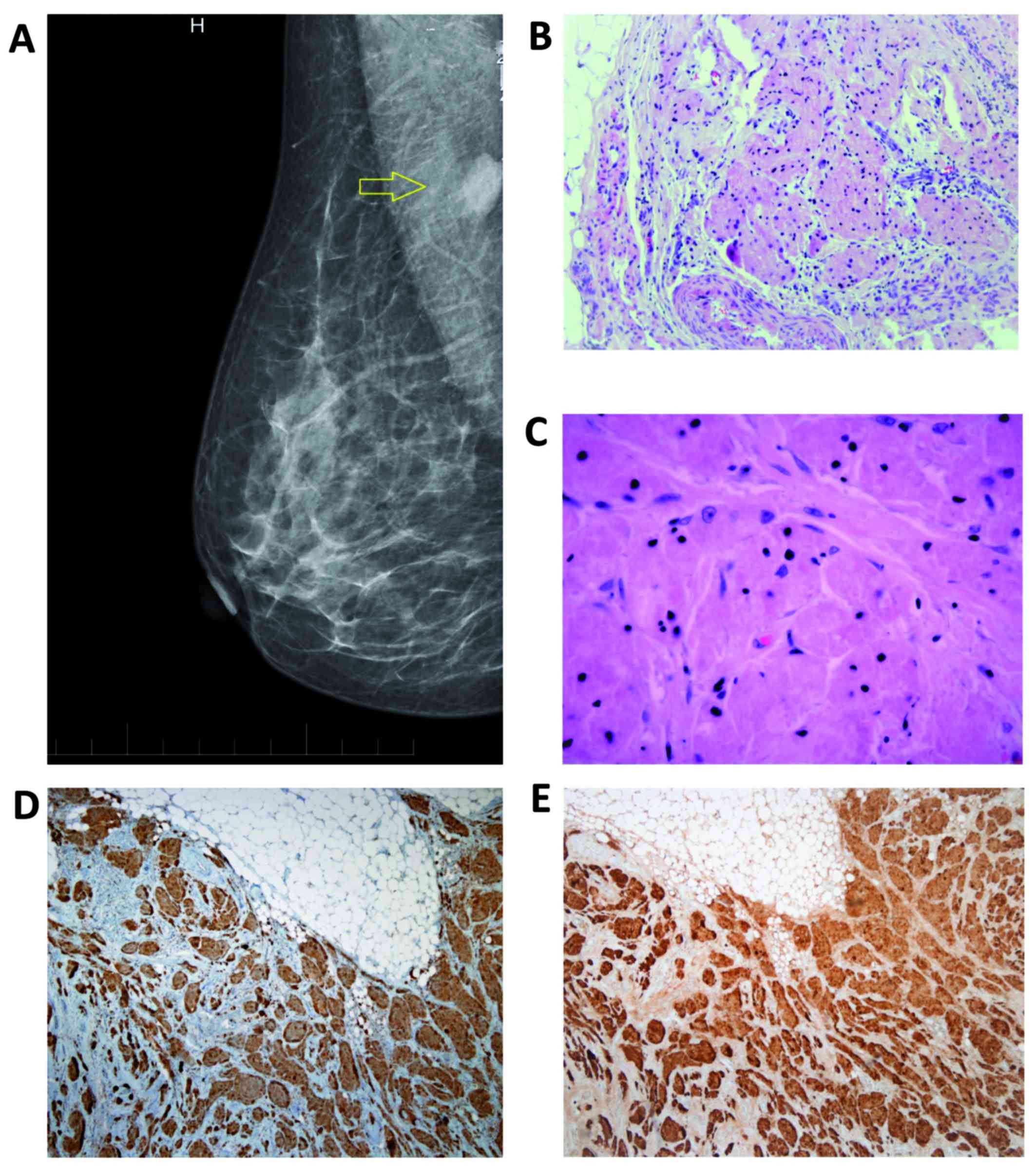
our Breast Unit in June, 2016. Mammography suggested a circumscribed, round, radiopaque lesion with unsharp contours in the right axillary region (Fig. 1A). Breast ultrasonography revealed an oval-shaped, low-echoic tumor of unclear aetiology. Infiltrating ductal carcinoma could not be excluded. A ultrasound guided large core-needle biopsy of the tumor was provided. Histologic examination of the core biopsy specimen pointed to benign form of GCT. Lumpectomy was performed and benign granular cell tumor was diagnosed by postoperative histopathologic examination (Fig. 1B-E). The surgically removed specimen was fixed using 10% neutral buffered formalin for 24 h. The fixed specimen was trimmed with a scalpel to fit into a tissue cassette. It was processed in an automated tissue processing machine (BenchMark XT; Ventana Medical Systems, Inc., Tucson, AZ, USA). The processing included dehydration, clearing, and embedding, in which the specimens were infiltrated with paraffin wax to create paraffin blocks. These blocks were cut with a rotary microtome (Leica RM2235, Leica Biosystems GmbH, Nussloch, Germany) to produce 5 µm sections. The sections were then stained with hematoxylin and eosin (H&E). Histological evaluation of the surgical specimens revealed tumor cells with a distinctive granular eosinophilic cytoplasm associated with typical nuclei, without increase in nuclear division or another signs of malignancy. Benign breast parenchyma and mature fat tissue was present between the tumor structures (Fig. 1B and C). Definitive diagnosis of GCT was established using immunohistochemistry (IHC). The immunostaining for S100 protein (mouse monoclonal antibody to S100; clone 4C4.9; cat. no. ab4066; 1:100 dilution, incubation for 30 min at 25°C; Abcam, Cambridge, UK) and CD68 (mouse monoclonal antibody, clone C68/684; cat. no. ab201340; 1:100 dilution, incubation for 30 min at 25°C; Abcam, Cambridge, UK) revealed positive results (Fig. 1D and E). IHC analysis of the tumor with anti-cytokeratins antibody (mouse monoclonal antibody to pan Cytokeratin; clone AE1/AE3 + 5D3; cat. no. ab86734; 1:200 dilution, incubation for 30 min at 37°C; Abcam, Cambridge, UK) showed negative results. There was also diffuse steroid receptors (estrogen and progesteron receptor) negativity. The post-operative course was uneventful and the patient was discharged home on post-operative day 7. The patient is now 4 months post-surgery and remains disease-free. Written informed consent was obtained from the patient.

Figure 1.

A circumscribed radiopaque lesion i with unsharp contour (arrow) in the right axilla in X-ray digital mammography (A). Granular cell tumor (hematoxilin and eosin staining): Nests of large cells with abundant granular eosinophilic cytoplasm and small nuclei (magnification, ×40) (B). Large, round and oval, slightly irregular cells with abundant granular eosinophilic cytoplasm with small, uniform, central nuclei, with inconspicuous nucleoli (magnification, ×200) (C). The immunostaining for CD68 (D) and S100 protein (E) revealed positive results (magnification, ×100).

Discussion

GCTs account for an incidence of 0.5% among soft tissue tumors (3–6). It may occur throughout the body, usually in the head and neck, skin or subcutaneous tissues of the trunk and upper extremities. Recently, Costa Almeida et al (7) published a case report of GCT on the upper limb and reviewed 21 patients with this GCT localization from the literature. When occurring in the breast, as it occurs in 5–8% of all cases of GCT, they present mostly as painless rounded nodules (4,6). GCT of the breast may mimic breast cancer both clinically and radiologically (8–10). These lesions have been defined as ranging from a round well-circumscribed mass to an indistinct or spiculated lesion on mammography (8,9). Microcalcifications are not normally a feature of GCTs. On ultrasound, GCTs can present as solid, poorly marginated lesions with marked posterior shadowing or as more benign-appearing well-circumscribed solid masses (8,9). MRI findings in a patient with GCT were described by Scaranelo et al (10). In contrast to clinical findings, mammography, ultrasonography and MRI positron emission tomography (PET) with 2-[fluorine-18] fluoro-2-deoxy-d-glucose (FDG) can correctly differentiate GCT from a malignant tumor (11). Fine-needle aspiration cytology and frozen section methods are inadequate for definitive diagnosis of GCT (12). The large core-needle biopsy did accurately predict a GCT in our patient.

The appearance of a GCT in axillary region is extremely rare. Aoyama et al (3), reported in their study of six cases of GCTs, a 54-year-old woman with a GCT in the left axillary cavity, which is very similar to our patient. Another patient with similar location of GCT in the upper outer quadrant of the right breast which appeared to be attached to the underlying pectoralis major muscle, was described by Patel et al (9). The mammographic appearence of the GCT was very similar to the patient from our case report. A pediatric GCT in a 15-year-old female in the right upper outer quadrant with no associated lymphadenopathy was recently refered by Heinzerling et al (13). Delaloye et al (14) reported a rare case of benign GCT of the breast associated with multiple similar lesions of the scalp, the right shoulder, the right flank, the abdominal wall and the vulva, treated with wide excision. Al-Ahmadie et al (15) documented in their report a GCT of the breast coexisting with an ipsilateral infiltrating ductal carcinoma, infiltrated each other. Coates et al (16) reported a case of a patient with a large infra-mammary fold GCT, the management of which required a multidisciplinary operative approach due to extensive chest wall invasion. Malignant forms of GCTs are very rare (1–3% of all GCT cases). Chen et al (6) and Akahane et al (12) described malignant GCTs in breast. Criteria for malignancy are not consistent; adjacent tissue and/or vascular invasion, high mitotic activity, and size >4–5 cm were discussed, but only the presence of metastases was accepted as explicit criterion (6,7). No data exist about the efficacy of adjuvant therapy in GCT treatment.

GCTs are macroscopically, solid, firm tumours with a yellowish-white cross sectional surface (10,11,13). The histogenesis of GCT remains uncertain, however the hypothesis of a neural or neuroectodermal origin is supported by the presence of the S-100 protein, typically expressed by these neoplastic cells, and by the similar ultrastructural features of the tumour cells and Schwann cells (Fig. 1E). On pathological examination they can be identified using both microscopic and immunohistochemical features. The cells have a distinctive granular eosinophilic cytoplasm associated with typical nuclei (Fig. 1B and C). Immunohistochemically they are positive for S100 protein, CD68 (Fig. 1D) and neuron specific endolase (NSE). Fine and Li (17) suggested interaction between expression of calretinin and the alpha-subunit of inhibin in granular cell tumors.

In conclusion, GCT of the breast is a usually benign disease of the breast which may mimic breast cancer both clinically and radiologically. Its presentation in axillary region is very rare. The definitive diagnosis is made by immunohistochemical examination. Clinicians should be aware of this finding in the differential diagnosis of breast and axillary masses to prevent overtreatment.

References

1 

Weber CO: Anatomische untersuchung einer hypertrophischen zunge nebst bemerkungen über die neubildung quergestreifter muskelfasern anatomical examination of a hypertrophic tongue as well as remarks on the new formation of transverse muscle fibers. Virchows Arch A Pathol Anat. 7:115–125. 1954. View Article : Google Scholar

2 

Abrikossoff AI: Weitere untersuchungen über myoblastenmyome. Virchows Arch Pathol Anat Physiol Klin Med. 280:723–740. 1931. View Article : Google Scholar

3 

Aoyama K, Kamio T, Hirano A, Seshimo A and Kameoka S: Granular cell tumors: A report of six cases. World J Surg Oncol. 10:2042012. View Article : Google Scholar : PubMed/NCBI

4 

Pergel A, Yucel AF, Karaca AS, Aydin I, Sahin DA and Demirbag N: A therapeutic and diagnostic dilemma: Granular cell tumor of the breast. Case Rep Med. 2011:9721682011. View Article : Google Scholar : PubMed/NCBI

5 

Gogas J, Markopoulos C, Kouskos E, Gogas H, Mantas D, Antonopoulou Z and Kontzoglou K: Granular cell tumor of the breast: A rare lesion resembling breast cancer. Eur J Gynaecol Oncol. 23:333–334. 2002.PubMed/NCBI

6 

Chen J, Wang L, Xu J, Pan T, Shen J, Hu W and Yuan X: Malignant granular cell tumor with breast metastasis: A case report and review of the literature. Oncol Lett. 4:63–66. 2012. View Article : Google Scholar : PubMed/NCBI

7 

Costa Almeida CE, Caroço T, Silva M and Albano MN: Abrikossoff's tumour on the upper limb: A rare presentation. BMJ Case Rep. 2017:pii: bcr-2017-222006. 2017.

8 

Yang WT, Edeiken-Monroe B, Sneige N and Fornage BD: Sonographic and mammographic appearances of granular cell tumors of the breast with pathological correlation. J Clin Ultrasound. 34:153–160. 2006. View Article : Google Scholar : PubMed/NCBI

9 

Patel A, Lefemine V, Yousuf SM and Abou-Samra W: Granular cell tumour of the pectoral muscle mimicking breast cancer. Cases J. 1:1422008. View Article : Google Scholar : PubMed/NCBI

10 

Scaranelo AM, Bukhanov K, Crystal P, Mulligan AM and O'Malley FP: Granular cell tumour of the breast: MRI findings and review of the literature. Br J Radiol. 80:970–974. 2007. View Article : Google Scholar : PubMed/NCBI

11 

Hoess C, Freitag K, Kolben M, Allgayer B, Laemmer-Skarke I, Nathrath WB, Avril N, Roemer W, Schwaiger M and Graeff H: FDG PET evaluation of granular cell tumor of the breast. J Nucl Med. 39:1398–1401. 1998.PubMed/NCBI

12 

Akahane K, Kato K, Ogiso S, Sakaguchi K, Hashimoto M, Ishikawa A, Kato T, Fuwa Y, Takahashi A and Kobayashi K: Malignant granular cell tumor of the breast: Case report and literature review. Breast Cancer. 22:317–323. 2015. View Article : Google Scholar : PubMed/NCBI

13 

Heinzerling NP, Koehler SM, Szabo S and Wagner AJ: Pediatric granular cell tumor of the breast: A case report and review of the literature. Case Rep Surg. 2015:5689402015.PubMed/NCBI

14 

Delaloye JF, Seraj F, Guillou L, Genton CY, Anciaux-Le Teno D, Schnyder P and De Grandi P: Granular cell tumor of the breast: A diagnostic pitfall. Breast. 11:316–319. 2002. View Article : Google Scholar : PubMed/NCBI

15 

Al-Ahmadie H, Hasselgren PO, Yassin R and Mutema G: Colocalized granular cell tumor and infiltrating ductal carcinoma of the breast. Arch Pathol Lab Med. 126:731–733. 2002.PubMed/NCBI

16 

Coates SJ, Mitchell K, Olorunnipa OB, DeSimone RA, Otterburn DM and Simmons RM: An unusual breast lesion: Granular cell tumor of the breast with extensive chest wall invasion. J Surg Oncol. 110:345–347. 2014. View Article : Google Scholar : PubMed/NCBI

17 

Fine SW and Li M: Expression of calretinin and the alpha-subunit of inhibin in granular cell tumors. Am J Clin Pathol. 119:259–264. 2003. View Article : Google Scholar : PubMed/NCBI

Related Articles

  • Abstract
  • View
  • Download
  • Twitter
Copy and paste a formatted citation
Spandidos Publications style
Jakubowska K, Kańczuga‑Koda L, Kisielewski W, Koda M and Famulski W: Squamous cell carcinoma of the breast as a clinical diagnostic challenge. Mol Clin Oncol 8: 582-586, 2018.
APA
Jakubowska, K., Kańczuga‑Koda, L., Kisielewski, W., Koda, M., & Famulski, W. (2018). Squamous cell carcinoma of the breast as a clinical diagnostic challenge. Molecular and Clinical Oncology, 8, 582-586. https://doi.org/10.3892/mco.2018.1581
MLA
Jakubowska, K., Kańczuga‑Koda, L., Kisielewski, W., Koda, M., Famulski, W."Squamous cell carcinoma of the breast as a clinical diagnostic challenge". Molecular and Clinical Oncology 8.4 (2018): 582-586.
Chicago
Jakubowska, K., Kańczuga‑Koda, L., Kisielewski, W., Koda, M., Famulski, W."Squamous cell carcinoma of the breast as a clinical diagnostic challenge". Molecular and Clinical Oncology 8, no. 4 (2018): 582-586. https://doi.org/10.3892/mco.2018.1581
Copy and paste a formatted citation
x
Spandidos Publications style
Jakubowska K, Kańczuga‑Koda L, Kisielewski W, Koda M and Famulski W: Squamous cell carcinoma of the breast as a clinical diagnostic challenge. Mol Clin Oncol 8: 582-586, 2018.
APA
Jakubowska, K., Kańczuga‑Koda, L., Kisielewski, W., Koda, M., & Famulski, W. (2018). Squamous cell carcinoma of the breast as a clinical diagnostic challenge. Molecular and Clinical Oncology, 8, 582-586. https://doi.org/10.3892/mco.2018.1581
MLA
Jakubowska, K., Kańczuga‑Koda, L., Kisielewski, W., Koda, M., Famulski, W."Squamous cell carcinoma of the breast as a clinical diagnostic challenge". Molecular and Clinical Oncology 8.4 (2018): 582-586.
Chicago
Jakubowska, K., Kańczuga‑Koda, L., Kisielewski, W., Koda, M., Famulski, W."Squamous cell carcinoma of the breast as a clinical diagnostic challenge". Molecular and Clinical Oncology 8, no. 4 (2018): 582-586. https://doi.org/10.3892/mco.2018.1581
Follow us
  • Twitter
  • LinkedIn
  • Facebook
About
  • Spandidos Publications
  • Careers
  • Cookie Policy
  • Privacy Policy
How can we help?
  • Help
  • Live Chat
  • Contact
  • Email to our Support Team