Spandidos Publications Logo
  • About
    • About Spandidos
    • Aims and Scopes
    • Abstracting and Indexing
    • Editorial Policies
    • Reprints and Permissions
    • Job Opportunities
    • Terms and Conditions
    • Contact
  • Journals
    • All Journals
    • Oncology Letters
      • Oncology Letters
      • Information for Authors
      • Editorial Policies
      • Editorial Board
      • Aims and Scope
      • Abstracting and Indexing
      • Bibliographic Information
      • Archive
    • International Journal of Oncology
      • International Journal of Oncology
      • Information for Authors
      • Editorial Policies
      • Editorial Board
      • Aims and Scope
      • Abstracting and Indexing
      • Bibliographic Information
      • Archive
    • Molecular and Clinical Oncology
      • Molecular and Clinical Oncology
      • Information for Authors
      • Editorial Policies
      • Editorial Board
      • Aims and Scope
      • Abstracting and Indexing
      • Bibliographic Information
      • Archive
    • Experimental and Therapeutic Medicine
      • Experimental and Therapeutic Medicine
      • Information for Authors
      • Editorial Policies
      • Editorial Board
      • Aims and Scope
      • Abstracting and Indexing
      • Bibliographic Information
      • Archive
    • International Journal of Molecular Medicine
      • International Journal of Molecular Medicine
      • Information for Authors
      • Editorial Policies
      • Editorial Board
      • Aims and Scope
      • Abstracting and Indexing
      • Bibliographic Information
      • Archive
    • Biomedical Reports
      • Biomedical Reports
      • Information for Authors
      • Editorial Policies
      • Editorial Board
      • Aims and Scope
      • Abstracting and Indexing
      • Bibliographic Information
      • Archive
    • Oncology Reports
      • Oncology Reports
      • Information for Authors
      • Editorial Policies
      • Editorial Board
      • Aims and Scope
      • Abstracting and Indexing
      • Bibliographic Information
      • Archive
    • Molecular Medicine Reports
      • Molecular Medicine Reports
      • Information for Authors
      • Editorial Policies
      • Editorial Board
      • Aims and Scope
      • Abstracting and Indexing
      • Bibliographic Information
      • Archive
    • World Academy of Sciences Journal
      • World Academy of Sciences Journal
      • Information for Authors
      • Editorial Policies
      • Editorial Board
      • Aims and Scope
      • Abstracting and Indexing
      • Bibliographic Information
      • Archive
    • International Journal of Functional Nutrition
      • International Journal of Functional Nutrition
      • Information for Authors
      • Editorial Policies
      • Editorial Board
      • Aims and Scope
      • Abstracting and Indexing
      • Bibliographic Information
      • Archive
    • International Journal of Epigenetics
      • International Journal of Epigenetics
      • Information for Authors
      • Editorial Policies
      • Editorial Board
      • Aims and Scope
      • Abstracting and Indexing
      • Bibliographic Information
      • Archive
    • Medicine International
      • Medicine International
      • Information for Authors
      • Editorial Policies
      • Editorial Board
      • Aims and Scope
      • Abstracting and Indexing
      • Bibliographic Information
      • Archive
  • Articles
  • Information
    • Information for Authors
    • Information for Reviewers
    • Information for Librarians
    • Information for Advertisers
    • Conferences
  • Language Editing
Spandidos Publications Logo
  • About
    • About Spandidos
    • Aims and Scopes
    • Abstracting and Indexing
    • Editorial Policies
    • Reprints and Permissions
    • Job Opportunities
    • Terms and Conditions
    • Contact
  • Journals
    • All Journals
    • Biomedical Reports
      • Information for Authors
      • Editorial Policies
      • Editorial Board
      • Aims and Scope
      • Abstracting and Indexing
      • Bibliographic Information
      • Archive
    • Experimental and Therapeutic Medicine
      • Information for Authors
      • Editorial Policies
      • Editorial Board
      • Aims and Scope
      • Abstracting and Indexing
      • Bibliographic Information
      • Archive
    • International Journal of Epigenetics
      • Information for Authors
      • Editorial Policies
      • Editorial Board
      • Aims and Scope
      • Abstracting and Indexing
      • Bibliographic Information
      • Archive
    • International Journal of Functional Nutrition
      • Information for Authors
      • Editorial Policies
      • Editorial Board
      • Aims and Scope
      • Abstracting and Indexing
      • Bibliographic Information
      • Archive
    • International Journal of Molecular Medicine
      • Information for Authors
      • Editorial Policies
      • Editorial Board
      • Aims and Scope
      • Abstracting and Indexing
      • Bibliographic Information
      • Archive
    • International Journal of Oncology
      • Information for Authors
      • Editorial Policies
      • Editorial Board
      • Aims and Scope
      • Abstracting and Indexing
      • Bibliographic Information
      • Archive
    • Medicine International
      • Information for Authors
      • Editorial Policies
      • Editorial Board
      • Aims and Scope
      • Abstracting and Indexing
      • Bibliographic Information
      • Archive
    • Molecular and Clinical Oncology
      • Information for Authors
      • Editorial Policies
      • Editorial Board
      • Aims and Scope
      • Abstracting and Indexing
      • Bibliographic Information
      • Archive
    • Molecular Medicine Reports
      • Information for Authors
      • Editorial Policies
      • Editorial Board
      • Aims and Scope
      • Abstracting and Indexing
      • Bibliographic Information
      • Archive
    • Oncology Letters
      • Information for Authors
      • Editorial Policies
      • Editorial Board
      • Aims and Scope
      • Abstracting and Indexing
      • Bibliographic Information
      • Archive
    • Oncology Reports
      • Information for Authors
      • Editorial Policies
      • Editorial Board
      • Aims and Scope
      • Abstracting and Indexing
      • Bibliographic Information
      • Archive
    • World Academy of Sciences Journal
      • Information for Authors
      • Editorial Policies
      • Editorial Board
      • Aims and Scope
      • Abstracting and Indexing
      • Bibliographic Information
      • Archive
  • Articles
  • Information
    • For Authors
    • For Reviewers
    • For Librarians
    • For Advertisers
    • Conferences
  • Language Editing
Login Register Submit
  • This site uses cookies
  • You can change your cookie settings at any time by following the instructions in our Cookie Policy. To find out more, you may read our Privacy Policy.

    I agree
Search articles by DOI, keyword, author or affiliation
Search
Advanced Search
presentation
Molecular and Clinical Oncology
Join Editorial Board Propose a Special Issue
Print ISSN: 2049-9450 Online ISSN: 2049-9469
Journal Cover
April-2018 Volume 8 Issue 4

Full Size Image

Sign up for eToc alerts
Recommend to Library

Journals

International Journal of Molecular Medicine

International Journal of Molecular Medicine

International Journal of Molecular Medicine is an international journal devoted to molecular mechanisms of human disease.

International Journal of Oncology

International Journal of Oncology

International Journal of Oncology is an international journal devoted to oncology research and cancer treatment.

Molecular Medicine Reports

Molecular Medicine Reports

Covers molecular medicine topics such as pharmacology, pathology, genetics, neuroscience, infectious diseases, molecular cardiology, and molecular surgery.

Oncology Reports

Oncology Reports

Oncology Reports is an international journal devoted to fundamental and applied research in Oncology.

Experimental and Therapeutic Medicine

Experimental and Therapeutic Medicine

Experimental and Therapeutic Medicine is an international journal devoted to laboratory and clinical medicine.

Oncology Letters

Oncology Letters

Oncology Letters is an international journal devoted to Experimental and Clinical Oncology.

Biomedical Reports

Biomedical Reports

Explores a wide range of biological and medical fields, including pharmacology, genetics, microbiology, neuroscience, and molecular cardiology.

Molecular and Clinical Oncology

Molecular and Clinical Oncology

International journal addressing all aspects of oncology research, from tumorigenesis and oncogenes to chemotherapy and metastasis.

World Academy of Sciences Journal

World Academy of Sciences Journal

Multidisciplinary open-access journal spanning biochemistry, genetics, neuroscience, environmental health, and synthetic biology.

International Journal of Functional Nutrition

International Journal of Functional Nutrition

Open-access journal combining biochemistry, pharmacology, immunology, and genetics to advance health through functional nutrition.

International Journal of Epigenetics

International Journal of Epigenetics

Publishes open-access research on using epigenetics to advance understanding and treatment of human disease.

Medicine International

Medicine International

An International Open Access Journal Devoted to General Medicine.

Journal Cover
April-2018 Volume 8 Issue 4

Full Size Image

Sign up for eToc alerts
Recommend to Library

  • Article
  • Citations
    • Cite This Article
    • Download Citation
    • Create Citation Alert
    • Remove Citation Alert
    • Cited By
  • Similar Articles
    • Related Articles (in Spandidos Publications)
    • Similar Articles (Google Scholar)
    • Similar Articles (PubMed)
  • Download PDF
  • Download XML
  • View XML
Case Report

Large-cell neuroendocrine carcinoma arising in the endometrium: A case report

  • Authors:
    • Jumpei Ogura
    • Yasushi Adachi
    • Koji Yasumoto
    • Akiharu Okamura
    • Hirofumi Nonogaki
    • Kazuyo Kakui
    • Koji Yamanoi
    • Koh Suginami
    • Takashi Koyama
    • Susumu Ikehara
  • View Affiliations / Copyright

    Affiliations: Department of Obstetrics and Gynecology, Toyooka Hospital, Toyooka, Hyogo 668-8501, Japan, Department of Diagnostic Pathology, Toyooka Hospital, Toyooka, Hyogo 668-8501, Japan, Department of Diagnostic Pathology, Kakogawa Central City Hospital, Kakogawa, Hyogo 675-8611, Japan, Department of Radiology, Toyooka Hospital, Toyooka, Hyogo 668‑8501, Japan, Professor Emeritus, Kansai Medical University, Hirakata, Osaka 573-1010, Japan
  • Pages: 571-574
    |
    Published online on: February 26, 2018
       https://doi.org/10.3892/mco.2018.1583
  • Expand metrics +
Metrics: Total Views: 0 (Spandidos Publications: | PMC Statistics: )
Metrics: Total PDF Downloads: 0 (Spandidos Publications: | PMC Statistics: )
Cited By (CrossRef): 0 citations Loading Articles...

This article is mentioned in:


Abstract

Large-cell neuroendocrine carcinoma (LCNEC) of the endometrium is an extremely rare, high-grade malignant tumor. We herein report a case of a rapidly growing LCNEC arising in the endometrium. A 52-year-old woman was referred to Toyooka Hospital (Tooyoka, Japan) due to genital bleeding in February 2016. There had been no abnormalities on a regular gynecological and physical examination 3 months prior to the consultation. Imaging (computed tomography and magnetic resonance imaging) and a pelvic examination revealed a tumor sized 16.9x8.4x7.8 mm occupying the intrauterine cavity and extending into the vaginal cavity. Multiple metastatic pelvic and paraaortic lymph nodes were also identified. Continuous bleeding from the tumor was observed, and a blood examination revealed anemia, which was likely due to that bleeding. Biopsy of the tumor was performed, and large atypical cells were identified. The tumor cells were negative for cytokeratin AE1/AE3 and chromogranin A, but positive for CD56 and synaptophysin. There was also an abundance of Ki-67-positive cells in the tumor, altogether suggesting that the tumor was an LCNEC. The patient succumbed to the disease 36 days after the first consultation. Based on the findings of the present case and previously published cases, LCNECs arising in the endometrium may progress rapidly and are associated with an unfavourable outcome. LCNEC should be included in the differential diagnosis in cases of rapidly growing tumors of the uterine corpus.

Introduction

Schwannomas are benign, slowly growing neoplasms composed of neoplastic Schwann cells (1). Melanocytic schwannoma (MS) is a rare schwannoma variant composed of melanin-producing cells with the ultrastructural characteristics of Schwann cells (2), accounting for ~1% of primary peripheral nerve sheath tumors (1,2). MS is primarily considered to be a benign tumor, with a relatively rarely reported propensity to metastasize (2,3); however, recent published literature suggests that MS must be reconsidered as a malignant neoplasm (4) with a greater potential to metastasize. The most common location of the tumor is in the nerve roots (5–7). MS is also encountered in extramedullary sites and the peripheral nervous system, but is particularly rare in intramedullary sites (8,9). There are only 8 reported cases of intramedullary MS (IMS) (1,8–14); we herein report the ninth IMS case in a 40-year-old man with a lesion located in the cervical cord, which was diagnosed based on the magnetic resonance imaging (MRI) and histopathological findings and treated with surgery.

Case report

A 40-year-old man presented in the Second Affiliated Hospital of Zhejiang University School of Medicine in March 2017 with left arm numbness that gradually worsened over a period of 4 months. The patient did not have any other neurological symptoms. Upon physical examination, no obvious deposition of pigment was found in the skin and mucosa. There was no previous history of a surgical procedure for the removal of MS. Upon neurological examination, the distal pinprick sensation in the left upper limb was slightly decreased. The muscle strength in all upper and lower limbs was unaffected (5/5) and the muscle tone was normal. All limb tendon reflexes were normal. No positive pathological reflexes were present bilaterally.

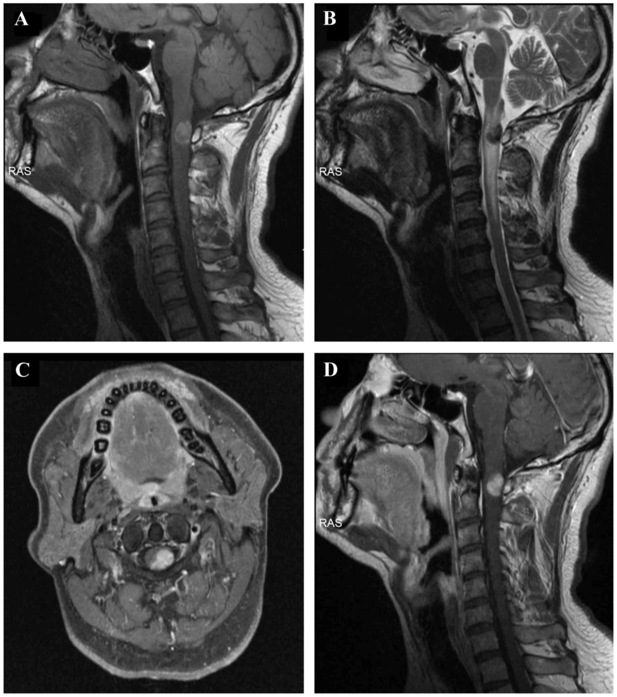
An MRI of the cervical spine revealed an intramedullary mass, sized 1.5×1.0 cm, within the spinal canal. The mass occupied 60% of the spinal canal at the level of C1-C2, with displacement of the spinal cord to the right side (Fig. 1A-D). The mass was T1 hyperintense (Fig. 1A) and T2 hypointense (Fig. 1B). Following enhancement, the mass was homogeneously enhanced (Fig. 1C and D). There was spinal cord expansion and edema above and below the mass, as evidenced by T2 hyperintensity (Fig. 1B). The radiological and clinical findings were consistent with melanoma, including primarily intramedullary melanoma or metastatic malignant melanoma. As primary tumors were not found anywhere on the skin of the limbs or the trunk, metastatic malignant melanoma was not considered in the initial diagnosis. A cavernous malformation with subacute hematoma was considered in the differential diagnosis; however, the history did not include a sudden onset, and the patient's symptoms gradually worsened over a relatively long period of time, which is not consistent with haemorrhagic manifestations. Finally, the preoperative diagnosis was a primarily intramedullary melanoma.

Figure 1.

The mass was pre-contrast (A) T1 hyperintense and (B) T2 hypointense. (C and D) The mass lesion, which appeared to be intramedullary at the level of C1-C2, exhibited homogenous enhancement.

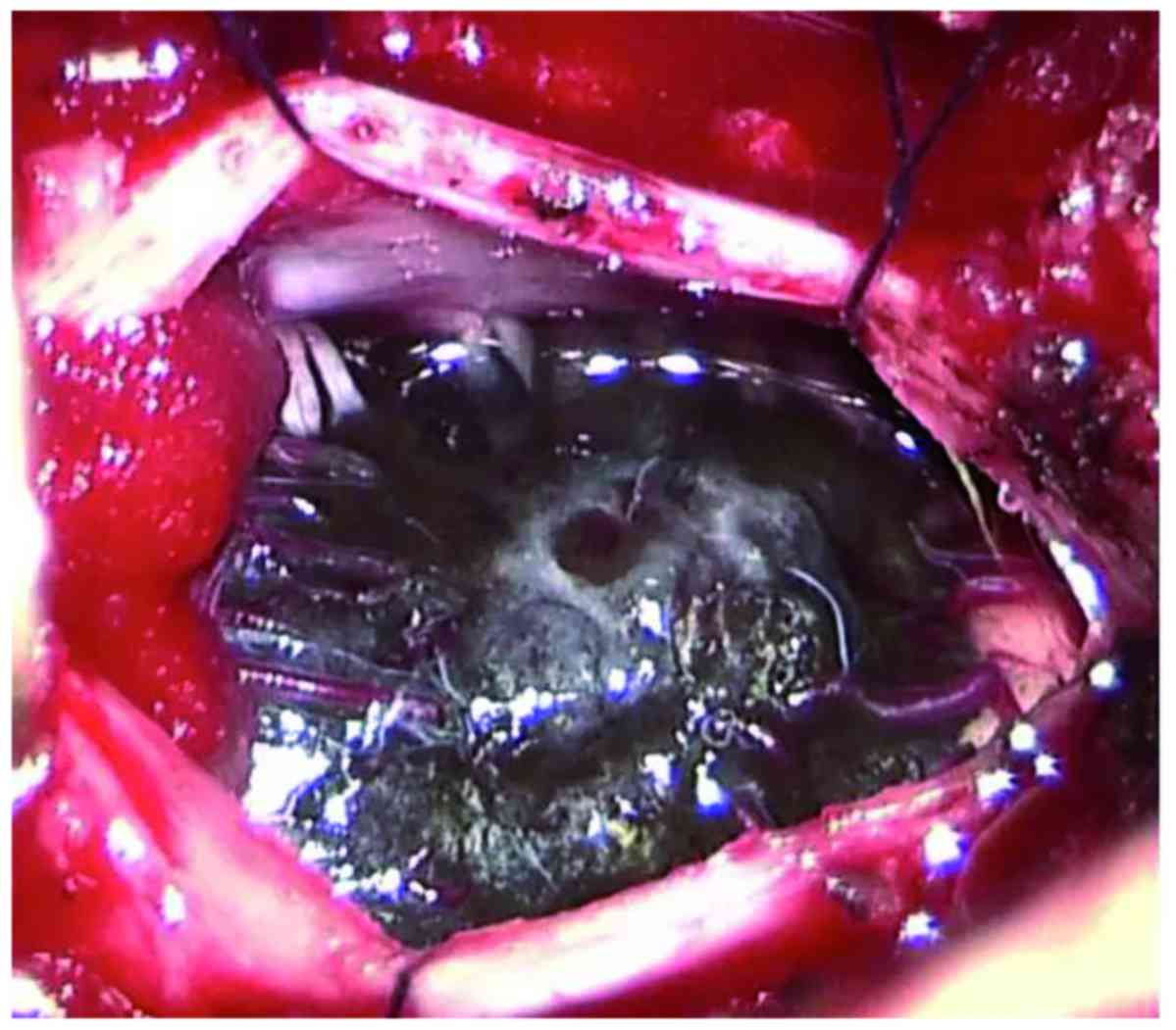
A C1 laminectomy was performed. The dura mater and arachnoid sheath were opened longitudinally and dark pigmentation was visible through the dura (Fig. 2). The dura was opened in the midline to expose an intramedullary tumor, with neurophysiological monitoring during surgery. The tumor invaded deeply into the spinal cord and had an unclear boundary. Spinal nerve roots were also invaded by the tumor. Due to the difficulty of complete removal and an intraoperative diagnosis of metastatic malignant melanoma by frozen section biopsy, the lesion was partially resected. The left arm numbness partially subsided 2 weeks later after the surgery.

Figure 2.

Intraoperative photograph showing an intramedullary lesion following opening of the dura mater.

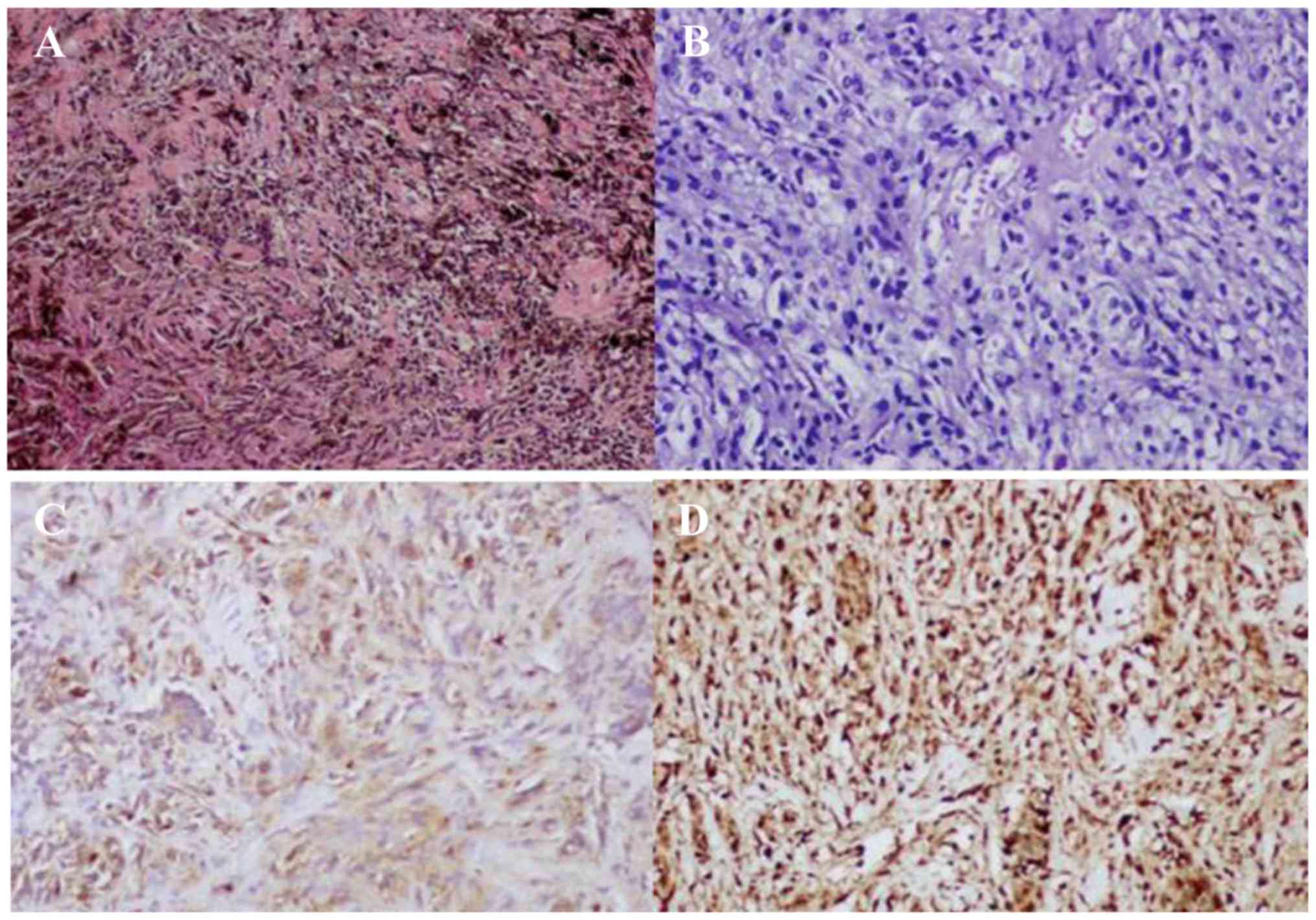
Sections stained with hematoxylin-eosin revealed that the tumor was composed of polygonal epithelioid and spindle-shaped cells, with abundant cytoplasm containing melanin granules (Fig. 3A). Depigmented sections revealed no obvious atypia of the tumor cell nuclei (Fig. 3B). Immunocytochemistry for human melanoma black 45 (Fig. 3C), p53, vimentin and S-100 (Fig. 3D) was positive, whereas the tumor cells were negative for melan-A and epithelial membrane antigen. The Ki-67 proliferative index was <1%. The pathological diagnosis was IMS. During the follow-up οn May 2017, the patient status was stable with the left arm numbness.

Figure 3.

(A-D) Light microscopy findings of melanotic schwannoma. (A) The tumor cells had abundant cytoplasm containing melanin granules and spindle-shaped cell nuclei; hematoxylin and eosin staining; magnification, ×40. (B) Following depigmentation, no obvious atypia of the nuclei was observed; magnification, ×10. Immunocytochemical examination revealed that the tumor cells were (C) human melanoma black 45-positive and (D) S-100-positive; magnification, ×10.

Written informed consent was obtained from the patient regarding the publication of the case details and associated images.

Discussion

MS is a tumor derived from progenitor neural crest cells that can differentiate into both Schwann cells and melanocytes, which is characterized by deposition of melanin in the Schwann cell cytoplasm (2). Theories for the production of melanin by these cells include neoplastic differentiation of neural crest cells into Schwann cells with melanogenetic properties, and melanocytic transformation of previously normal Schwann cells (1). The presence of psammoma bodies is typical of the psammomatous variant of MS. Approximately 50% of psammomatous MS are part of the Carney syndrome (along with myxomas, skin pigmentation and endocrine tumors or overactivity) (15). The most frequent sites of MS are the dorsal spinal nerve roots, sympathetic chain, acoustic nerve, cerebellum and orbit (10). IMS is particularly rare and, to the best of our knowledge, this is the ninth case reported to date (1,8–14) (Table I).

Table I.

Summary of the intramedullary melanotic schwannoma cases reported in the literature.

Table I.

Summary of the intramedullary melanotic schwannoma cases reported in the literature.

Case no.AuthorsSexAge, yearsLocationTreatmentOutcomeFollow-up(Refs.)
1Solomon et alMale69Caudal medulla and C3Gross total removal––(11)
2Marchese et alFemale72C4-C6Partial removalFunctional recovery–(12)
3Sola-Pérez et alFemale63C7-T1Needle aspiration––(13)
4Acciarri et alFemale44T2-T3Gross total removalPartial neurological recovery–(14)
5Santaguida et alMale35C4-C5Gross total removalPartial neurological recoveryRecurrence at 2 years, radiotherapy, and repeatresection at 4 years(1)
6Mouchaty et alFemale56ConusGross total removalPartial neurological recoveryNo recurrence at 12 months(9)
7Hoover et alFemale62T11Gross total removalGood neurological recoveryNo recurrence at 10 months(8)
8Mohamed et al  Male43T9-T10Gross total removalGood neurological recoveryNo recurrence at 12 months(10)
9Present caseMale40C1-C2Partial removalPartial recovery–

MRI is currently the optimal diagnostic modality for evaluating lesions of the spinal cord. Melanin may be associated with shortened T1 and T2 relaxation times due to its content of paramagnetic free radicals; thus, melanotic lesions appear hyperintense on T1-weighted and hypointense on T2-weighted images (16). By contrast, non-melanotic tumors are hypointense on T1-weighted and hyperintense on T2-weighted images, which helps differentiate melanotic from non-melanotic lesions. However, subacute hematoma is difficult to differentiate from melanotic lesions, as subacute hematoma may display similar characteristics on MRI (17). In the present case, a cavernous malformation with subacute hematoma was also initially considered on T1- and T2-weighted images. However, considering the patient's medical history, the onset of the symptoms was not sudden but rather a chronic process; thus, subacute hematoma was not considered in the initial preoperative diagnosis. Following administration of gadolinium, the tumor typically exhibited homogeneous enhancement; however, enhancement may be heterogeneous in the presence of haemorrhage within the lesion (10). A diagnosis of intramedullary schwannoma may be confidently made when there is continuity of the intramedullary lesion with a contrast-enhanced thickened spinal root (18); however, no obvious enhancing thickened spinal root involvement was observed in the present IMS case.

From the 8 previously reported cases (Table I), the main treatment for IMS is gross total removal. Approximately 10% of MS cases are reported to exhibit an aggressive clinical course, with local recurrence and metastasis (1,16). Due to the rarity of IMS, the role of radiotherapy has not been established. The fifth documented case (Table I) of a patient with IMS recurred 2 years after initial gross total removal. Subsequently, the patient received radiotherapy, but the tumor progressed 2 years after radiotherapy and the patient was again treated with surgery. Considering the recurrence potential of IMS, annual follow-up of patients with IMS with MRI is required (8). In the present case, the lesion was partially resected, due to the difficulty of complete removal, and an intraoperative diagnosis of metastatic malignant melanoma was made by frozen section biopsy. Due to the rarity of IMS, intraoperative frozen section diagnosis is challenging without immunohistochemical examination.

In conclusion, MRI is the preferred method for evaluating lesions of the spinal cord. A standard IMS would typically be T1 hyperintense, T2 hypointense and homogeneously enhanced. However, although IMS has these characteristic MRI features, preoperative diagnosis as well as intraoperative frozen section diagnosis are challenging due to the rarity of this tumor. Correct diagnosis is crucial for management planning; therefore, immunohistochemical examination is warranted. In addition, careful follow-up is required for all IMS patients, particularly when the mass cannot be completely resected.

References

1 

Santaguida C, Sabbagh AJ, Guiot MC and Del Maestro RF: Aggressive intramedullary melanotic schwannoma: Case report. Neurosurgery. 55:14302004. View Article : Google Scholar : PubMed/NCBI

2 

Zhang HY, Yang GH, Chen HJ, Wei B, Ke Q, Guo H, Ye L, Bu H, Yang K and Zhang YH: Clinicopathological, immunohistochemical and ultrastructural study of 13 cases of melanotic schwannoma. Chin Med J (Engl). 118:1451–1461. 2005.PubMed/NCBI

3 

Vallat-Decouvelaere AV, Wassef M, Lot G, Catala M, Moussalam M, Caruel N and Mikol J: Spinal melanotic schwannoma: A tumour with poor prognosis. Histopathology. 35:558–566. 1999. View Article : Google Scholar : PubMed/NCBI

4 

Torres-Mora J, Dry S, Li X, Binder S, Amin M and Folpe AL: Malignant melanotic schwannian tumor: A clinicopathologic, immunohistochemical and gene expression profiling study of 40 cases, with a proposal for the reclassification of ‘melanotic schwannoma’. Am J Surg Pathol. 38:94–105. 2014. View Article : Google Scholar : PubMed/NCBI

5 

De Cerchio L, Contratti F and Fraioli MF: Dorsal dumb-bell melanotic schwannoma operated on by posterior and anterior approach: Case report and a review of the literature. Eur Spine J. 15 Suppl 5:S664–S669. 2006. View Article : Google Scholar

6 

Er U, Kazanci A, Eyriparmak T, Yigitkanli K and Senveli E: Melanotic schwannoma. J Clin Neurosci. 14:676–678. 2007. View Article : Google Scholar : PubMed/NCBI

7 

Martin-Reay DG, Shattuck MC and Guthrie FW Jr: Psammomatous melanotic schwannoma: An additional component of Carney's complex. Report of a case. Am J Clin Pathol. 95:484–489. 1991. View Article : Google Scholar : PubMed/NCBI

8 

Hoover JM, Bledsoe JM, Giannini C and Krauss WE: Intramedullary melanotic schwannoma. Rare Tumors. 4:e32012. View Article : Google Scholar : PubMed/NCBI

9 

Mouchaty H, Conti R, Buccoliero AM and Conti P: Intramedullary melanotic schwannoma of the conus medullaris: A case report. Spinal Cord. 46:703–706. 2008. View Article : Google Scholar : PubMed/NCBI

10 

Mohamed M, Panos S, Baborie A, Das K and Pillay R: Atypical benign melanotic thoracic intradural schwannoma. Br J Neurosurg. 28:411–413. 2014. View Article : Google Scholar : PubMed/NCBI

11 

Solomon RA, Handler MS, Sedelli RV and Stein BM: Intramedullary melanotic schwannoma of the cervicomedullary junction. Neurosurgery. 20:36–38. 1987. View Article : Google Scholar : PubMed/NCBI

12 

Marchese MJ and McDonald JV: Intramedullary melanotic schwannoma of the cervical spinal cord: Report of a case. Surg Neurol. 33:353–355. 1990. View Article : Google Scholar : PubMed/NCBI

13 

Sola-Pérez J, Pérez-Guillermo M, Bas-Bernal A, Giménez-Bascuñana A and Montes-Clavero C: Melanocytic schwannoma: The cytologic aspect in fine-needle aspiration cytology (FNAC): Report of a case located in the spinal cord. Diagn Cytopathol. 11:291–296. 1994. View Article : Google Scholar : PubMed/NCBI

14 

Acciarri N, Padovani R and Riccioni L: Intramedullary melanotic schwannoma. Report of a case and review of the literature. Br J Neurosurg. 13:322–325. 1999. View Article : Google Scholar : PubMed/NCBI

15 

Shields LB, Glassman SD, Raque GH and Shields CB: Malignant psammomatous melanotic schwannoma of the spine: A component of carney complex. Surg Neurol Int. 2:1362011. View Article : Google Scholar : PubMed/NCBI

16 

Tawk RG, Tan D, Mechtler L and Fenstermaker RA: Melanotic schwannoma with drop metastases to the caudal spine and high expression of CD117 (c-kit). J Neurooncol. 71:151–156. 2005. View Article : Google Scholar : PubMed/NCBI

17 

Höllinger P, Godoy N and Sturzenegger M: Magnetic resonance imaging findings in isolated spinal psammomatous melanotic schwannoma. J Neurol. 246:1100–1102. 1999. View Article : Google Scholar : PubMed/NCBI

18 

Colosimo C, Cerase A, Denaro L, Maira G and Greco R: Magnetic resonance imaging of intramedullary spinal cord schwannomas. Report of two cases and review of the literature. J Neurosurg. 99 Suppl 1:S114–S117. 2003.

Related Articles

  • Abstract
  • View
  • Download
  • Twitter
Copy and paste a formatted citation
Spandidos Publications style
Ogura J, Adachi Y, Yasumoto K, Okamura A, Nonogaki H, Kakui K, Yamanoi K, Suginami K, Koyama T, Ikehara S, Ikehara S, et al: Large-cell neuroendocrine carcinoma arising in the endometrium: A case report. Mol Clin Oncol 8: 571-574, 2018.
APA
Ogura, J., Adachi, Y., Yasumoto, K., Okamura, A., Nonogaki, H., Kakui, K. ... Ikehara, S. (2018). Large-cell neuroendocrine carcinoma arising in the endometrium: A case report. Molecular and Clinical Oncology, 8, 571-574. https://doi.org/10.3892/mco.2018.1583
MLA
Ogura, J., Adachi, Y., Yasumoto, K., Okamura, A., Nonogaki, H., Kakui, K., Yamanoi, K., Suginami, K., Koyama, T., Ikehara, S."Large-cell neuroendocrine carcinoma arising in the endometrium: A case report". Molecular and Clinical Oncology 8.4 (2018): 571-574.
Chicago
Ogura, J., Adachi, Y., Yasumoto, K., Okamura, A., Nonogaki, H., Kakui, K., Yamanoi, K., Suginami, K., Koyama, T., Ikehara, S."Large-cell neuroendocrine carcinoma arising in the endometrium: A case report". Molecular and Clinical Oncology 8, no. 4 (2018): 571-574. https://doi.org/10.3892/mco.2018.1583
Copy and paste a formatted citation
x
Spandidos Publications style
Ogura J, Adachi Y, Yasumoto K, Okamura A, Nonogaki H, Kakui K, Yamanoi K, Suginami K, Koyama T, Ikehara S, Ikehara S, et al: Large-cell neuroendocrine carcinoma arising in the endometrium: A case report. Mol Clin Oncol 8: 571-574, 2018.
APA
Ogura, J., Adachi, Y., Yasumoto, K., Okamura, A., Nonogaki, H., Kakui, K. ... Ikehara, S. (2018). Large-cell neuroendocrine carcinoma arising in the endometrium: A case report. Molecular and Clinical Oncology, 8, 571-574. https://doi.org/10.3892/mco.2018.1583
MLA
Ogura, J., Adachi, Y., Yasumoto, K., Okamura, A., Nonogaki, H., Kakui, K., Yamanoi, K., Suginami, K., Koyama, T., Ikehara, S."Large-cell neuroendocrine carcinoma arising in the endometrium: A case report". Molecular and Clinical Oncology 8.4 (2018): 571-574.
Chicago
Ogura, J., Adachi, Y., Yasumoto, K., Okamura, A., Nonogaki, H., Kakui, K., Yamanoi, K., Suginami, K., Koyama, T., Ikehara, S."Large-cell neuroendocrine carcinoma arising in the endometrium: A case report". Molecular and Clinical Oncology 8, no. 4 (2018): 571-574. https://doi.org/10.3892/mco.2018.1583
Follow us
  • Twitter
  • LinkedIn
  • Facebook
About
  • Spandidos Publications
  • Careers
  • Cookie Policy
  • Privacy Policy
How can we help?
  • Help
  • Live Chat
  • Contact
  • Email to our Support Team