Spandidos Publications Logo
  • About
    • About Spandidos
    • Aims and Scopes
    • Abstracting and Indexing
    • Editorial Policies
    • Reprints and Permissions
    • Job Opportunities
    • Terms and Conditions
    • Contact
  • Journals
    • All Journals
    • Oncology Letters
      • Oncology Letters
      • Information for Authors
      • Editorial Policies
      • Editorial Board
      • Aims and Scope
      • Abstracting and Indexing
      • Bibliographic Information
      • Archive
    • International Journal of Oncology
      • International Journal of Oncology
      • Information for Authors
      • Editorial Policies
      • Editorial Board
      • Aims and Scope
      • Abstracting and Indexing
      • Bibliographic Information
      • Archive
    • Molecular and Clinical Oncology
      • Molecular and Clinical Oncology
      • Information for Authors
      • Editorial Policies
      • Editorial Board
      • Aims and Scope
      • Abstracting and Indexing
      • Bibliographic Information
      • Archive
    • Experimental and Therapeutic Medicine
      • Experimental and Therapeutic Medicine
      • Information for Authors
      • Editorial Policies
      • Editorial Board
      • Aims and Scope
      • Abstracting and Indexing
      • Bibliographic Information
      • Archive
    • International Journal of Molecular Medicine
      • International Journal of Molecular Medicine
      • Information for Authors
      • Editorial Policies
      • Editorial Board
      • Aims and Scope
      • Abstracting and Indexing
      • Bibliographic Information
      • Archive
    • Biomedical Reports
      • Biomedical Reports
      • Information for Authors
      • Editorial Policies
      • Editorial Board
      • Aims and Scope
      • Abstracting and Indexing
      • Bibliographic Information
      • Archive
    • Oncology Reports
      • Oncology Reports
      • Information for Authors
      • Editorial Policies
      • Editorial Board
      • Aims and Scope
      • Abstracting and Indexing
      • Bibliographic Information
      • Archive
    • Molecular Medicine Reports
      • Molecular Medicine Reports
      • Information for Authors
      • Editorial Policies
      • Editorial Board
      • Aims and Scope
      • Abstracting and Indexing
      • Bibliographic Information
      • Archive
    • World Academy of Sciences Journal
      • World Academy of Sciences Journal
      • Information for Authors
      • Editorial Policies
      • Editorial Board
      • Aims and Scope
      • Abstracting and Indexing
      • Bibliographic Information
      • Archive
    • International Journal of Functional Nutrition
      • International Journal of Functional Nutrition
      • Information for Authors
      • Editorial Policies
      • Editorial Board
      • Aims and Scope
      • Abstracting and Indexing
      • Bibliographic Information
      • Archive
    • International Journal of Epigenetics
      • International Journal of Epigenetics
      • Information for Authors
      • Editorial Policies
      • Editorial Board
      • Aims and Scope
      • Abstracting and Indexing
      • Bibliographic Information
      • Archive
    • Medicine International
      • Medicine International
      • Information for Authors
      • Editorial Policies
      • Editorial Board
      • Aims and Scope
      • Abstracting and Indexing
      • Bibliographic Information
      • Archive
  • Articles
  • Information
    • Information for Authors
    • Information for Reviewers
    • Information for Librarians
    • Information for Advertisers
    • Conferences
  • Language Editing
Spandidos Publications Logo
  • About
    • About Spandidos
    • Aims and Scopes
    • Abstracting and Indexing
    • Editorial Policies
    • Reprints and Permissions
    • Job Opportunities
    • Terms and Conditions
    • Contact
  • Journals
    • All Journals
    • Biomedical Reports
      • Information for Authors
      • Editorial Policies
      • Editorial Board
      • Aims and Scope
      • Abstracting and Indexing
      • Bibliographic Information
      • Archive
    • Experimental and Therapeutic Medicine
      • Information for Authors
      • Editorial Policies
      • Editorial Board
      • Aims and Scope
      • Abstracting and Indexing
      • Bibliographic Information
      • Archive
    • International Journal of Epigenetics
      • Information for Authors
      • Editorial Policies
      • Editorial Board
      • Aims and Scope
      • Abstracting and Indexing
      • Bibliographic Information
      • Archive
    • International Journal of Functional Nutrition
      • Information for Authors
      • Editorial Policies
      • Editorial Board
      • Aims and Scope
      • Abstracting and Indexing
      • Bibliographic Information
      • Archive
    • International Journal of Molecular Medicine
      • Information for Authors
      • Editorial Policies
      • Editorial Board
      • Aims and Scope
      • Abstracting and Indexing
      • Bibliographic Information
      • Archive
    • International Journal of Oncology
      • Information for Authors
      • Editorial Policies
      • Editorial Board
      • Aims and Scope
      • Abstracting and Indexing
      • Bibliographic Information
      • Archive
    • Medicine International
      • Information for Authors
      • Editorial Policies
      • Editorial Board
      • Aims and Scope
      • Abstracting and Indexing
      • Bibliographic Information
      • Archive
    • Molecular and Clinical Oncology
      • Information for Authors
      • Editorial Policies
      • Editorial Board
      • Aims and Scope
      • Abstracting and Indexing
      • Bibliographic Information
      • Archive
    • Molecular Medicine Reports
      • Information for Authors
      • Editorial Policies
      • Editorial Board
      • Aims and Scope
      • Abstracting and Indexing
      • Bibliographic Information
      • Archive
    • Oncology Letters
      • Information for Authors
      • Editorial Policies
      • Editorial Board
      • Aims and Scope
      • Abstracting and Indexing
      • Bibliographic Information
      • Archive
    • Oncology Reports
      • Information for Authors
      • Editorial Policies
      • Editorial Board
      • Aims and Scope
      • Abstracting and Indexing
      • Bibliographic Information
      • Archive
    • World Academy of Sciences Journal
      • Information for Authors
      • Editorial Policies
      • Editorial Board
      • Aims and Scope
      • Abstracting and Indexing
      • Bibliographic Information
      • Archive
  • Articles
  • Information
    • For Authors
    • For Reviewers
    • For Librarians
    • For Advertisers
    • Conferences
  • Language Editing
Login Register Submit
  • This site uses cookies
  • You can change your cookie settings at any time by following the instructions in our Cookie Policy. To find out more, you may read our Privacy Policy.

    I agree
Search articles by DOI, keyword, author or affiliation
Search
Advanced Search
presentation
Molecular and Clinical Oncology
Join Editorial Board Propose a Special Issue
Print ISSN: 2049-9450 Online ISSN: 2049-9469
Journal Cover
October-2018 Volume 9 Issue 4

Full Size Image

Sign up for eToc alerts
Recommend to Library

Journals

International Journal of Molecular Medicine

International Journal of Molecular Medicine

International Journal of Molecular Medicine is an international journal devoted to molecular mechanisms of human disease.

International Journal of Oncology

International Journal of Oncology

International Journal of Oncology is an international journal devoted to oncology research and cancer treatment.

Molecular Medicine Reports

Molecular Medicine Reports

Covers molecular medicine topics such as pharmacology, pathology, genetics, neuroscience, infectious diseases, molecular cardiology, and molecular surgery.

Oncology Reports

Oncology Reports

Oncology Reports is an international journal devoted to fundamental and applied research in Oncology.

Experimental and Therapeutic Medicine

Experimental and Therapeutic Medicine

Experimental and Therapeutic Medicine is an international journal devoted to laboratory and clinical medicine.

Oncology Letters

Oncology Letters

Oncology Letters is an international journal devoted to Experimental and Clinical Oncology.

Biomedical Reports

Biomedical Reports

Explores a wide range of biological and medical fields, including pharmacology, genetics, microbiology, neuroscience, and molecular cardiology.

Molecular and Clinical Oncology

Molecular and Clinical Oncology

International journal addressing all aspects of oncology research, from tumorigenesis and oncogenes to chemotherapy and metastasis.

World Academy of Sciences Journal

World Academy of Sciences Journal

Multidisciplinary open-access journal spanning biochemistry, genetics, neuroscience, environmental health, and synthetic biology.

International Journal of Functional Nutrition

International Journal of Functional Nutrition

Open-access journal combining biochemistry, pharmacology, immunology, and genetics to advance health through functional nutrition.

International Journal of Epigenetics

International Journal of Epigenetics

Publishes open-access research on using epigenetics to advance understanding and treatment of human disease.

Medicine International

Medicine International

An International Open Access Journal Devoted to General Medicine.

Journal Cover
October-2018 Volume 9 Issue 4

Full Size Image

Sign up for eToc alerts
Recommend to Library

  • Article
  • Citations
    • Cite This Article
    • Download Citation
    • Create Citation Alert
    • Remove Citation Alert
    • Cited By
  • Similar Articles
    • Related Articles (in Spandidos Publications)
    • Similar Articles (Google Scholar)
    • Similar Articles (PubMed)
  • Download PDF
  • Download XML
  • View XML
Case Report

Radiation‑induced glioblastoma with rhabdoid characteristics following treatment for medulloblastoma: A case report and review of the literature

  • Authors:
    • Ying Wang
    • Shujun Song
    • Xiaoming Su
    • Jihua Wu
    • Zhuojie Dai
    • Di Cui
    • Ye Reng
    • Jingjing Fan
    • Yulong Shen
    • Qingqin Wu
    • Zongye Wang
  • View Affiliations / Copyright

    Affiliations: Department of Radiotherapy, 306th Hospital of PLA, Beijing 100101, P.R. China, Center for Special Medicine and Experimental Research, 306th Hospital of PLA, Beijing 100101, P.R. China, Department of Pathology, 306th Hospital of PLA, Beijing 100101, P.R. China
  • Pages: 415-418
    |
    Published online on: August 20, 2018
       https://doi.org/10.3892/mco.2018.1703
  • Expand metrics +
Metrics: Total Views: 0 (Spandidos Publications: | PMC Statistics: )
Metrics: Total PDF Downloads: 0 (Spandidos Publications: | PMC Statistics: )
Cited By (CrossRef): 0 citations Loading Articles...

This article is mentioned in:


Abstract

Glioblastoma multiforme (GBM) is a high‑grade glioma that may develop from several other central nervous system tumors after radiation therapy. We herein report a case of GBM occurring 8 years after radiation therapy for medulloblastoma. The secondary tumor was histologically distinctly different from the primary tumor. Previously reported cases indicate that GBM induced by radiation therapy is associated with a highly aggressive clinical course with a high risk of early recurrence and poor prognosis. In addition, histological examination revealed that the tumor cells exhibited characteristics of both GBM and rhabdoid tumor cells. The diverse pathological characteristics of GBM may reflect the potential effects of radiation therapy on the tumor.

Introduction

As the survival of patients with brain tumors has increased with the advances in radiotherapy or intensive chemotherapy, the risk of secondary brain tumors has also increased significantly. Glioblastoma multiforme (GBM) is a high-grade glioma that may develop from several other tumors, such as medulloblastoma (MB), germinomas and Burkitt's lymphoma, after radiation therapy (1). The first case of radiation-induced GBM developing from MB following radiotherapy of the central nervous system (CNS) was reported by Kleriga et al in 1978 (2). Several similar cases have since been reported. The majority of radiation-induced glioblastomas (RIGs) may develop after a long latency period, and appear more frequently in the same location as the irradiation field. Furthermore, previous studies on RIGs report a more aggressive course and poorer prognosis compared with typical GBM.

Glioblastoma with rhabdoid characteristics was first described by Wyatt-Ashmead in 2001 (3) as an extremely rare occurrence. An increasing number of previous reports indicated that glioblastoma with rhabdoid components exhibited a highly aggressive course and often tended to occur in younger patients compared with typical GBM.

We herein report a case of RIG with rhabdoid characteristics in a 13-year-old boy who had received CNS radiotherapy for MB of the posterior fossa 8 years earlier.

Case report

A 4-year-old boy was first admitted to the hospital in April 2009 with headache, vomiting and gait disturbance. None of his family members had any known genetic diseases predisposing to cancer. Magnetic resonance imaging (MRI) of brain revealed an enhanced space-occupying lesion in the fourth ventricle. This lesion was completely resected, and was diagnosed as MB in April 2009 by postoperative histological examination. Postoperative radiotherapy was performed, with a dose of 30.6 Gy delivered to the craniospinal region and an additional delivery of 54 Gy to the posterior fossa after craniospinal irradiation. Follow-up MRI or CT scans were regularly performed thereafter, and revealed no evidence of recurrence over the next 8 years.

On January 2017, the patient was readmitted with weakness of the distal left limbs without objective loss of sensation, gait disturbance and ipsilateral angular salivation. 
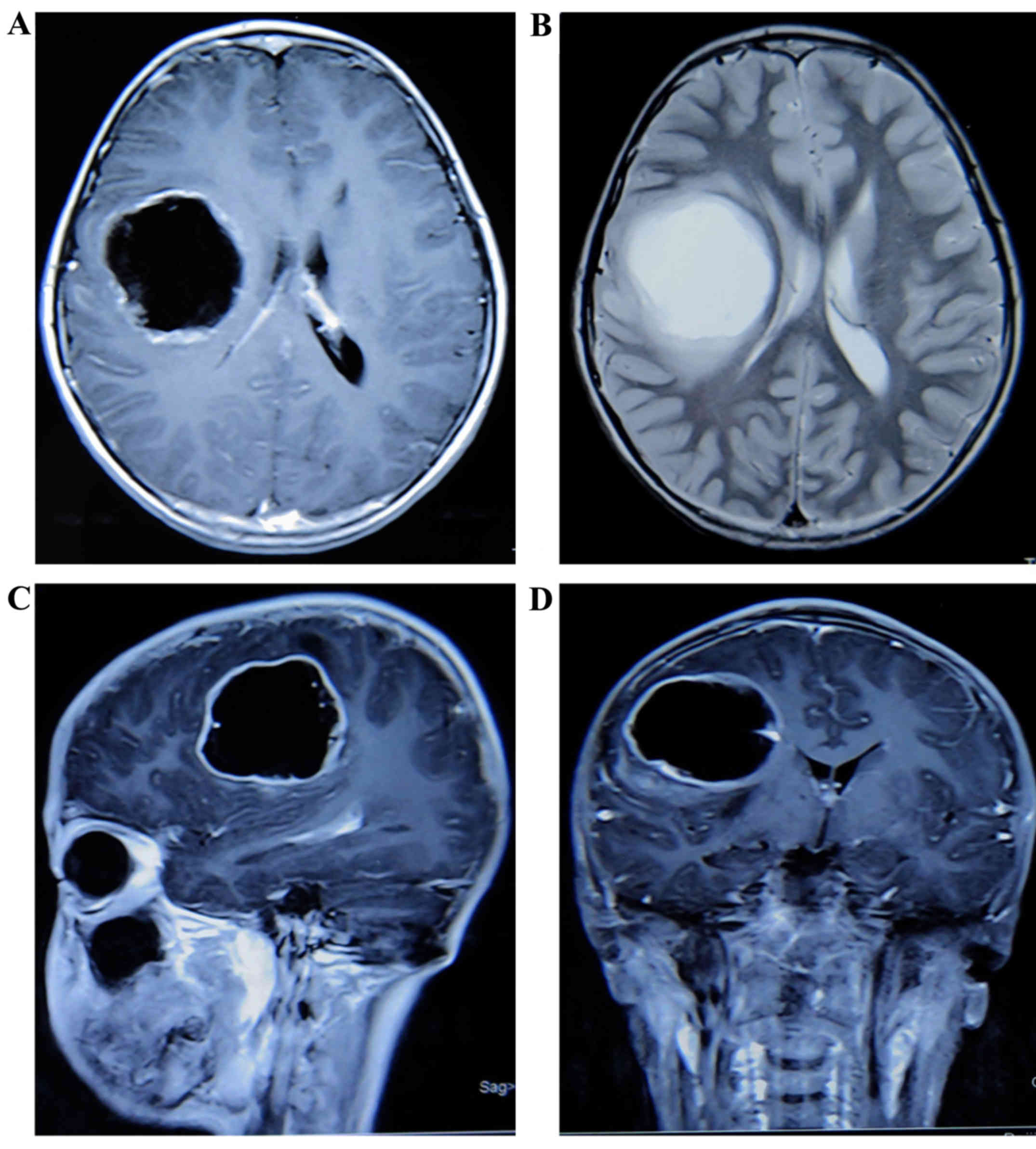
Subsequently, a brain MRI revealed a quasi-circular cystic lesion with a significantly enhanced capsule in the right frontal lobe. The lesion was poorly circumscribed, without obvious peritumoral edema. The cystic component displayed inhomogeneous low signal on T1-weighted images, and inhomogeneous hyperintense signal on T2-weighted images. On February 2017, the patient underwent surgical resection, and the subsequent histological diagnosis was GBM. Intraoperatively, the main component of the lesion was cystic, and the wall of the cystic lesion exhibited inhomogeneous thickness. After surgery, the weakness of the left limbs worsened and 2 months later the patient was readmitted with paroxysmal headache. A brain MRI revealed multiple enhanced tissues adjacent to the tumor bed in the right frontal lobe (Fig. 1). Subsequently, the patient received 4 cycles of chemotherapy with nimotuzumab, bevacizumab and irinotecan (dosage records unavailable), following which there remained no evidence, from regular reexaminations, to indicate tumor progression.

Figure 1.

Magnetic resonance imaging of the brain in 2017. (A) Axial T1-weighted post-gadolinium image showing an inhomogeneous low-signal quasi-circular cystic lesion in the right frontal lobe. (B) Axial T2-weighted image showing inhomogeneous hyperintense signal of the cystic component. (C) Sagittal plane. (D) Coronary plane.

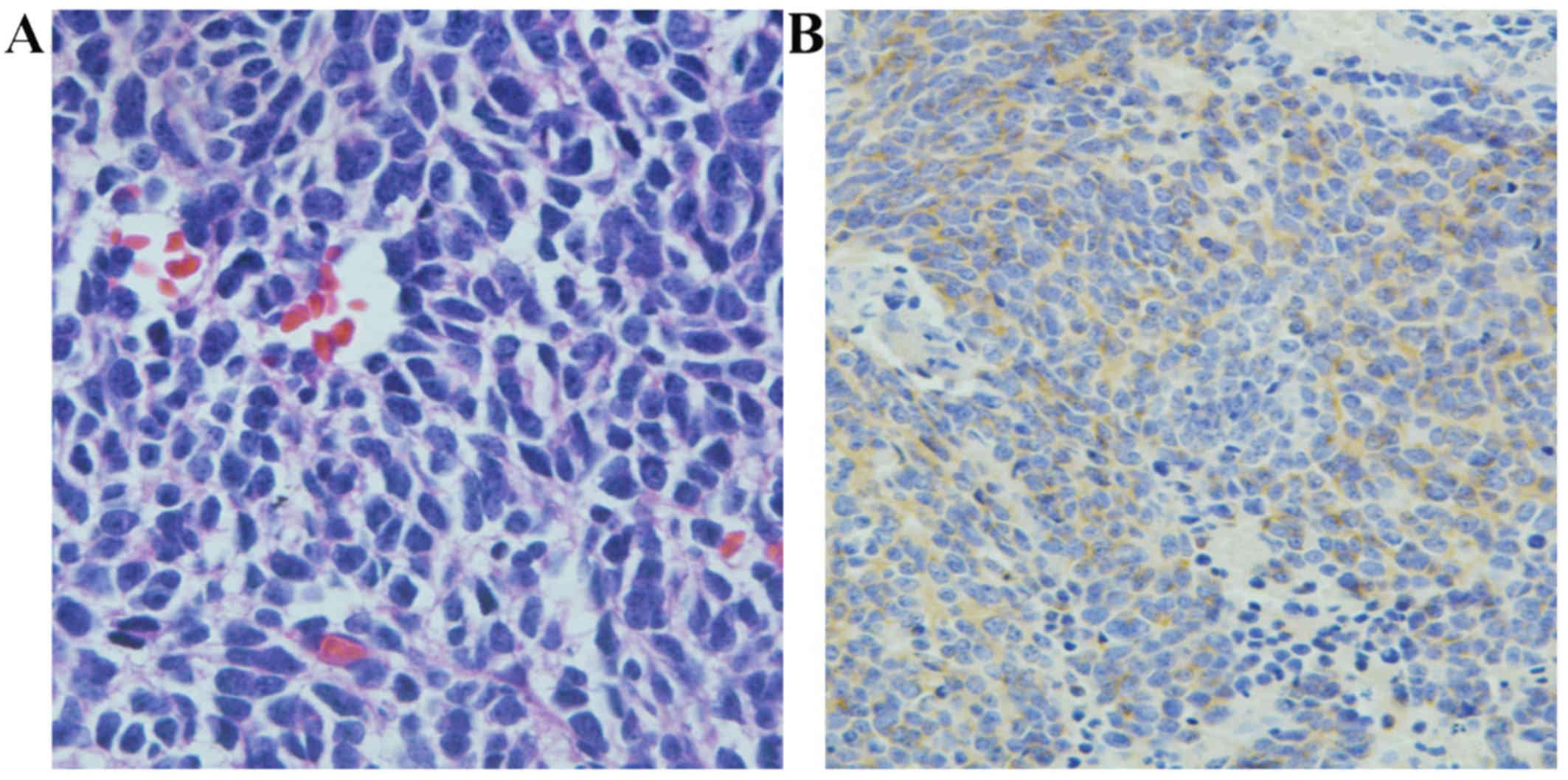
The first diagnosed tumor displayed characteristics typical of MB: Pleomorphic tumor cells with sparse cytoplasm, hyperchromatic nuclei, extensive presence of apoptosis/necrosis and mitotic figures, with Homer-Wright rosettes (Fig. 2A). Immunohistochemical examination revealed expression of synaptophysin (Syn) in the cytoplasm of some MB cells (Fig. 2B). However, neurofilament protein, S-100 and vimentin (Vim) were not expressed in the tumor tissue.

Figure 2.

Microscopic characteristics of medulloblastoma. (A) The original tumor was composed of tumor cells with sparse cytoplasm, hyperchromatic nuclei, extensive apoptosis/necrosis and mitotic figures, with typical characteristics of medulloblastomas (hematoxylin and eosin staining; magnification, ×400). (B) Immunohistochemical staining showing that the tumor cells were strongly positive for synaptophysin (magnification, ×200).

Histological examination of the secondary tumor revealed that the neoplastic cells exhibited characteristics of both glioblastoma and rhabdoid tumor cells. The majority of the tumor cells exhibited microvascular proliferation, obvious atypia, abundant cytoplasm, extensive apoptosis and mitotic figures, as well as patchy necrosis. A few cells were characterized by eccentric nuclei with prominent nucleoli. Furthermore, scattered giant cells were identified. Immunohistochemically, the tumor cells were strongly positive for glial fibrillary acidic protein (GFAP), Vim and Olig-2. In addition, S-100, p53, isocitrate dehydrogenase 1 and α-thalassemia/mental retardation syndrome X-linked (ATRX) were also expressed by these tumor cells, and smooth muscle actin (SMA) was focally positive, reflecting the loss of integrase interactor-1 (INI-1) protein. However, there were no observations indicating primitive neuroectodermal tumor in this case. The molecular pathology revealed ATRX mutation and O6-methylguanine-DNA-methyltransferase promoter methylation. Examination by fluorescence in situ hybridization demonstrated that there was no loss of heterozygosity on chromosome 1p19q. The Ki-67 index (Ki 67-positive cells) was ~30–40% (Fig. 3). Unfortunately, the patient died 1 year following the secondary recurrence.
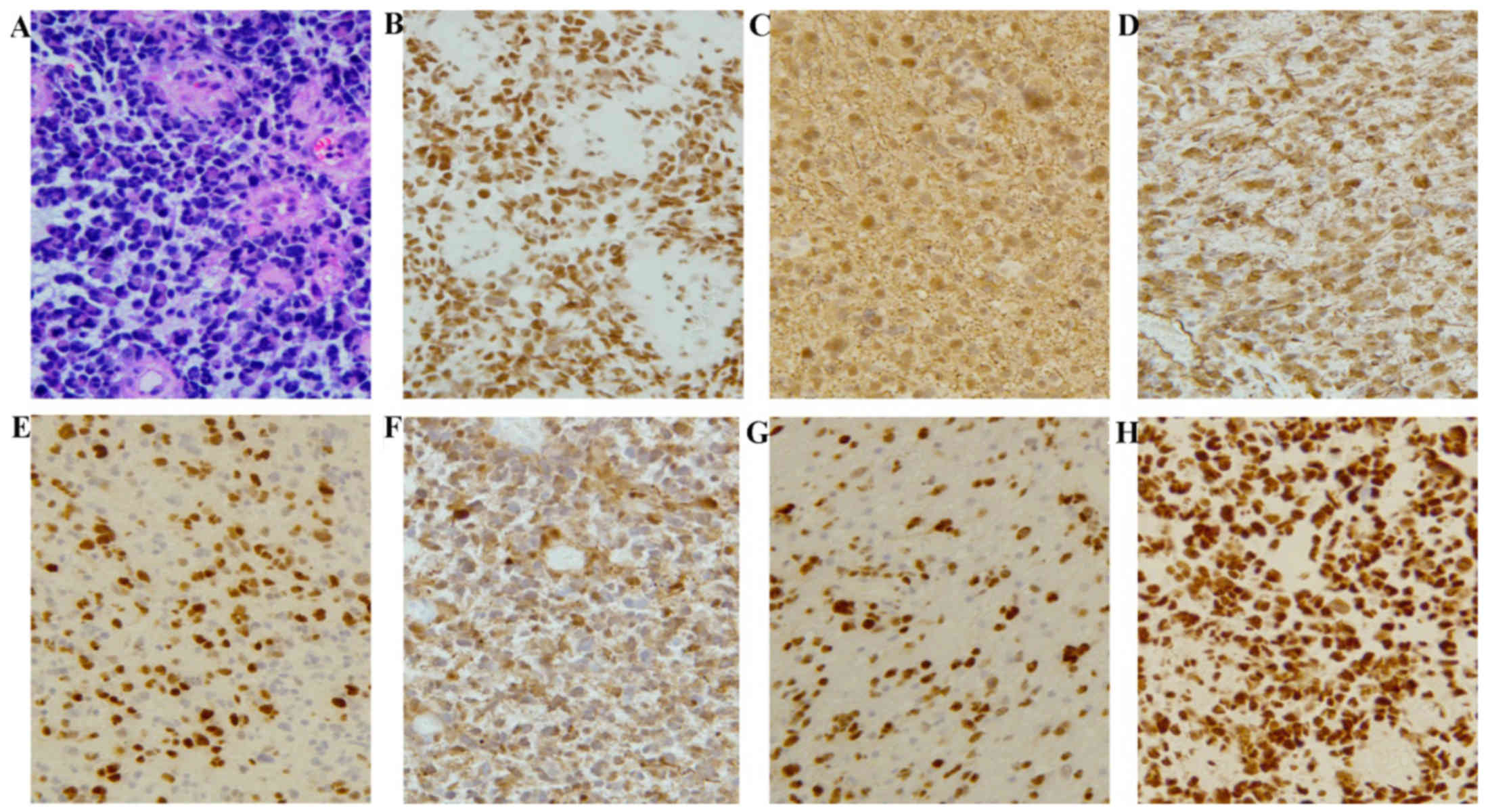
Figure 3.

Histological and immunohistochemical characteristics of glioblastoma multiforme. (A) The histological characteristics of the secondary tumor were obviously different from those of the original tumor. Glioblastoma cells exhibited microvascular proliferation, obvious atypia, abundant cytoplasm, extensive apoptosis/necrosis and mitotic figures, as well as patchy necrosis. A few cells were characterized by eccentrically located nuclei with prominent nucleoli (hematoxylin and eosin staining; magnification, ×200). (B) The tumor cell bearing characteristics of rhabdoid cells exhibited loss of the INI-1 protein compared with the surrounding INI-1-expressing cells (magnification, ×200). (C-H) Immunohistochemically, the tumor cells highly expressed (C) GFAP, (D) vimentin and (E) Olig-2. (F) S-100, (G) p53, and (H) ATRX were also expressed in those tumor cells (magnification, ×200). INI-1, integrase interactor 1; GFAP, glial fibrillary acidic protein; ATRX, α-thalassemia/mental retardation syndrome X-linked.

Discussion

Glioblastoma is a high-grade glioma that may develop as a secondary tumor following radiation therapy. As reported in the first animal experiment conducted by Haymaker et al in 1972 (4), glioblastoma was induced by radiation in a monkey. Over the last 45 years, ~296 cases of radiation-induced brain tumors, such as meningiomas, sarcomas and gliomas, have been reported. The criteria of radiation-induced glioblastoma established by Cahan et al (5) were as follows: i) The phenotypes of the primary and secondary tumors were distinctly different; ii) the site of the secondary tumor was within the field of irradiation; iii) the secondary tumor should be histologically diagnosed; iv) there should be a prolonged latency period between the treatment of the primary and the appearance of the secondary tumor. The case in our report was diagnosed as glioblastoma by histological examination 8 years after radiotherapy for MB.

In a study by Minniti et al (6), the risk of secondary brain tumor following conservative surgery and radiotherapy for pituitary adenoma was 2.4% in 20 years. Similar to these results, scientists in Margaret Hospital reported that the estimated cumulative risk for RIG was 1.7–2.7% at 15 years after radiation therapy (7). RIG may develop from primary tumors such as hematological malignancies, MB, pituitary adenoma, low-grade glioma and craniopharyngioma. As demonstrated by previous data, MB accounts for 12.8% of primary tumors (8).

The majority of radiation-induced brain tumors (RIBTs) are reported in children who were aged <10 years when they initially received radiotherapy. The first case of RBIT reported by Kleriga et al (2), was a patient treated for MB at the age of 10 months and developed a malignant astrocytoma in the same location 11 years after radiotherapy. You et al reported 5 cases of secondary brain tumors; all these cases were irradiated at an age of <10 years (1). The age of patients who develop RIGs is frequently lower compared with that of spontaneous high-grade gliomas. The case of RIG reported herein had a history of radiotherapy for MB at the age of 4 years. These findings indicate that patient age at first radiotherapy may be an important factor in the development of RIBTs. Another factor affecting the development of RIGs is irradiation volume. The incidence of RIGs increased with increasing irradiation volume. According to a review of patients with secondary brain tumors induced by radiation therapy, the proportion of radiation-induced malignant gliomas was 75% among patients receiving initial radiotherapy of craniospinal or whole-brain fields (7). Our patient was treated with craniospinal irradiation at first admission and the secondary tumor occurred 8 years later within the radiation field.

A study by Yang et al (9) reported that the secondary tumor exhibited strong immunoreactivity for both GFAP and p53. These histological characteristics were also described in a case reported by Donson et al (10). Although p53 mutations were not detected, strong immunoreactivity for p53 is frequently associated with gene mutations of p53. p53 gene mutations have been identified in radiation-induced tumors (11,12); thus, they may play a relevant role in the development of RIGs. However, the association between p53 gene mutations and the occurrence of secondary tumors remains largely unexplored.

In the present case, in addition to the strong expression of GFAP and p53, Vim, Syn and epithelial membrane antigen (EMA) were also expressed. As previously reported, Vim, SMA, EMA and Syn were mostly expressed in the secondary epithelioid/rhabdoid glioblastoma (13,14). Furthermore, focal loss of the INI-1 protein was observed in rhabdoid tumor cells, which were characterized by eccentric nuclei with prominent nucleoli. Focal loss of the INI-1 protein was also observed in cases reported by Sugimoto et al (15). Similar findings were also reported by a study on four cases of rhabdoid glioblastoma (16). Unfortunately, all these cases had a poor prognosis, with a median survival from the time of diagnosis of rhabdoid glioblastoma of only ~4.9 months (16,17).

The present case represents a rare occurrence of RIG with the characteristics of rhabdoid glioblastoma following CNS radiotherapy. Due to the extremely poor prognosis, this type of tumor should be further investigated and more studies are required to determine its relevance to irradiation.

Acknowledgements

Not applicable.

Funding

This manuscript was supported by the National Keypoint Research and Invention Program (grant no. 2016YFC0105705).

Availability of data and materials

All data generated during this study are included in this published article.

Authors' contributions

YW, SJS, ZW and XS conceived and designed the study. ZW, YW and ZD drafted the manuscript. JW, DC, YR, JF, QW and YS critically revised the manuscript for content. All authors approved the manuscript for submission.

Ethics approval and consent to participate

Not applicable.

Patient consent for publication

The patient's parents provided written informed consent for the publication of the case details and associated images.

Competing interests

The authors declare that they have no competing interests.

References

1 

You SH, Lyu CJ, Kim DS and Suh CO: Second primary brain tumors following cranial irradiation for pediatric solid brain tumors. Child's Nerv Syst. 29:1865–1870. 2013. View Article : Google Scholar

2 

Kleriga E, Sher JH, Nallainathan SK, Stein SC and Sacher M: Development of cerebellar malignant astrocytoma at site of a medulloblastoma treated 11 years earlier. Case report. J Neurosurg. 49:445–449. 1978. View Article : Google Scholar : PubMed/NCBI

3 

Wyatt-Ashmead J, Kleinschmidt-De Masters BK, Hill DA, Mierau GW, McGavran L, Thompson SJ and Foreman NK: Rhabdoid glioblastoma. Clin Neuropathol. 20:248–255. 2001.PubMed/NCBI

4 

Haymaker W, Rubinstein LJ and Miquel J: Brain tumors in irradiated monkeys. Acta neuropathologica. 20:267–277. 1972. View Article : Google Scholar : PubMed/NCBI

5 

Cahan WG, Woodard HQ, Higinbotham NL, Stewart FW and Coley BL: Sarcoma arising in irradiated bone: Report of eleven cases. 1948. Cancer. 82:8–34. 1998. View Article : Google Scholar : PubMed/NCBI

6 

Minniti G, Traish D, Ashley S, Gonsalves A and Brada M: Risk of second brain tumor after conservative surgery and radiotherapy for pituitary adenoma: Update after an additional 10 years. J Clin Endocrinol Metab. 90:800–804. 2005. View Article : Google Scholar : PubMed/NCBI

7 

Paulino AC, Mai WY, Chintagumpala M, Taher A and Teh BS: Radiation-induced malignant gliomas: is there a role for reirradiation? Int J Radiat Oncol Biol Phys. 71:1381–1387. 2008. View Article : Google Scholar : PubMed/NCBI

8 

Yamanaka R, Hayano A and Kanayama T: Radiation-induced gliomas: A comprehensive review and meta-analysis. Neurosurg Rev. 5:2016.(Epub ahead of print).

9 

Yang SY, Wang KC, Cho BK, Kim YY, Lim SY, Park SH, Kim IH and Kim SK: Radiation-induced cerebellar glioblastoma at the site of a treated medulloblastoma: Case report. J Neurosurg. 102 4 Suppl:1–422. 2005.

10 

Donson AM, Erwin NS, Kleinschmidt-DeMasters BK, Madden JR, Addo-Yobo SO and Foreman NK: Unique molecular characteristics of radiation-induced glioblastoma. J Neuropathol Exp Neurol. 66:740–749. 2007. View Article : Google Scholar : PubMed/NCBI

11 

Gessi M, Maderna E, Guzzetti S, Cefalo G, Massimino M, Solero CL, Finocchiaro G and Pollo B: Radiation-induced glioblastoma in a medulloblastoma patient: a case report with molecular features. Neuropathology. 28:633–639. 2008.PubMed/NCBI

12 

Brachman DG, Hallahan DE, Beckett MA, Yandell DW and Weichselbaum RR: p53 gene mutations and abnormal retinoblastoma protein in radiation-induced human sarcomas. Cancer Res. 51:6393–6396. 1991.PubMed/NCBI

13 

Nagai S, Kurimoto M, Ishizawa S, Hayashi N, Hamada H, Kamiyama H and Endo S: A rare astrocytic tumor with rhabdoid features. Brain Tumor Pathol. 26:19–24. 2009. View Article : Google Scholar : PubMed/NCBI

14 

Mutou J, Hirose Y, Ikeda E, Yoshida K, Nakazato Y and Kawase T: Malignant brain tumor with rhabdoid features in an adult. Neurol Med Chir (Tokyo). 51:449–454. 2011. View Article : Google Scholar : PubMed/NCBI

15 

Sugimoto K, Ideguchi M, Kimura T, Kajiwara K, Imoto H, Sadahiro H, Ishii A, Kawano H, Ikeda E and Suzuki M: Epithelioid/rhabdoid glioblastoma: A highly aggressive subtype of glioblastoma. Brain Tumor Pathol. 33:137–146. 2016. View Article : Google Scholar : PubMed/NCBI

16 

Babu R, Hatef J, McLendon RE, Cummings TJ, Sampson JH, Friedman AH and Adamson C: Clinicopathological characteristics and treatment of rhabdoid glioblastoma. J Neurosurg. 119:412–419. 2013. View Article : Google Scholar : PubMed/NCBI

17 

Byeon SJ, Cho HJ, Baek HW, Park CK, Choi SH, Kim SH, Kim HK and Park SH: Rhabdoid glioblastoma is distinguishable from classical glioblastoma by cytogenetics and molecular genetics. Hum Pathol. 45:611–620. 2014. View Article : Google Scholar : PubMed/NCBI

Related Articles

  • Abstract
  • View
  • Download
  • Twitter
Copy and paste a formatted citation
Spandidos Publications style
Wang Y, Song S, Su X, Wu J, Dai Z, Cui D, Reng Y, Fan J, Shen Y, Wu Q, Wu Q, et al: Radiation‑induced glioblastoma with rhabdoid characteristics following treatment for medulloblastoma: A case report and review of the literature. Mol Clin Oncol 9: 415-418, 2018.
APA
Wang, Y., Song, S., Su, X., Wu, J., Dai, Z., Cui, D. ... Wang, Z. (2018). Radiation‑induced glioblastoma with rhabdoid characteristics following treatment for medulloblastoma: A case report and review of the literature. Molecular and Clinical Oncology, 9, 415-418. https://doi.org/10.3892/mco.2018.1703
MLA
Wang, Y., Song, S., Su, X., Wu, J., Dai, Z., Cui, D., Reng, Y., Fan, J., Shen, Y., Wu, Q., Wang, Z."Radiation‑induced glioblastoma with rhabdoid characteristics following treatment for medulloblastoma: A case report and review of the literature". Molecular and Clinical Oncology 9.4 (2018): 415-418.
Chicago
Wang, Y., Song, S., Su, X., Wu, J., Dai, Z., Cui, D., Reng, Y., Fan, J., Shen, Y., Wu, Q., Wang, Z."Radiation‑induced glioblastoma with rhabdoid characteristics following treatment for medulloblastoma: A case report and review of the literature". Molecular and Clinical Oncology 9, no. 4 (2018): 415-418. https://doi.org/10.3892/mco.2018.1703
Copy and paste a formatted citation
x
Spandidos Publications style
Wang Y, Song S, Su X, Wu J, Dai Z, Cui D, Reng Y, Fan J, Shen Y, Wu Q, Wu Q, et al: Radiation‑induced glioblastoma with rhabdoid characteristics following treatment for medulloblastoma: A case report and review of the literature. Mol Clin Oncol 9: 415-418, 2018.
APA
Wang, Y., Song, S., Su, X., Wu, J., Dai, Z., Cui, D. ... Wang, Z. (2018). Radiation‑induced glioblastoma with rhabdoid characteristics following treatment for medulloblastoma: A case report and review of the literature. Molecular and Clinical Oncology, 9, 415-418. https://doi.org/10.3892/mco.2018.1703
MLA
Wang, Y., Song, S., Su, X., Wu, J., Dai, Z., Cui, D., Reng, Y., Fan, J., Shen, Y., Wu, Q., Wang, Z."Radiation‑induced glioblastoma with rhabdoid characteristics following treatment for medulloblastoma: A case report and review of the literature". Molecular and Clinical Oncology 9.4 (2018): 415-418.
Chicago
Wang, Y., Song, S., Su, X., Wu, J., Dai, Z., Cui, D., Reng, Y., Fan, J., Shen, Y., Wu, Q., Wang, Z."Radiation‑induced glioblastoma with rhabdoid characteristics following treatment for medulloblastoma: A case report and review of the literature". Molecular and Clinical Oncology 9, no. 4 (2018): 415-418. https://doi.org/10.3892/mco.2018.1703
Follow us
  • Twitter
  • LinkedIn
  • Facebook
About
  • Spandidos Publications
  • Careers
  • Cookie Policy
  • Privacy Policy
How can we help?
  • Help
  • Live Chat
  • Contact
  • Email to our Support Team