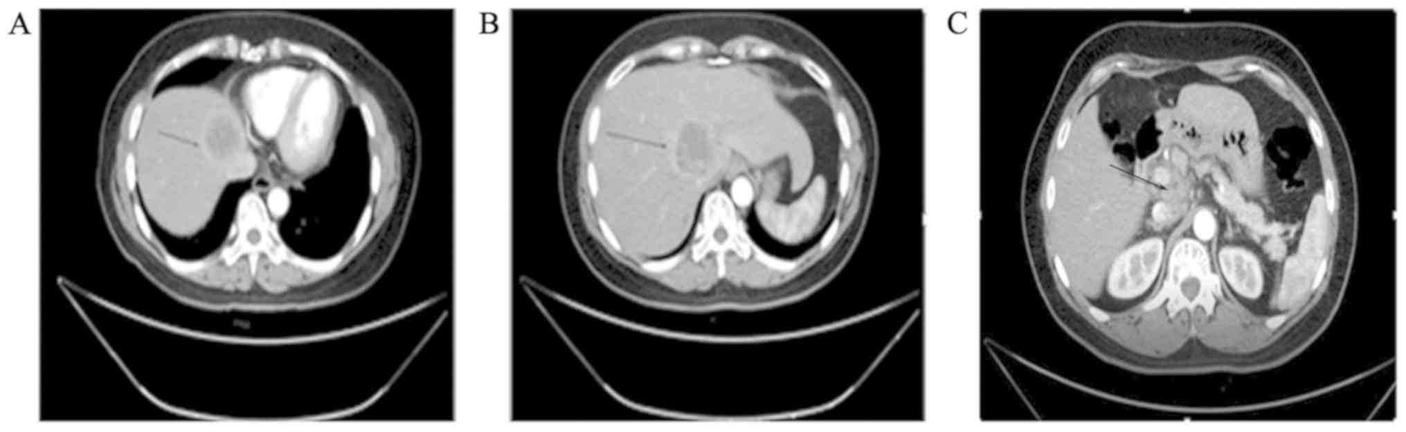
|
1
|
Jain A, Singla S, Jagdeesh KS and
Vishnumurthy HY: Mixed adenoneuroendocrine carcinoma of cecum: A
rare entity. J Clin Imaging Sci. 3(10)2013.PubMed/NCBI View Article : Google Scholar
|
|
2
|
Shimada N, Miwa S, Arai T, Kitagawa N,
Akita S, Iinuma N and Ishii K: Cystic mixed adenoneuroendocrine
carcinoma of the pancreas: A case report. Int J Surg Case Rep.
52:1–4. 2018.PubMed/NCBI View Article : Google Scholar
|
|
3
|
Kwok CM: Mixed adenoneuroendocrine
carcinoma of the stomach. Case Rep Gastroenterol. 9:241–245.
2015.PubMed/NCBI View Article : Google Scholar
|
|
4
|
Yamauchi H, Sakurai S, Nakazawa N, Yoshida
T, Tabe Y, Saitoh K, Fukasawa T, Kiriyama S, Naitoh H and Kuwano H:
A case of mixed adenoneuroendocrine carcinoma of the stomach with
focal intestinal metaplasia and hypergastrinemia. Int Surg.
100:562–567. 2015.PubMed/NCBI View Article : Google Scholar
|
|
5
|
Minakawa K, Oka K, Nihei T, Sando N,
Oikawa H, Toda J, Hosokawa Y, Matsumoto T and Yanagisawa A:
Pancreatic endocrine tumor with partial acinar cell
differentiation. APMIS. 114:720–725. 2006.PubMed/NCBI View Article : Google Scholar
|
|
6
|
Kadhim MM, Jespersen ML, Pilegaard HK,
Nordsmark M and Villadsen GE: Mixed adenoneuroendocrine carcinoma
is a rare but important tumour found in the oesophagus. Case Rep
Gastrointest Med. 2016(9542687)2016.PubMed/NCBI View Article : Google Scholar
|
|
7
|
Ginori A, Lo Bello G, Vassallo L and
Tripodi SA: Amphicrine carcinoma of the ampullary region. Tumori.
101:e70–e72. 2015.PubMed/NCBI View Article : Google Scholar
|
|
8
|
Gurzu S, Kadar Z, Bara T, Bara T Jr,
Tamasi A, Azamfirei L and Jung I: Mixed adenoneuroendocrine
carcinoma of gastrointestinal tract: Report of two cases. World J
Gastroenterol. 21:1329–1333. 2015.PubMed/NCBI View Article : Google Scholar
|
|
9
|
Onishi I, Kitagawa H, Harada K, Maruzen S,
Sakai S, Makino I, Hayashi H, Nakagawara H, Tajima H, Takamura H,
et al: Intraductal papillary neoplasm of the bile duct accompanying
biliary mixed adenoneuroendocrine carcinoma. World J Gastroenterol.
19:3161–3164. 2013.PubMed/NCBI View Article : Google Scholar
|
|
10
|
Niederle MB, Hackl M, Kaserer K and
Niederle B: Gastroenteropancreatic neuroendocrine tumours: The
current incidence and staging based on the WHO and European
neuroendocrine tumour society classification: An analysis based on
prospectively collected parameters. Endocr Relat Cancer.
17:909–918. 2010.PubMed/NCBI View Article : Google Scholar
|
|
11
|
Tang Q, Zhou Z, Chen J, Di M, Ji J, Yuan
W, Liu Z, Wu L, Zhang X, Li K and Shu X: Correlation of metastasis
characteristics with prognosis in gastric mixed adenoneuroendocrine
carcinoma: Two case reports. Medicine (Baltimore).
96(e9189)2017.PubMed/NCBI View Article : Google Scholar
|
|
12
|
La Rosa S, Marando A, Sessa F and Capella
C: Mixed Adenoneuroendocrine carcinomas (MANECs) of the
gastrointestinal tract: An update. Cancers (Basel). 4:11–30.
2012.PubMed/NCBI View Article : Google Scholar
|
|
13
|
Volante M, Rindi G and Papotti M: The grey
zone between pure (neuro)endocrine and non-(neuro)endocrine
tumours: A comment on concepts and classification of mixed
exocrine-endocrine neoplasms. Virchows Arch. 449:499–506.
2006.PubMed/NCBI View Article : Google Scholar
|
|
14
|
Furlan D, Cerutti R, Genasetti A, Pelosi
G, Uccella S, La Rosa S and Capella C: Microallelotyping defines
the monoclonal or the polyclonal origin of mixed and collision
endocrine-exocrine tumors of the gut. Lab Invest. 83:963–971.
2003.PubMed/NCBI View Article : Google Scholar
|
|
15
|
Paniz Mondolfi AE, Slova D, Fan W, Attiyeh
FF, Afthinos J, Reidy J, Pang Y and Theise ND: Mixed
adenoneuroendocrine carcinoma (MANEC) of the gallbladder: A
possible stem cell tumor? Pathol Int. 61:608–614. 2011.PubMed/NCBI View Article : Google Scholar
|
|
16
|
Vanacker L, Smeets D, Hoorens A, Teugels
E, Algaba R, Dehou MF, De Becker A, Lambrechts D and De Greve J:
Mixed adenoneuroendocrine carcinoma of the colon: Molecular
pathogenesis and treatment. Anticancer Res. 34:5517–5521.
2014.PubMed/NCBI
|
|
17
|
Zhang W, Xiao W, Ma H, Sun M, Chen H and
Zheng S: Neuroendocrine liver metastasis in gastric mixed
adenoneuroendocrine carcinoma with trilineage cell differentiation:
A case report. Int J Clin Exp Pathol. 7:6333–6338. 2014.PubMed/NCBI
|
|
18
|
Lee EJ, Park SM, Maeng L, Lee A and Kim
KM: Composite glandular-endocrine cell carcinomas of the stomach:
Clinicopathologic and methylation study. APMIS. 113:569–576.
2005.PubMed/NCBI View Article : Google Scholar
|
|
19
|
Levi Sandri GB, Carboni F, Valle M, Visca
P and Garofalo A: Mixed adenoneuroendocrine gastric carcinoma: A
case report and review of the literature. J Gastric Cancer.
14:63–66. 2014.PubMed/NCBI View Article : Google Scholar
|
|
20
|
Scardoni M, Vittoria E, Volante M, Rusev
B, Bersani S, Mafficini A, Gottardi M, Giandomenico V, Malleo G,
Butturini G, et al: Mixed adenoneuroendocrine carcinomas of the
gastrointestinal tract: Targeted next-generation sequencing
suggests a monoclonal origin of the two components.
Neuroendocrinology. 100:310–316. 2014.PubMed/NCBI View Article : Google Scholar
|
|
21
|
Kim JJ, Kim JY, Hur H, Cho YK and Han SU:
Clinicopathologic significance of gastric adenocarcinoma with
neuroendocrine features. J Gastric Cancer. 11:195–199.
2011.PubMed/NCBI View Article : Google Scholar
|
|
22
|
Lee JH, Kim HW, Kang DH, Choi CW, Park SB
and Kim SH: A gastric composite tumor with an adenocarcinoma and a
neuroendocrine carcinoma: A case report. Clin Endosc. 46:280–283.
2013.PubMed/NCBI View Article : Google Scholar
|
|
23
|
Li Y, Yau A, Schaeffer D, Magliocco A, Gui
X, Urbanski S, Waghray R, Owen D and Gao ZH: Colorectal
glandular-neuroendocrine mixed tumor: Pathologic spectrum and
clinical implications. Am J Surg Pathol. 35:413–425.
2011.PubMed/NCBI View Article : Google Scholar
|
|
24
|
Delle Fave G, Kwekkeboom DJ, Van Cutsem E,
Rindi G, Kos-Kudla B, Knigge U, Sasano H, Tomassetti P, Salazar R
and Ruszniewski P: ENETS Consensus Guidelines for the management of
patients with gastroduodenal neoplasms. Neuroendocrinology.
95:74–87. 2012.PubMed/NCBI View Article : Google Scholar
|
|
25
|
Van Laethem JL, Carneiro F, Ducreux M,
Messman H, Lordick F, Ilson DH, Allum WH, Haustermans K, Lepage C,
Matysiak-Budnik T, et al: The multidisciplinary management of
gastro-oesophageal junction tumours: European society of digestive
oncology (ESDO): Expert discussion and report from the 16th ESMO
world congress on gastrointestinal cancer, barcelona. Dig Liver
Dis. 48:1283–1289. 2016.PubMed/NCBI View Article : Google Scholar
|