Spandidos Publications Logo
  • About
    • About Spandidos
    • Aims and Scopes
    • Abstracting and Indexing
    • Editorial Policies
    • Reprints and Permissions
    • Job Opportunities
    • Terms and Conditions
    • Contact
  • Journals
    • All Journals
    • Oncology Letters
      • Oncology Letters
      • Information for Authors
      • Editorial Policies
      • Editorial Board
      • Aims and Scope
      • Abstracting and Indexing
      • Bibliographic Information
      • Archive
    • International Journal of Oncology
      • International Journal of Oncology
      • Information for Authors
      • Editorial Policies
      • Editorial Board
      • Aims and Scope
      • Abstracting and Indexing
      • Bibliographic Information
      • Archive
    • Molecular and Clinical Oncology
      • Molecular and Clinical Oncology
      • Information for Authors
      • Editorial Policies
      • Editorial Board
      • Aims and Scope
      • Abstracting and Indexing
      • Bibliographic Information
      • Archive
    • Experimental and Therapeutic Medicine
      • Experimental and Therapeutic Medicine
      • Information for Authors
      • Editorial Policies
      • Editorial Board
      • Aims and Scope
      • Abstracting and Indexing
      • Bibliographic Information
      • Archive
    • International Journal of Molecular Medicine
      • International Journal of Molecular Medicine
      • Information for Authors
      • Editorial Policies
      • Editorial Board
      • Aims and Scope
      • Abstracting and Indexing
      • Bibliographic Information
      • Archive
    • Biomedical Reports
      • Biomedical Reports
      • Information for Authors
      • Editorial Policies
      • Editorial Board
      • Aims and Scope
      • Abstracting and Indexing
      • Bibliographic Information
      • Archive
    • Oncology Reports
      • Oncology Reports
      • Information for Authors
      • Editorial Policies
      • Editorial Board
      • Aims and Scope
      • Abstracting and Indexing
      • Bibliographic Information
      • Archive
    • Molecular Medicine Reports
      • Molecular Medicine Reports
      • Information for Authors
      • Editorial Policies
      • Editorial Board
      • Aims and Scope
      • Abstracting and Indexing
      • Bibliographic Information
      • Archive
    • World Academy of Sciences Journal
      • World Academy of Sciences Journal
      • Information for Authors
      • Editorial Policies
      • Editorial Board
      • Aims and Scope
      • Abstracting and Indexing
      • Bibliographic Information
      • Archive
    • International Journal of Functional Nutrition
      • International Journal of Functional Nutrition
      • Information for Authors
      • Editorial Policies
      • Editorial Board
      • Aims and Scope
      • Abstracting and Indexing
      • Bibliographic Information
      • Archive
    • International Journal of Epigenetics
      • International Journal of Epigenetics
      • Information for Authors
      • Editorial Policies
      • Editorial Board
      • Aims and Scope
      • Abstracting and Indexing
      • Bibliographic Information
      • Archive
    • Medicine International
      • Medicine International
      • Information for Authors
      • Editorial Policies
      • Editorial Board
      • Aims and Scope
      • Abstracting and Indexing
      • Bibliographic Information
      • Archive
  • Articles
  • Information
    • Information for Authors
    • Information for Reviewers
    • Information for Librarians
    • Information for Advertisers
    • Conferences
  • Language Editing
Spandidos Publications Logo
  • About
    • About Spandidos
    • Aims and Scopes
    • Abstracting and Indexing
    • Editorial Policies
    • Reprints and Permissions
    • Job Opportunities
    • Terms and Conditions
    • Contact
  • Journals
    • All Journals
    • Biomedical Reports
      • Information for Authors
      • Editorial Policies
      • Editorial Board
      • Aims and Scope
      • Abstracting and Indexing
      • Bibliographic Information
      • Archive
    • Experimental and Therapeutic Medicine
      • Information for Authors
      • Editorial Policies
      • Editorial Board
      • Aims and Scope
      • Abstracting and Indexing
      • Bibliographic Information
      • Archive
    • International Journal of Epigenetics
      • Information for Authors
      • Editorial Policies
      • Editorial Board
      • Aims and Scope
      • Abstracting and Indexing
      • Bibliographic Information
      • Archive
    • International Journal of Functional Nutrition
      • Information for Authors
      • Editorial Policies
      • Editorial Board
      • Aims and Scope
      • Abstracting and Indexing
      • Bibliographic Information
      • Archive
    • International Journal of Molecular Medicine
      • Information for Authors
      • Editorial Policies
      • Editorial Board
      • Aims and Scope
      • Abstracting and Indexing
      • Bibliographic Information
      • Archive
    • International Journal of Oncology
      • Information for Authors
      • Editorial Policies
      • Editorial Board
      • Aims and Scope
      • Abstracting and Indexing
      • Bibliographic Information
      • Archive
    • Medicine International
      • Information for Authors
      • Editorial Policies
      • Editorial Board
      • Aims and Scope
      • Abstracting and Indexing
      • Bibliographic Information
      • Archive
    • Molecular and Clinical Oncology
      • Information for Authors
      • Editorial Policies
      • Editorial Board
      • Aims and Scope
      • Abstracting and Indexing
      • Bibliographic Information
      • Archive
    • Molecular Medicine Reports
      • Information for Authors
      • Editorial Policies
      • Editorial Board
      • Aims and Scope
      • Abstracting and Indexing
      • Bibliographic Information
      • Archive
    • Oncology Letters
      • Information for Authors
      • Editorial Policies
      • Editorial Board
      • Aims and Scope
      • Abstracting and Indexing
      • Bibliographic Information
      • Archive
    • Oncology Reports
      • Information for Authors
      • Editorial Policies
      • Editorial Board
      • Aims and Scope
      • Abstracting and Indexing
      • Bibliographic Information
      • Archive
    • World Academy of Sciences Journal
      • Information for Authors
      • Editorial Policies
      • Editorial Board
      • Aims and Scope
      • Abstracting and Indexing
      • Bibliographic Information
      • Archive
  • Articles
  • Information
    • For Authors
    • For Reviewers
    • For Librarians
    • For Advertisers
    • Conferences
  • Language Editing
Login Register Submit
  • This site uses cookies
  • You can change your cookie settings at any time by following the instructions in our Cookie Policy. To find out more, you may read our Privacy Policy.

    I agree
Search articles by DOI, keyword, author or affiliation
Search
Advanced Search
presentation
Molecular and Clinical Oncology
Join Editorial Board Propose a Special Issue
Print ISSN: 2049-9450 Online ISSN: 2049-9469
Journal Cover
March-2021 Volume 14 Issue 3

Full Size Image

Sign up for eToc alerts
Recommend to Library

Journals

International Journal of Molecular Medicine

International Journal of Molecular Medicine

International Journal of Molecular Medicine is an international journal devoted to molecular mechanisms of human disease.

International Journal of Oncology

International Journal of Oncology

International Journal of Oncology is an international journal devoted to oncology research and cancer treatment.

Molecular Medicine Reports

Molecular Medicine Reports

Covers molecular medicine topics such as pharmacology, pathology, genetics, neuroscience, infectious diseases, molecular cardiology, and molecular surgery.

Oncology Reports

Oncology Reports

Oncology Reports is an international journal devoted to fundamental and applied research in Oncology.

Experimental and Therapeutic Medicine

Experimental and Therapeutic Medicine

Experimental and Therapeutic Medicine is an international journal devoted to laboratory and clinical medicine.

Oncology Letters

Oncology Letters

Oncology Letters is an international journal devoted to Experimental and Clinical Oncology.

Biomedical Reports

Biomedical Reports

Explores a wide range of biological and medical fields, including pharmacology, genetics, microbiology, neuroscience, and molecular cardiology.

Molecular and Clinical Oncology

Molecular and Clinical Oncology

International journal addressing all aspects of oncology research, from tumorigenesis and oncogenes to chemotherapy and metastasis.

World Academy of Sciences Journal

World Academy of Sciences Journal

Multidisciplinary open-access journal spanning biochemistry, genetics, neuroscience, environmental health, and synthetic biology.

International Journal of Functional Nutrition

International Journal of Functional Nutrition

Open-access journal combining biochemistry, pharmacology, immunology, and genetics to advance health through functional nutrition.

International Journal of Epigenetics

International Journal of Epigenetics

Publishes open-access research on using epigenetics to advance understanding and treatment of human disease.

Medicine International

Medicine International

An International Open Access Journal Devoted to General Medicine.

Journal Cover
March-2021 Volume 14 Issue 3

Full Size Image

Sign up for eToc alerts
Recommend to Library

  • Article
  • Citations
    • Cite This Article
    • Download Citation
    • Create Citation Alert
    • Remove Citation Alert
    • Cited By
  • Similar Articles
    • Related Articles (in Spandidos Publications)
    • Similar Articles (Google Scholar)
    • Similar Articles (PubMed)
  • Download PDF
  • Download XML
  • View XML
Article

Dermatofibrosarcoma protuberans of the breast: A case study

  • Authors:
    • Dionysios Dimas
    • Ioannis Boutas
    • Anastasios Potiris
    • Nektarios Koufopoulos
    • Dimitrios Balalis
    • Kyparissia Sitara
    • Fotios Danglis
    • Ioannis Misitzis
  • View Affiliations / Copyright

    Affiliations: Breast Unit, Athens Medical Center, Psychiko Clinic, Athens 11525, Greece, Third Department of Obstetrics and Gynecology, Attikon University Hospital, National and Kapodistrian University of Athens, Athens 12462, Greece, Second Department of Pathology, Attikon University Hospital, National and Kapodistrian University of Athens, Athens 12462, Greece, Department of Surgery, Saint Savvas Anti‑Cancer Hospital, Athens 11521, Greece, Surgical Pathology Department, National and Kapodistrian University of Athens, Athens 15125, Greece
  • Article Number: 50
    |
    Published online on: January 20, 2021
       https://doi.org/10.3892/mco.2021.2212
  • Expand metrics +
Metrics: Total Views: 0 (Spandidos Publications: | PMC Statistics: )
Metrics: Total PDF Downloads: 0 (Spandidos Publications: | PMC Statistics: )
Cited By (CrossRef): 0 citations Loading Articles...

This article is mentioned in:


Abstract

Dermatofibrosarcoma protuberans (DFSP) is a superficial mesenchymal neoplasm that originates from the dermal fibroblasts and tends to be locally aggressive. Although infrequent, it is one of the most common cutaneous sarcomas. It mainly affects young and middle‑aged patients 20 to 50 years old. Any area of skin may be involved, but the most common sites of presentation are the trunk and extremities. DFSP of the breast is extremely rare. It classically presents as a nodular, exophytic, cutaneous mass, though initially it can manifest as a flat plague and can show persistent but slow growth for many years. Due to increased risk of local recurrence, the standard of care for localized disease is surgical excision with adequate margins. Wide local excision is the most common technique used, but as an alternative Mohs micrographic surgery has emerged as a procedure that offers lower local recurrence rates. Metastases are rare but have been previously reported. In such patients, treatment with imatinib or radiotherapy can be considered. The current case presents a 52‑year‑old lady with DFSP of the breast that was successfully managed by the Breast Unit of Athens Medical Center‑Psychiko Clinic.

Introduction

Dermatofibrosarcoma was first described as progressive recurrent dermatofibroma in 1924 by Darier and Ferrand. Subsequently, in 1925 Hoffmann referred to the disease as dermatofibrosarcoma protuberans (DFSP) (1). It is a very rare neoplasm of the breast. Dermatofibrosarcoma of the breast presents with a slow growing pattern, initially in the dermis and then invading the subcutaneous tissue (2). Usually, DFSP is characterized as low to intermediate grade neoplasm with high recurrence rate but rarely metastasises (3). The majority of DFSP presents as low-grade tumours, but in 5-10% of cases the tumour contains fibrosarcomatosous cells that upgrade the neoplasm in intermediate grade with a higher aggressivity (4). Mainly affects young and middle-aged patients 20 to 50 years old and as the lesion progresses, it appears more and more protuberant (1). Surgery is the Gold Standard with particular attention to obtain free margins in order to reduce the recurrence rate (5). Wide local excision (WLE) with minimum 3 cm free margins or Mohs micrographic surgery (MMS) are recommended. We present a rare case of dermatofibrosarcoma protuberans in the right breast of a 52-year-old female patient.

Case report

A 52-year-old patient presented to our Breast Unit due to a skin lesion of her right breast that had slow but persistent growth during the past 15 years. The physical examination revealed a light-reddish, delineated exophytic, nodular cutaneous mass 3x4 cm in the upper outer quadrant of the right breast. No other finding from the breasts or axillary lymphadenopathy was noted. She had an unremarkable personal and family history.

Mammography revealed a dense, broad-based, well circumscribed, cutaneous lesion. There was no associated intramammary mass or microcalcifications. The lesion was evident on previous mammograms, presenting a small growth rate.

In order to establish a diagnosis, a core-needle biopsy was suggested which the patient denied. Subsequently, she underwent surgical excision under local anesthesia. On gross examination, there was a firm, grey-like mass 3.5 cm in diameter, with irregular borders and extension into the subcutaneous tissue. 
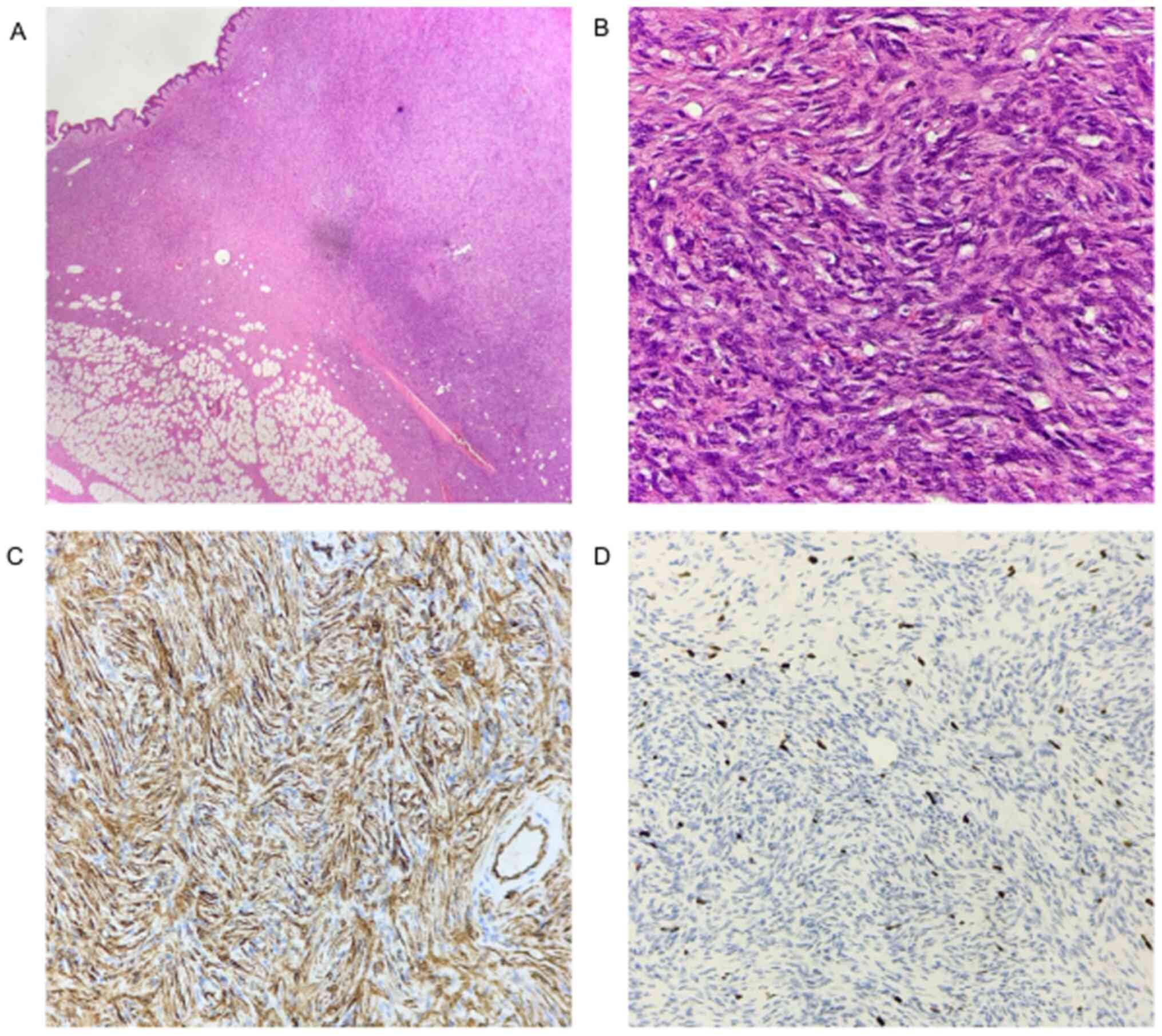
On microscopic examination, the tumor consisted of monomorphic spindle-like cells arranged in storiform architectural pattern. The nuclei were hyperchromatic, elongated, with little pleomorphism and low mitotic activity (Fig. 1A and 1B). Immunohistochemically as Fig. 1C and 1D show, the tumor cells showed diffuse staining for CD34 and negative staining for CD68, while Ki67 was 15%.

Figure 1

Histological and Immunohistochemical analysis of the resected tumor. (A) Hematoxylin-eosin staining with diffuse infiltration of subcutaneous fat. Magnification, x4. (B) Hematoxylin-eosin staining with storiform or whorled growth pattern, minimal cytological atypia and low mitotic activity. Magnification, x40. (C) Immunohistochemical analysis with CD34 positive (+) staining in spindled tumor cells (Magnification, x20) and (D) Immunohistochemical analysis with Ki67 (Mib-1) positive (+) in ~10% of tumor cells (Magnification, x20).

Based on histological and immunohistochemical findings, the mass was eventually diagnosed as dermatofibrosarcoma protuberans. As the lateral and deep margins were focally involved and in order to minimize the chance of local recurrence, a wide re-excision of 3 cm healthy margins was performed.

Discussion

Dermatofibrosarcoma protuberans typically presents as a painless, skin-coloured or yellow to brown nodular, exophytic, nodular cutaneous mass (6,7). It tends to be well-circumscribed or has subtle areas of microlobulation, while it can vary in size (0.5->5 cm) (8). The lesion is usually centered in the dermis, but it can invade the subcutaneous tissue (9). It has an indolent clinical course typically for years before the patients seek for medical assistance.

Mammographically, DFSP presents as a dense mass. Preoperative ultrasound examination or MRI offer limited information about the degree of subcutaneous infiltration and can be helpful only in selected cases (6,10). Diagnosis is made histologically, either with a core-needle or an excisional biopsy (7,10).

DFSP may appear histopathological similar to benign fibrous histiocytoma but arises as a subcutaneous mass, infiltrating and spreading along the surrounding tissue and fascia with radial extensions of tumor in large distances (2). This growth pattern might be confusing since both advanced breast cancer and squamous skin cancer follow the same pattern. However, it rarely disseminates systematically and metastasizes (3).

The etiology is not completely known. A history of previous trauma has been suggested as predisposing factor in approximately 10-20% of the cases. The reported cases included lesions in surgical scars, burns, tattoo skin regions and even bacillus Calmette-Guerin vaccination scar (11). But, the vast majority of cases arise from the rearrangement of chromosomes 17 and 22, creating a supernumerary ring chromosome composed of hybrid material derived from t (17;22). This translocation leads to a continuous activation of platelet derived growth factor receptor β-protein tyrosine kinase due to a fusion of PDGFB gene with collagen Type Ia1 gene (COL1A1) (12). Interestingly, this PDGFB continuous loop is thought to be the main reason for the sensitivity that this tumor has to imatinib, which is a tyrosine kinase inhibitor. What this rearrangement fails to predict is the metastatic potential of the tumor. There is increasing evidence that positive immunoreactivity with CD34 and D2-40 antibody might be the most accurate predictor for the latter (13). Moreover, increased age, high mitotic index and increased cellularity are predictors of poor clinical outcome (2). In our case, the patient had no previous scar or trauma in the right breast and immunochemistry showed a positive staining with CD34 and a low mitotic index.

As far as treatment is concerned, surgical excision is the standard of care, since DFSPs are resistant to chemotherapy and radiotherapy (5). The exact margin of resection is still unknown, but due to histologically tumour-free margins varying significantly from clinically-free ones, an excision with no less than 2-3 cm with skin, subcutaneous tissue and fascia included is wide accepted. Mohs Micrographic Surgery allows the extent of excision to be customized to the microscopic extent of tumour and results in better tridimensional margin control. In a series of 29 patients, this technique resulted in 93.4% cure rate after the first surgery and low recurrence rates at least short-term (14). MMS offers lower but comparable local recurrence rates compared to WLE (5,15). As a result, MMS is currently advised as the method of choice for the treatment of DFSP (7,15). Setbacks of this procedure include longer operative time, more complex defect closure techniques and limited surgical expertise (7,15,16). However, MMS has yet to be widely diffused and many centers-including ours-use as a primary mode of treatment standard surgical techniques (WLE) and histopathological procedures (7,17). Elective lymph node resection has no additional benefit in prognosis of the disease (18). Adjuvant external irradiation is used in cases with adverse prognostic factors such as high mitotic index and positive margins.

DFSP tends to be locally aggressive (19). Systemic dissemination is rare (1-4%) and usually occurs after multiple local recurrences (20,21). Metastases are hematogenous, with the lungs being the most common site (21). Extensive initial staging workup is not done routinely and is essential only for patients with suspected systemic disease (21). As there have been recurrences reported after 5 years from the initial diagnosis, a long-term follow-up is warranted. Diagnosis of DFSP in breast is challenging because it is rare and imitates a wide range of benign and malignant breast lesions and requires an increased readiness for differentiating and managing it properly. We present this case to recommend that obstetricians and gynecologists should be aware of it and refer to a breast unit. Though a few treatments have been suggested, wide local excision or Mohs micrographic surgery remain the standard of care. To minimize recurrences, a personalized follow-up plan should be applied due to the lack of an existing evidence-based guideline.

Acknowledgements

Not applicable.

Funding

No funding was received.

Availability of data and materials

Not applicable.

Authors' contribution

DD and IB were major contributors in writing the manuscript. AP was a major contributor in manuscript writing and figure editing. NK performed the histological examination. DB and KS performed the literature review. FD and IM contributed to manuscript editing. All authors read and approved the final version of the manuscript.

Ethics approval and consent to participate

Not applicable.

Patient consent for publication

The patient gave a written informed consent allowing the publication of histological findings and case presentation as long as anonymity is preserved.

Competing interests

The authors declare that the they have no competing interests.

References

1 

Cottier O, Fiche M, Meuwly JY and Delaloye JF: Dermatofibrosarcoma presenting as a nodule in the breast of a 75-year-old woman: A case report. J Med Case Rep. 5(503)2011.PubMed/NCBI View Article : Google Scholar

2 

Bowne WB, Antonescu CR, Leung DH, Katz SC, Hawkins WG, Woodruff JM, Brennan MF and Lewis JJ: Dermatofibrosarcoma protuberans: A clinicopathologic analysis of patients treated and followed at a single institution. Cancer. 88:2711–2720. 2000.PubMed/NCBI

3 

Dragoumis DM, Katsohi LA, Amplianitis IK and Tsiftsoglou AP: Late local recurrence of dermatofibrosarcoma protuberans in the skin of female breast. World J Surg Oncol. 8(48)2010.PubMed/NCBI View Article : Google Scholar

4 

Woo KJ, Bang SI, Mun GH, Oh KS, Pyon JK and Lim SY: Long-term outcomes of surgical treatment for dermatofibrosarcoma protuberans according to width of gross resection margin. J Plast Reconstr Aesthet Surg. 69:395–401. 2016.PubMed/NCBI View Article : Google Scholar

5 

Loghdey MS, Varma S, Rajpara SM, Al-Rawi H, Perks G and Perkins W: Mohs micrographic surgery for dermatofibrosarcoma protuberans (DFSP): A single-centre series of 76 patients treated by frozen-section Mohs micrographic surgery with a review of the literature. J Plast Reconstr Aesthet Surg. 67:1315–1321. 2014.PubMed/NCBI View Article : Google Scholar

6 

Lee SJ, Mahoney MC and Shaughnessy E: Dermatofibrosarcoma protuberans of the breast: Imaging features and review of the literature. AJR Am J Roentgenol. 193:W64–W69. 2009.PubMed/NCBI View Article : Google Scholar

7 

Saiag P, Grob JJ, Lebbe C, Malvehy J, del Marmol V, Pehamberger H, Peris K, Stratigos A, Middelton M, Basholt L, et al: Diagnosis and treatment of dermatofibrosarcoma protuberans. European consensus-based interdisciplinary guideline. Eur J Cancer. 51:2604–2608. 2015.PubMed/NCBI View Article : Google Scholar

8 

Taylor HB and Helwig EB: Dermatofibrosarcoma protuberans. A study of 115 cases. Cancer. 15:717–725. 1962.PubMed/NCBI View Article : Google Scholar

9 

Bague S and Folpe AL: Dermatofibrosarcoma protuberans presenting as a subcutaneous mass: A clinicopathological study of 15 cases with exclusive or near-exclusive subcutaneous involvement. Am J Dermatopathol. 30:327–232. 2008.PubMed/NCBI View Article : Google Scholar

10 

Ugurel S, Kortmann RD, Mohr P, Mentzel T, Garbe C and Breuninger H: Short German guidelines: Dermatofibrosarcoma protuberans. J Dtsch Dermatol Ges. 6 (Suppl 1):S17–S18. 2008.PubMed/NCBI View Article : Google Scholar : (In English, German).

11 

Stamatakos M, Fyllos A, Siafogianni A, Ntzeros K, Tasiopoulou G, Rozis M and Kontzoglou K: Dermatofibrosarcoma protuberans: A rare entity and review of the literature. J Buon. 19:34–41. 2014.PubMed/NCBI

12 

Nakanishi G, Lin SN, Asagoe K, Suzuki N, Matsuo A, Tanaka R, Makino E, Fujimoto W and Iwatsuki K: A novel fusion gene of collagen type I alpha 1 (exon 31) and platelet-derived growth factor B-chain (exon 2) in dermatofibrosarcoma protuberans. Eur J Dermatol. 17:217–219. 2007.PubMed/NCBI View Article : Google Scholar

13 

Sadullahoglu C, Dere Y, Atasever TR, Oztop MT and Karaaslan O: The Role of CD34 and D2-40 in the differentiation of dermatofibroma and dermatofibrosarcoma protuberans. Turk Patoloji Derg. 1:223–227. 2017.PubMed/NCBI View Article : Google Scholar

14 

Snow SN, Gordon EM, Larson PO, Bagheri MM, Bentz ML and Sable DB: Dermatofibrosarcoma protuberans: A report on 29 patients treated by Mohs micrographic surgery with long-term follow-up and review of the literature. Cancer. 101:28–38. 2004.PubMed/NCBI View Article : Google Scholar

15 

Malan M, Xuejingzi W and Quan SJ: The efficacy of Mohs micrographic surgery over the traditional wide local excision surgery in the cure of dermatofibrosarcoma protuberans. Pan Afr Med J. 33(297)2019.PubMed/NCBI View Article : Google Scholar

16 

Meguerditchian AN, Wang J, Lema B, Kraybill WG, Zeitouni NC and Kane JM III: Wide excision or Mohs micrographic surgery for the treatment of primary dermatofibrosarcoma protuberans. Am J Clin Oncol. 33:300–303. 2010.PubMed/NCBI View Article : Google Scholar

17 

Maji S, Paul MJ and Sen S: Dermatofibrosarcoma protuberans of the breast-a rare entity. Indian J Surg Oncol. 9:351–354. 2018.PubMed/NCBI View Article : Google Scholar

18 

Llombart B, Serra-Guillen C, Monteagudo C, Lopez Guerrero JA and Sanmartin O: Dermatofibrosarcoma protuberans: A comprehensive review and update on diagnosis and management. Semin Diagn Pathol. 30:13–28. 2013.PubMed/NCBI View Article : Google Scholar

19 

Popov P, Bohling T, Asko-Seljavaara S and Tukiainen E: Microscopic margins and results of surgery for dermatofibrosarcoma protuberans. Plast Reconstr Surg. 119:1779–1784. 2007.PubMed/NCBI View Article : Google Scholar

20 

Pohlodek K, Meciarova I, Grossmann P and Kinkor Z: Dermatofibrosarcoma protuberans of the breast: A case report. Oncol Lett. 14:993–998. 2017.PubMed/NCBI View Article : Google Scholar

21 

Rutgers EJ, Kroon BB, Albus-Lutter CE and Gortzak E: Dermatofibrosarcoma protuberans: Treatment and prognosis. Eur J Surg Oncol. 18:241–248. 1992.PubMed/NCBI

Related Articles

  • Abstract
  • View
  • Download
  • Twitter
Copy and paste a formatted citation
Spandidos Publications style
Dimas D, Boutas I, Potiris A, Koufopoulos N, Balalis D, Sitara K, Danglis F and Misitzis I: Dermatofibrosarcoma protuberans of the breast: A case study. Mol Clin Oncol 14: 50, 2021.
APA
Dimas, D., Boutas, I., Potiris, A., Koufopoulos, N., Balalis, D., Sitara, K. ... Misitzis, I. (2021). Dermatofibrosarcoma protuberans of the breast: A case study. Molecular and Clinical Oncology, 14, 50. https://doi.org/10.3892/mco.2021.2212
MLA
Dimas, D., Boutas, I., Potiris, A., Koufopoulos, N., Balalis, D., Sitara, K., Danglis, F., Misitzis, I."Dermatofibrosarcoma protuberans of the breast: A case study". Molecular and Clinical Oncology 14.3 (2021): 50.
Chicago
Dimas, D., Boutas, I., Potiris, A., Koufopoulos, N., Balalis, D., Sitara, K., Danglis, F., Misitzis, I."Dermatofibrosarcoma protuberans of the breast: A case study". Molecular and Clinical Oncology 14, no. 3 (2021): 50. https://doi.org/10.3892/mco.2021.2212
Copy and paste a formatted citation
x
Spandidos Publications style
Dimas D, Boutas I, Potiris A, Koufopoulos N, Balalis D, Sitara K, Danglis F and Misitzis I: Dermatofibrosarcoma protuberans of the breast: A case study. Mol Clin Oncol 14: 50, 2021.
APA
Dimas, D., Boutas, I., Potiris, A., Koufopoulos, N., Balalis, D., Sitara, K. ... Misitzis, I. (2021). Dermatofibrosarcoma protuberans of the breast: A case study. Molecular and Clinical Oncology, 14, 50. https://doi.org/10.3892/mco.2021.2212
MLA
Dimas, D., Boutas, I., Potiris, A., Koufopoulos, N., Balalis, D., Sitara, K., Danglis, F., Misitzis, I."Dermatofibrosarcoma protuberans of the breast: A case study". Molecular and Clinical Oncology 14.3 (2021): 50.
Chicago
Dimas, D., Boutas, I., Potiris, A., Koufopoulos, N., Balalis, D., Sitara, K., Danglis, F., Misitzis, I."Dermatofibrosarcoma protuberans of the breast: A case study". Molecular and Clinical Oncology 14, no. 3 (2021): 50. https://doi.org/10.3892/mco.2021.2212
Follow us
  • Twitter
  • LinkedIn
  • Facebook
About
  • Spandidos Publications
  • Careers
  • Cookie Policy
  • Privacy Policy
How can we help?
  • Help
  • Live Chat
  • Contact
  • Email to our Support Team