Introduction
From the early stages of breast cancer development to dissemination, the tumor microenvironment plays an important role in tumor growth, it influences the antitumor immune response and is regulated by immunogenetic signaling (1,2). Mononuclear immune cells that infiltrate invasive carcinomatous tissue are known as tumor-infiltrating lymphocytes (TILs). The TIL count is an indicator of the immune system status in primary breast cancer (3). Several studies of preoperative drug therapy suggest a strong relationship between the number of TILs and the therapeutic effects of the drug on breast cancer. Moreover, Denkert et al (4) reported that the pathological complete response rate after neoadjuvant chemotherapy (an anthracycline-plus-taxane combination, with or without carboplatin) was significantly higher in the high TIL group compared with that in the low TIL group. In 2014, the International TILs Working Group created a guideline for the evaluation of the TIL count (5). Kurozumi et al (6) showed that TIL grade (low, intermediate and high), evaluated using the International TILs Working Group guideline, was a powerful predictor of response to neoadjuvant chemotherapy with trastuzumab in human epidermal growth factor receptor 2 (HER2)-positive breast cancer.
In routine clinical practice, the intrinsic subtype of the primary tumor is key to deciding the most suitable treatment regimens for breast cancer. On the basis of previous studies, it was hypothesized that the role of TILs as a prognostic factor in breast tumors would depend on the intrinsic subtype. Several retrospective studies have suggested the possibility of TILs as a prognostic factor in estrogen receptor (ER)-negative breast cancer (6-9). In addition, a large, pooled analysis investigating the role of TILs in prognosis, confirmed that a high TIL grade was a powerful prognostic factor in patients with early-stage triple-negative breast cancer (9). However, the prognostic value of TILs in ER-positive and HER2-negative breast cancer remains unclear.
The aim of the present study was to examine TIL count in pretreatment primary breast cancer tissues, in order to identify the clinicopathological characteristics and prognostic power of the number of TILs in ER-positive and HER2-negative breast cancer.
Patients and methods
Background of enrolled patients
A total of 65 patients with ER-positive and HER2-negative breast cancer who had undergone breast-conserving surgery or modified radical mastectomy between July 2003 and December 2004 at Gunma University Hospital (Gunma, Japan) were enrolled in the present study. Male breast cancer patients were not included in the study. None of the patients received neoadjuvant treatment. The median age of the patients was 53 years (range, 34-86 years). Evaluation of pathological tumor size, nodal status and presence of lymphovascular invasion were performed as previously reported by Tokiniwa et al (10). The present study was approved by the Institutional Review Board of Gunma University Hospital (reference no. 2016-003) and was conducted according to the tenets of the Declaration of Helsinki. All patients consented to participate in the present study via the opt-out system.
Immunohistochemistry
ER, progesterone receptor (PgR) and HER2 status was confirmed by pathology reports. Briefly, buffered formalin-fixed paraffin-embedded surgical specimens were cut into 4-µm sections and prepared for immunohistochemistry for ER, PgR and HER2. Immunostaining was performed using antibodies against ER (clone 1D5); PgR (clone PgR636); and HER2 (clone cerbB-2) (all from Dako; Agilent Technologies, Inc.). ER- and PgR-positive status was determined by a nuclear staining rate ≥1% (11). HER2 immunostaining expression was scored as 0, 1+, 2+ and 3+, stratified according to staining intensity of the cell membrane. In cases with a HER2 score of 2+, a fluorescence in situ hybridization (FISH) assay was also performed. HER2-positive status was determined as a HER2 score 3+ or HER2 score 2+ plus FISH-positive assay.
The methods of immunostaining and evaluation for Ki67 have previously been reported by Tokiniwa et al (10). Immunohistochemical examination was conducted using 4-µm sections obtained from formalin-fixed, paraffin-embedded blocks of surgical specimens. Immunostaining was performed on the sections using an EnVision system (Dako; Agilent Technologies, Inc.). Following deparaffinization with xylene and hydration with downgraded ethanol, the sections were heated in an autoclave for 20 min with Target Retrieval Solution, pH 9.0 (cat. no. S2367; Dako; Agilent Technologies, Inc.). Processing and staining of the slides, including blocking step, primary and secondary antigen-antibody reactions, were performed with Dako Autostainer (Dako; Agilent Technologies, Inc.), in accordance with the manufacturer's guidelines. Quenching endogenous peroxidase activity was carried out by incubating the specimens for 5 min at room temperature with peroxidase blocking reagent (cat. no. S2003; Dako; Agilent Technologies, Inc.) included as part of the kit. The Ki67 primary antibody (clone MIB-1; Dako; Agilent Technologies, Inc.) was diluted 1:150 with Antigen Dilution Solution (Dako; Agilent Technologies, Inc.). Secondary detection was performed with the EnVision System Hrp Mouse kit (cat. no. K4007; Dako; Agilent Technologies, Inc.). Diaminobenzidine tetrahydrochloride was used as the chromogen; the sections were counterstained for 30 sec at room temperature with Meyer's hematoxylin. The percentage of Ki67-positive cancer cells in the surgical specimens were evaluated under a light microscope at a magnification of x200-400.
Patients with PgR-positive, low nuclear grade (grade 1 and 2) and a low Ki67 index score (labeling index ≤10%) were determined to have luminal A-like type breast cancer according to our previous study (12). In all other patients, the cancer was classified as luminal B-like type.
Evaluation of TILs
The percentage of stromal-TILs (str-TILs) was evaluated using H&E-stained slides of the surgical specimens under a light microscope at a x200-400 magnification. The str-TILs were defined as mononuclear cells localized in the stromal tissue of breast cancer. The str-TIL count was categorized according to the International TILs Working Group guideline into three grades (5) as follows: low (0-10%), intermediate (10-40%), or high (40-90%). The denominator used to determine the TIL grade was the area of stromal tissue (5). Scoring of str-TILs was performed by two evaluators (AK and SK). The significance of the correlation between the results of the two evaluators was assessed using the κ value, and the results for the two evaluators were significantly concordant (κ-value=0.70).
Statistical analysis
The association of str-TIL grade with several clinicopathological factors was assessed using Fisher's exact tests. For the correlation between str-TIL grade and prognosis, Kaplan-Meier curves of recurrence-free survival (RFS) were drawn using the log-rank test. RFS was defined as the interval from the day of surgery to the day of initial locoregional and/or distant breast cancer relapse in the follow-up term. P<0.05 was considered to indicate statistically significant differences. Statistical analyses were performed mainly using SPSS v22.0 (IBM Corp.).
Results
Patient characteristics
The clinicopathological characteristics of all 65 ER-positive and HER2-negative patients are summarized in Table I. A total of 48 patients (73.8%) underwent breast-conserving surgery, and 32 (49.2%) received sentinel lymph node biopsy only. A total of 10 patients (15.4%) received adjuvant chemotherapy. The PgR positivity rate was 86.2% and 43.3% of the patients had a Ki67 index score ≤10%. A total of 21 patients (32.3%) were classified as having luminal A-like type breast cancer.
 |
Table I
Patient characteristics (n=65).
|
Table I
Patient characteristics (n=65).
| Characteristics |
No. of patients |
% |
| Age range (years) |
|
|
| 34-40 |
3 |
4.6 |
| 41-59 |
36 |
55.4 |
| ≥60 |
26 |
40.0 |
| Pathological T status in TNM classification |
|
|
| T1 |
30 |
46.2 |
| T2 |
30 |
46.2 |
| T3 |
2 |
3.1 |
| T4 |
3 |
4.6 |
| Stage |
|
|
| I |
20 |
30.8 |
| II |
40 |
61.5 |
| III |
5 |
7.7 |
| Nuclear grade |
|
|
| 1-2 |
52 |
80.0 |
| 3 |
13 |
20.0 |
| Pathological nodal status |
|
|
| Negative |
44 |
67.7 |
| Positive |
21 |
32.3 |
| Lymphovascular invasion |
|
|
| Negative |
25 |
38.5 |
| Positive |
40 |
61.5 |
| Type of breast surgery |
|
|
| Breast-conserving surgery |
48 |
73.8 |
| Mastectomy |
17 |
26.2 |
| Axillary surgery |
|
|
| Sentinel lymph node biopsy only |
32 |
49.2 |
| Axillary lymph node dissection |
33 |
50.8 |
| Adjuvant chemotherapy |
|
|
| Yes |
10 |
15.4 |
| No |
55 |
84.6 |
Association of str-TILs with clinicopathological factors
The distribution of str-TILs by grade (low, intermediate and high) is shown in Fig. 1. The distribution of str-TILs was as follows: Low, 51 patients (78.5%); intermediate, 11 patients (16.9%); and high, 3 patients (4.6%). The association between TIL count and the clinicopathological factors is shown in Table II. No patients in the high-grade str-TIL group had luminal A-like type breast cancer. The Ki67 levels in the high-grade str-TIL group were significantly higher compared with those in the low str-TIL group (P=0.023). No statistically significant association was found between str-TIL count and other clinicopathological factors, such as PgR expression (P=0.30), tumor size (P=1.00), nodal status (P=0.54), nuclear grade (P=0.47), lymphovascular invasion (P=1.00) or intrinsic subtype (P=0.54).
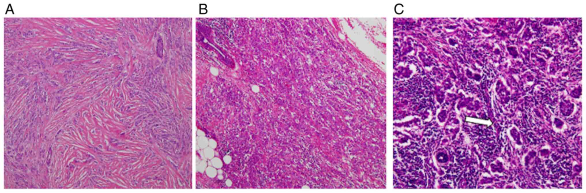 |
Figure 1
Distribution of tumor-infiltrating lymphocytes in estrogen receptor-positive and human epidermal growth factor receptor 2-negative breast cancer. H&E-stained sections showing (A) low, (B) intermediate and (C) high levels of tumor-infiltrating lymphocytes (arrow) in the stromal tissue of breast cancer (magnification, x200 for all images).
|
|
Table II
Association between TIL count and clinicopathological factors.
|
Table II
Association between TIL count and clinicopathological factors.
| |
TIL count, n (%) |
Significance |
| Clinicopathological factors |
Low |
Intermediate |
High |
Total no. |
P-value |
| Ki67 |
|
|
|
|
0.023 |
| ≤10% |
22 (84.6) |
4 (15.4) |
0 (0.0) |
26 |
|
| >10% and <30% |
16 (72.7) |
6 (27.3) |
0 (0.0) |
22 |
|
| ≥30% |
13 (76.5) |
1 (5.9) |
3 (17.6) |
17 |
|
| Progesterone receptor |
|
|
|
|
0.19 |
| Positive |
46 (82.1) |
8 (14.3) |
2 (3.6) |
56 |
|
| Negative |
5 (55.6) |
3 (33.3) |
1 (11.1) |
9 |
|
| Pathological T status in TNM classification |
|
|
|
|
0.87 |
| T1 and T2 |
47 (78.3) |
10 (16.7) |
3 (5.0) |
60 |
|
| T3 and T4 |
4 (80.0) |
1 (20.0) |
0 (0.0) |
5 |
|
| Nodal status |
|
|
|
|
0.46 |
| Positive |
34 (77.3) |
7 (15.9) |
3 (6.8) |
44 |
|
| Negative |
17 (81.0) |
4 (19.0) |
0 (0.0) |
21 |
|
| Nuclear grade |
|
|
|
|
0.65 |
| 3 |
9 (69.2) |
3 (23.1) |
1 (7.7) |
13 |
|
| 1 and 2 |
42 (80.8) |
8 (15.4) |
2 (3.8) |
52 |
|
| Lymphovascular invasion |
|
|
|
|
0.68 |
| Positive |
30 (75.0) |
8 (20.0) |
2 (5.0) |
40 |
|
| Negative |
21 (84.0) |
3 (12.0) |
1 (4.0) |
25 |
|
| Intrinsic subtype |
|
|
|
|
0.41 |
| Luminal A-like |
18 (85.7) |
3 (14.3) |
0 (0.0) |
21 |
|
| Luminal B-like |
33 (75.0) |
8 (18.2) |
3 (6.8) |
44 |
|
Prognostic analysis of str-TILs in luminal B-like breast cancer
The median RFS was 65 months (range, 10-158 months; Fig. S1). No patient with intermediate or high-grade str-TIL count suffered a relapse. However, str-TIL count was not a significant prognostic factor in all patients (Fig. 2 and Table SI). RFS was significantly worse in patients with luminal B-like cancer compared with that in patients with luminal A-like cancer (log-rank χ2=5.27, P=0.022), and no patient with luminal A-like cancer developed recurrence of breast cancer (Fig. S2). In addition, no patient with luminal B-like breast cancer and intermediate or high str-TIL count developed recurrence of breast cancer. The RFS of patients with intermediate or high-grade str-TIL count was generally longer compared with that of patients with low-grade str-TIL count, although the difference was not considered as statistically significant (log-rank χ2=3.23, P=0.072; Fig. 3).
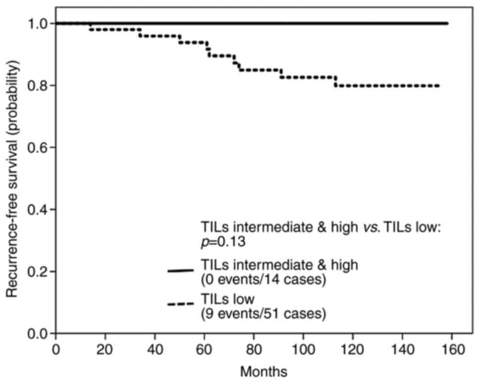 |
Figure 2
Survival curves of the patients with breast cancer stratified by TIL count in ER-positive and HER2-negative breast cancers. TIL count was not found to be a significant prognostic factor for the patients with ER-positive and HER2-negative breast cancers. TILs, tumor-infiltrating lymphocytes; ER, estrogen receptor; HER2, human epidermal growth factor receptor 2.
|
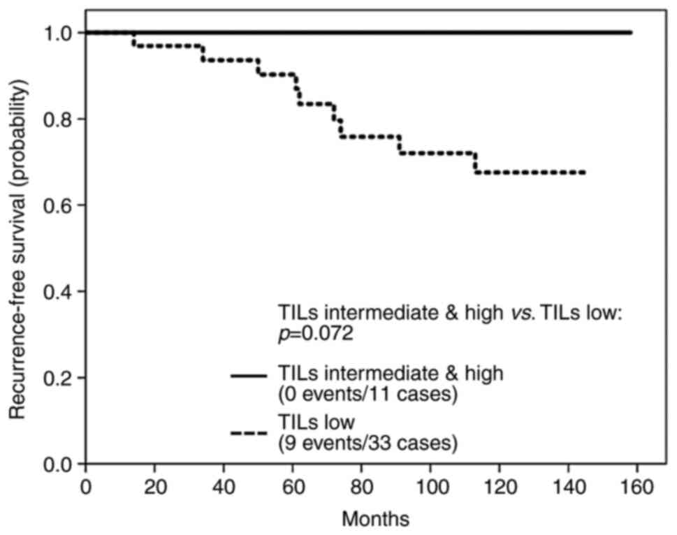 |
Figure 3
Survival curves of breast cancer patients stratified by TIL count in luminal B-like breast cancer. Among patients with luminal B-like breast cancer, the recurrence-free survival of patients with intermediate or high levels of TILs was generally longer compared with that in patients with low levels of TILs. TILs, tumor-infiltrating lymphocytes.
|
Discussion
The present study found that only 4.6% of ER-positive and HER2-negative patients were classified into the high-grade str-TIL group, and str-TIL count was not a powerful prognostic factor in all patients with ER-positive and HER2-negative breast cancer.
Several retrospective studies have suggested the potential use of TIL count as a prognostic factor in ER-negative breast cancer (6-9). Moreover, Denkert et al (9) confirmed that a high TIL grade was a powerful prognostic factor in a large number of patients with early-stage triple-negative breast cancer. In our previous study, ~30% of ER-negative patients had a high TIL count, which was a powerful prognostic marker. On the other hand, our previous study demonstrated that 3.1% of hormonal receptor-positive and HER2-negative patients had a high-grade TIL count according to the international guidelines (8). In this previous study, TIL grade was not identified as a significant prognostic factor in ER-positive breast cancer, similar to the results of the present study.
Previous studies suggested that ER potentially activated tumor immunosuppression (13,14). However, for the treatment of ER-positive breast cancer, the relationship between TIL count and response to endocrine therapy remains unclear. Tumor mutational burden is an important factor associated with tumor immunity and response to immune therapy (15). A high tumor mutation burden is associated with poor outcomes in ER-positive cancer (16,17). In addition, a previous study suggested that TIL count in ER-positive cancer is morphologically more heterogeneous compared with that in ER-negative cases (18). This intratumor heterogeneity may affect the difference in the prognostic power of TIL count between ER-positive and ER-negative cancer types.
In the present study, there was a significant association between high levels of Ki67 expression and a high str-TIL count. Fujimoto et al (19) suggested the possibility that the composition of immune cells may vary between breast cancers with high and low levels of Ki67 expression. Moreover, Xu et al (20) demonstrated that high tumor mutational burden was associated with high levels of Ki67 expression. High microsatellite instability and aberrant mismatch deficiency cause a high mutational burden in cancer (21). Microsatellite instability is known as a predictive biomarker of antitumor immune response and response to immune-checkpoint inhibitors (22,23). These findings suggest that the antitumor immune response may be increased in aggressive tumors with high levels of Ki67 expression.
In the present study, none of the patients with luminal B-like breast cancer and intermediate or high-grade str-TIL count developed recurrence during follow-up. Luminal B-like tumors generally have a worse outcome compared with luminal A-like-subtype tumors, even with the administration of hormonal therapy (24). The biological differences between these subtypes may be caused by the intracellular signaling pathways associated with estrogen among ER-positive breast cancer cells (25,26). However, the difference in molecular mechanisms between luminal A- and luminal B-like subtypes remains unclear. In routine practice, adjuvant hormone therapy alone is selected for patients with luminal A-like subtype and chemo-endocrine therapy for patients with luminal B-like subtype. Several clinical studies demonstrated that TIL count was correlated with response to chemotherapy mainly in patients with ER-negative breast cancer (27,28). However, for the use of TILs based on the subtypes of the ER-positive patients as luminal A and B subtypes, the biological evidence is unclear. In the future, TIL count may become a useful biomarker for determining the molecular mechanisms of tumor immunity in ER-positive breast tumors with potential clinical relevance for patients with luminal-type breast cancer.
To confirm the potential role of TIL count in breast cancer prognosis, the characteristics of immune cells constituting TILs will have to be examined in more detail. Cytotoxic T cells, which attack cancer cells, are positive for CD8, and T-reg cells, which inhibit immune responses to cancer cells, are positive for CD4, CD25 and FOXP3. How TIL count impacts patient response to treatment should also be determined, as CD8-positive T lymphocytes are associated with the therapeutic efficacy of chemotherapy (3).
In conclusion, it was herein confirmed that there is a significant association between TIL count and the occurrence of aggressive breast tumors with high levels of Ki67 expression in ER-positive and HER2-negative breast cancer. The survival of patients with intermediate or high-grade str-TIL count tends to be superior to that of patients with low-grade str-TIL count, although the difference was not found to be statistically significant. However, there were several limitations to the present study. First, the number of the enrolled patients was small. Second, this study is a retrospective trial. Therefore, further prospective and large-scale clinical trials are necessary to confirm the value of TIL count as a prognostic factor for patients with ER-positive/HER2-negative breast cancer. Moreover, additional functional studies will be necessary to determine how TIL count controls antitumor immune response and immune-checkpoint systems in ER-positive breast cancer.
Supplementary Material
Box-and-whisker plot; interquartile range of recurrence-free survival.
Survival curves of the patients with luminal A- and B-like breast cancers. A significant difference in survival rate was observed between the luminal A- and B-like subtypes.
Results of the multivariate analysis of the clinicopathological factors that influence breast cancer recurrence.
Acknowledgements
The authors gratefully acknowledge the work of our research technician, Kumiko Sudo.
Funding
No funding was received.
Availability of data and materials
The datasets generated and/or analyzed during the present study are not publicly available due to the regulations of the Institutional Review Board of Gunma University Hospital, but are available from the corresponding author on reasonable request.
Authors' contributions
All the authors participated in the design of the study. CH and SK mainly performed image acquisition and statistical analyses. AK and SK performed histological examinations. KM, YN, MO, SO and RY assisted in collecting the clinical information. BHK, TM, TO, JH, KS and TF contributed to the theoretical organization of the manuscript. SK and TF have seen and can confirm the authenticity of the raw data. All the authors have read and approved the final manuscript.
Ethics approval and consent to participate
The present study was conducted according to the tenets of the Declaration of Helsinki, and the study protocol was approved by the Institutional Review Board of Gunma University Hospital (registration no. 2016-003).
Patient consent for publication
Not applicable.
Competing interests
Takaaki Fujii received research funding from Eisai Co., Ltd. All other authors declare that they have no competing interests.
References
|
1
|
Bates JP, Derakhshandeh R, Jones L and Webb TJ: Mechanisms of immune evasion in breast cancer. BMC Cancer. 18(556)2018.PubMed/NCBI View Article : Google Scholar
|
|
2
|
Tower H, Ruppert M and Britt K: The immune microenvironment of breast cancer progression. Cancers (Basel). 11(1375)2019.PubMed/NCBI View Article : Google Scholar
|
|
3
|
Kurozumi S, Fujii T, Matsumoto H, Inoue K, Kurosumi M, Horiguchi J and Kuwano H: Significance of evaluating tumor-infiltrating lymphocytes (TILs) and programmed cell death-ligand 1 (PD-L1) expression in breast cancer. Med Mol Morphol. 50:185–194. 2017.PubMed/NCBI View Article : Google Scholar
|
|
4
|
Denkert C, von Minckwitz G, Brase JC, Sinn BV, Gade S, Kronenwett R, Pfitzner BM, Salat C, Loi S, Schmitt WD, et al: Tumor-infiltrating lymphocytes and response to neoadjuvant chemotherapy with or without carboplatin in human epidermal growth factor receptor 2-positive and triple-negative primary breast cancers. J Clin Oncol. 33:983–991. 2015.PubMed/NCBI View Article : Google Scholar
|
|
5
|
Salgado R, Denkert C, Demaria S, Sirtaine N, Klauschen F, Pruneri G, Wienert S, Van den Eynden G, Baehner FL, Penault-Llorca F, et al: The evaluation of tumor-infiltrating lymphocytes (TILs) in breast cancer: Recommendations by an International TILs Working Group 2014. Ann Oncol. 26:259–271. 2015.PubMed/NCBI View Article : Google Scholar
|
|
6
|
Kurozumi S, Inoue K, Matsumoto H, Fujii T, Horiguchi J, Oyama T, Kurosumi M and Shirabe K: Prognostic utility of tumor-infiltrating lymphocytes in residual tumor after neoadjuvant chemotherapy with trastuzumab for HER2-positive breast cancer. Sci Rep. 9(1583)2019.PubMed/NCBI View Article : Google Scholar
|
|
7
|
Loi S, Michiels S, Salgado R, Sirtaine N, Jose V, Fumagalli D, Kellokumpu-Lehtinen PL, Bono P, Kataja V, Desmedt C, et al: Tumor infiltrating lymphocytes are prognostic in triple negative breast cancer and predictive for trastuzumab benefit in early breast cancer: Results from the FinHER trial. Ann Oncol. 25:1544–1550. 2014.PubMed/NCBI View Article : Google Scholar
|
|
8
|
Kurozumi S, Matsumoto H, Kurosumi M, Inoue K, Fujii T, Horiguchi J, Shirabe K, Oyama T and Kuwano H: Prognostic significance of tumour-infiltrating lymphocytes for oestrogen receptor-negative breast cancer without lymph node metastasis. Oncol Lett. 17:2647–2656. 2019.PubMed/NCBI View Article : Google Scholar
|
|
9
|
Denkert C, von Minckwitz G, Darb-Esfahani S, Lederer B, Heppner BI, Weber KE, Budczies J, Huober J, Klauschen F, Furlanetto J, et al: Tumour-infiltrating lymphocytes and prognosis in different subtypes of breast cancer: A pooled analysis of 3771 patients treated with neoadjuvant therapy. Lancet Oncol. 19:40–50. 2018.PubMed/NCBI View Article : Google Scholar
|
|
10
|
Tokiniwa H, Horiguchi J, Takata D, Kikuchi M, Rokutanda N, Nagaoka R, Sato A, Odawara H, Tozuka K, Oyama T and Takeyoshi I: Topoisomerase II alpha expression and the Ki-67 labeling index correlate with prognostic factors in estrogen receptor-positive and human epidermal growth factor type-2-negative breast cancer. Breast Cancer. 4:309–314. 2012.PubMed/NCBI View Article : Google Scholar
|
|
11
|
Hammond ME, Hayes DF, Dowsett M, Allred DC, Hagerty KL, Badve S, Fitzgibbons PL, Francis G, Goldstein NS, Hayes M, et al: American society of clinical oncology/college of American pathologists guideline recommendations for immunohistochemical testing of estrogen and progesterone receptors in breast cancer. J Clin Oncol. 28:2784–2795. 2010.PubMed/NCBI View Article : Google Scholar
|
|
12
|
Kurozumi S, Matsumoto H, Hayashi Y, Tozuka K, Inoue K, Horiguchi J, Takeyoshi I, Oyama T and Kurosumi M: Power of PgR expression as a prognostic factor for ER-positive/HER2-negative breast cancer patients at intermediate risk classified by the Ki67 labeling index. BMC Cancer. 17(354)2017.PubMed/NCBI View Article : Google Scholar
|
|
13
|
Svoronos N, Perales-Puchalt A, Allegrezza MJ, Rutkowski MR, Payne KK, Tesone AJ, Nguyen JM, Curiel TJ, Cadungog MG, Singhal S, et al: Tumor cell-independent estrogen signaling drives disease progression through mobilization of myeloid-derived suppressor cells. Cancer Discov. 7:72–85. 2017.PubMed/NCBI View Article : Google Scholar
|
|
14
|
Montagna E, Vingiani A, Maisonneuve P, Cancello G, Contaldo F, Pruneri G and Colleoni M: Unfavorable prognostic role of tumor-infiltrating lymphocytes in hormone-receptor positive, HER2 negative metastatic breast cancer treated with metronomic chemotherapy. Breast. 34:83–88. 2017.PubMed/NCBI View Article : Google Scholar
|
|
15
|
Goodman AM, Kato S, Bazhenova L, Patel SP, Frampton GM, Miller V, Stephens PJ, Daniels GA and Kurzrock R: Tumor mutational burden as an independent predictor of response to immunotherapy in diverse cancers. Mol Cancer Ther. 16:2598–2608. 2017.PubMed/NCBI View Article : Google Scholar
|
|
16
|
Pereira B, Chin SF, Rueda OM, Vollan HK, Provenzano E, Bardwell HA, Pugh M, Jones L, Russell R, Sammut SJ, et al: The somatic mutation profiles of 2,433 breast cancers refines their genomic and transcriptomic landscapes. Nat Commun. 7(11479)2016.PubMed/NCBI View Article : Google Scholar
|
|
17
|
Haricharan S, Bainbridge MN, Scheet P and Brown PH: Somatic mutation load of estrogen receptor-positive breast tumors predicts overall survival: An analysis of genome sequence data. Breast Cancer Res Treat. 146:211–220. 2014.PubMed/NCBI View Article : Google Scholar
|
|
18
|
Heindl A, Sestak I, Naidoo K, Cuzick J, Dowsett M and Yuan Y: Relevance of spatial heterogeneity of immune infiltration for predicting risk of recurrence after endocrine therapy of ER+ breast cancer. J Natl Cancer Inst. 110(10.1093/jnci/djx137)2018.PubMed/NCBI View Article : Google Scholar
|
|
19
|
Fujimoto Y, Watanabe T, Hida AI, Higuchi T, Miyagawa Y, Ozawa H, Bun A, Fukui R, Sata A, Imamura M, et al: Prognostic significance of tumor-infiltrating lymphocytes may differ depending on Ki67 expression levels in estrogen receptor-positive/HER2-negative operated breast cancers. Breast Cancer. 26:738–747. 2019.PubMed/NCBI View Article : Google Scholar
|
|
20
|
Xu J, Guo X, Jing M and Sun T: Prediction of tumor mutation burden in breast cancer based on the expression of ER, PR, HER-2, and Ki-67. Onco Targets Ther. 11:2269–2275. 2019.PubMed/NCBI View Article : Google Scholar
|
|
21
|
Le DT, Uram JN, Wang H, Bartlett BR, Kemberling H, Eyring AD, Skora AD, Luber BS, Azad NS, Laheru D, et al: PD-1 blockade in tumors with mismatch-repair deficiency. N Engl J Med. 372:2509–2520. 2015.PubMed/NCBI View Article : Google Scholar
|
|
22
|
Dudley JC, Lin MT, Le DT and Eshleman JR: Microsatellite instability as a biomarker for PD-1 blockade. Clin Cancer Res. 22:813–820. 2016.PubMed/NCBI View Article : Google Scholar
|
|
23
|
Marabelle A, Le DT, Ascierto PA, Di Giacomo AM, De Jesus-Acosta A, Delord JP, Geva R, Gottfried M, Penel N, Hansen AR, et al: Efficacy of pembrolizumab in patients with noncolorectal high microsatellite instability/mismatch repair-deficient cancer: Results from the phase II KEYNOTE-158 study. J Clin Oncol. 38:1–10. 2020.PubMed/NCBI View Article : Google Scholar
|
|
24
|
Kurozumi S, Yamaguchi Y, Matsumoto H, Kurosumi M, Hayashi SI, Fujii T, Horiguchi J, Shirabe K and Inoue K: Utility of Ki67 labeling index, cyclin D1 expression, and ER-activity level in postmenopausal ER-positive and HER2-negative breast cancer with neoadjuvant chemo-endocrine therapy. PLoS One. 14(e0217279)2019.PubMed/NCBI View Article : Google Scholar
|
|
25
|
Kurozumi S, Matsumoto H, Inoue K, Tozuka K, Hayashi Y, Kurosumi M, Oyama T, Fujii T, Horiguchi J and Kuwano H: Impact of combining the progesterone receptor and preoperative endocrine prognostic index (PEPI) as a prognostic factor after neoadjuvant endocrine therapy using aromatase inhibitors in postmenopausal ER positive and HER2 negative breast cancer. PLoS One. 13(e0201846)2018.PubMed/NCBI View Article : Google Scholar
|
|
26
|
Kurozumi S, Yamaguchi Y, Matsumoto H, Inoue K, Kurosumi M, Oyama T, Horiguchi J, Fujii T and Shirabe K: Comparing protein and mRNA expressions of the human epidermal growth factor receptor family in estrogen receptor-positive breast cancer. Med Mol Morphol. 52:90–98. 2019.PubMed/NCBI View Article : Google Scholar
|
|
27
|
Savas P, Salgado R, Denkert C, Sotiriou C, Darcy PK, Smyth MJ and Loi S: Clinical relevance of host immunity in breast cancer: From TILs to the clinic. Nat Rev Clin Oncol. 13:228–241. 2016.PubMed/NCBI View Article : Google Scholar
|
|
28
|
Kurozumi S, Inoue K, Matsumoto H, Fujii T, Horiguchi J, Oyama T, Kurosumi M and Shirabe K: Clinicopathological values of PD-L1 expression in HER2-positive breast cancer. Sci Rep. 9(16662)2019.PubMed/NCBI View Article : Google Scholar
|


