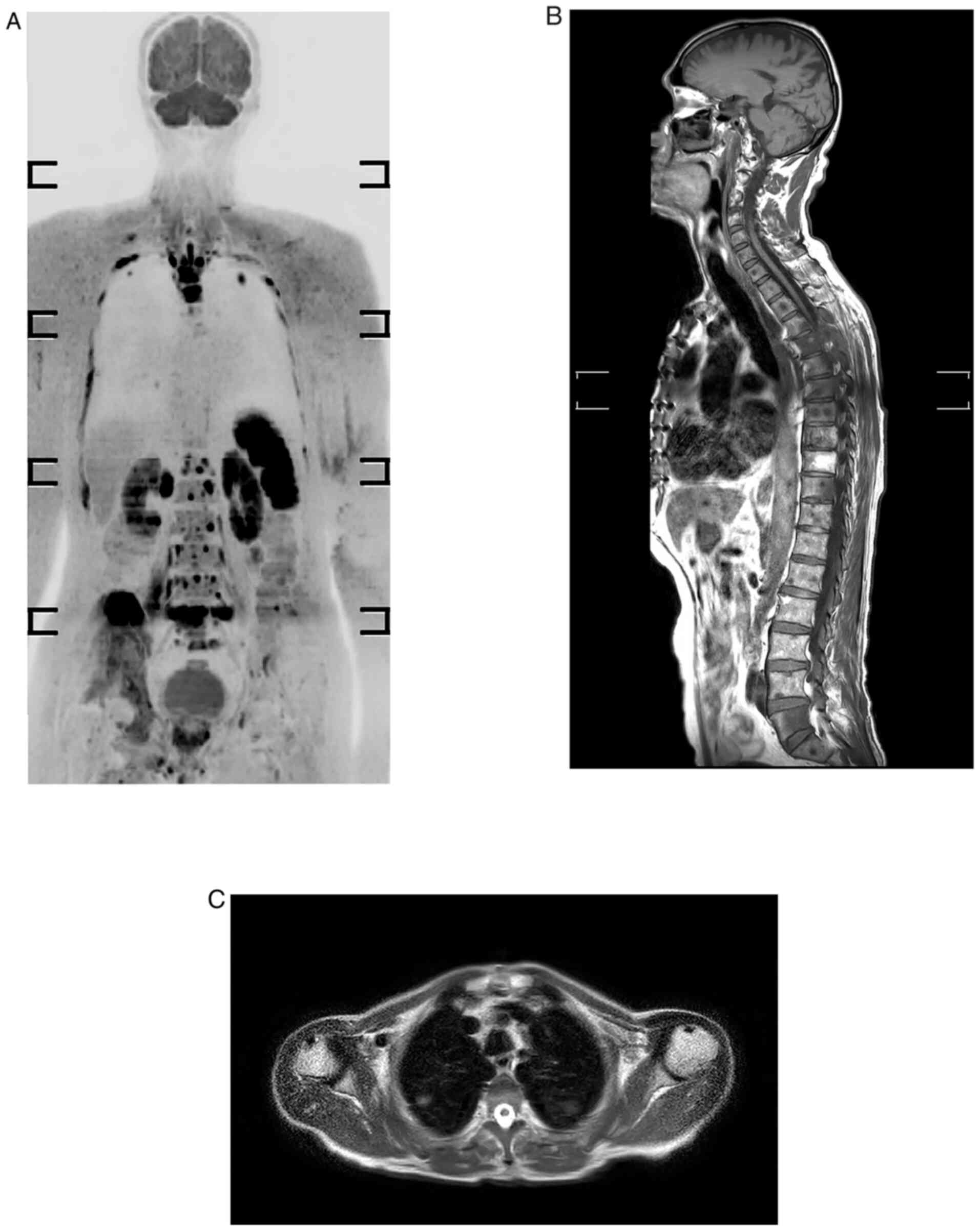
|
1
|
Guan X: Cancer metastases: Challenges and
opportunities. Acta Pharm Sin B. 5:402–418. 2015.PubMed/NCBI View Article : Google Scholar
|
|
2
|
Heindel W, Gübitz R, Vieth V, Weckesser M,
Schober O and Schäfers M: The diagnostic imaging of bone
metastases. Dtsch Arztebl Int. 111:741–747. 2014.PubMed/NCBI View Article : Google Scholar
|
|
3
|
Imam K and Bluemke DA: MR imaging in the
evaluation of hepatic metastases. Magn Reson Imaging Clin N Am.
8:741–756. 2000.PubMed/NCBI
|
|
4
|
Guimarães MD, Noschang J, Teixeira SR,
Santos MK, Lederman HM, Tostes V, Kundra V, Oliveira AD, Hochhegger
B and Marchiori E: Whole-body MRI in pediatric patients with
cancer. Cancer Imaging. 17(6)2017.PubMed/NCBI View Article : Google Scholar
|
|
5
|
Schlemmer HP, Schäfer J, Pfannenberg C,
Radny P, Korchidi S, Müller-Horvat C, Nägele T, Tomaschko K,
Fenchel M and Claussen CD: Fast whole-body assessment of metastatic
disease using a novel magnetic resonance imaging system: Initial
experiences. Invest Radiol. 40:64–71. 2005.PubMed/NCBI View Article : Google Scholar
|
|
6
|
Godinho MV, Lopes FPPL and Costa FM:
Whole-body magnetic resonance imaging for the assessment of
metastatic breast cancer. Cancer Manag Res. 10:6743–6756.
2018.PubMed/NCBI View Article : Google Scholar
|
|
7
|
Lauenstein TC and Semelka RC: Emerging
techniques: Whole-body screening and staging with MRI. J Magn Reson
Imaging. 24:489–498. 2006.PubMed/NCBI View Article : Google Scholar
|
|
8
|
Schmidt GP, Haug AR, Schoenberg SO and
Reiser MF: Whole-body MRI and PET-CT in the management of cancer
patients. Eur Radiol. 16:1216–1225. 2006.PubMed/NCBI View Article : Google Scholar
|
|
9
|
Bisschop C, de Heer EC, Brouwers AH,
Hospers GAP and Jalving M: Rational use of 18F-FDG
PET/CT in patients with advanced cutaneous melanoma: A systematic
review. Crit Rev Oncol Hematol. 153(103044)2020.PubMed/NCBI View Article : Google Scholar
|
|
10
|
Li B, Li Q, Nie W and Liu S: Diagnostic
value of whole-body diffusion-weighted magnetic resonance imaging
for detection of primary and metastatic malignancies: A
meta-analysis. Eur J Radiol. 83:338–344. 2014.PubMed/NCBI View Article : Google Scholar
|
|
11
|
Nievelstein RA and Littooij AS: Whole-Body
MRI in Pediatric Oncology. Img Ped Oncol. 107–135. 2019.
|
|
12
|
Pasoglou V, Michoux N, Tombal B and
Lecouvet F: Optimising TNM staging of patients with prostate cancer
using WB-MRI. J Belg Soc Radiol. 100(101)2016.PubMed/NCBI View Article : Google Scholar
|
|
13
|
Kwee TC, Takahara T, Ochiai R, Nievelstein
RA and Luijten PR: Diffusion-weighted whole-body imaging with
background body signal suppression (DWIBS): Features and potential
applications in oncology. Eur Radiol. 18:1937–1952. 2008.PubMed/NCBI View Article : Google Scholar
|
|
14
|
Puls R, Kühn JP, Ewert R and Hosten N:
Whole-body magnetic resonance imaging for staging of lung cancer.
Front Radiat Ther Oncol. 42:46–54. 2010.PubMed/NCBI View Article : Google Scholar
|
|
15
|
Akay S, Kocaoglu M, Emer O, Battal B and
Arslan N: Diagnostic accuracy of whole-body diffusion-weighted
magnetic resonance imaging with 3.0 T in detection of primary and
metastatic neoplasms. J Med Imaging Radiat Oncol. 57:274–282.
2013.PubMed/NCBI View Article : Google Scholar
|
|
16
|
Kachewar SG: Using DWIBS MRI technique as
an alternative to bone scan or PET scan for whole-body imaging in
oncology patients. Acta Radiol. 52(788)2011.PubMed/NCBI View Article : Google Scholar
|
|
17
|
Schmidt GP, Reiser MF and Baur-Melnyk A:
Whole-body MRI for the staging and follow-up of patients with
metastasis. Eur J Radiol. 70:393–400. 2009.PubMed/NCBI View Article : Google Scholar
|
|
18
|
Schmidt GP, Baur-Melnyk A, Haug A,
Heinemann V, Bauerfeind I, Reiser MF and Schoenberg SO:
Comprehensive imaging of tumor recurrence in breast cancer patients
using whole-body MRI at 1.5 and 3 T compared to FDG-PET-CT. Eur J
Radiol. 65:47–58. 2008.PubMed/NCBI View Article : Google Scholar
|
|
19
|
Sohaib SA, Koh DM, Barbachano Y, Parikh J,
Husband JE, Dearnaley DP, Horwich A and Huddart R: Prospective
assessment of MRI for imaging retroperitoneal metastases from
testicular germ cell tumours. Clin Radiol. 64:362–367.
2009.PubMed/NCBI View Article : Google Scholar
|
|
20
|
Ciliberto M, Maggi F, Treglia G, Padovano
F, Calandriello L, Giordano A and Bonomo L: Comparison between
whole-body MRI and Fluorine-18-Fluorodeoxyglucose PET or PET/CT in
oncology: A systematic review. Radiol Oncol. 47:206–218.
2013.PubMed/NCBI View Article : Google Scholar
|
|
21
|
Barchetti F, Stagnitti A, Megna V, Al
Ansari N, Marini A, Musio D, Monti ML, Barchetti G, Tombolini V,
Catalano C and Panebianco V: Unenhanced whole-body MRI versus
PET-CT for the detection of prostate cancer metastases after
primary treatment. Eur Rev Med Pharmacol Sci. 20:3770–3776.
2016.PubMed/NCBI
|
|
22
|
Li B, Li Q, Chen C, Guan Y and Liu S: A
systematic review and meta-analysis of the accuracy of
diffusion-weighted MRI in the detection of malignant pulmonary
nodules and masses. Acad Radiol. 21:21–29. 2014.PubMed/NCBI View Article : Google Scholar
|
|
23
|
Regier M, Schwarz D, Henes FO, Groth M,
Kooijman H, Begemann PG and Adam G: Diffusion-weighted MR-imaging
for the detection of pulmonary nodules at 1.5 Tesla:
Intraindividual comparison with multidetector computed tomography.
J Med Imaging Radiat Oncol. 55:266–274. 2011.PubMed/NCBI View Article : Google Scholar
|
|
24
|
Goda HH, Abd Elkareem HA, Ahmed EA,
Megally HI, Khalaf MI, Taha AM and Mohamed HEG: Whole body
diffusion-weighted MRI in detection of metastasis and lymphoma: A
prospective longitudinal clinical study. Egypt J Radiol Nucl Med.
51:1–2. 2020.
|
|
25
|
Eissawy MG, Saadawy AMI, Farag K, Akl T
and Kamr WH: Accuracy and diagnostic value of diffusion-weighted
whole body imaging with background body signal suppression (DWIBS)
in metastatic breast cancer. Egypt J Radiol Nucl Med.
52(74)2021.
|
|
26
|
Iwamura H, Kaiho Y, Ito J, Anan G, Satani
N, Matsuura T, Tamura R, Murakami K, Koyama K and Sato M:
Evaluation of tumor viability for primary and bone metastases in
metastatic castration-resistant prostate cancer using whole-body
magnetic resonance imaging. Case Rep Urol.
2018(4074378)2018.PubMed/NCBI View Article : Google Scholar
|
|
27
|
Usuda K, Iwai S, Yamagata A, Iijima Y,
Motono N, Matoba M, Doai M, Yamada S, Ueda Y, Hirata K, et al:
Diffusion-weighted whole-body imaging with background suppression
(DWIBS) is effective and economical for detection of metastasis or
recurrence of lung cancer. Thorac Cancer. 12:676–684.
2021.PubMed/NCBI View Article : Google Scholar
|
|
28
|
Paruthikunnan SM, Kadavigere R and
Karegowda LH: Accuracy of whole-body DWI for metastases screening
in a diverse group of malignancies: Comparison with conventional
cross-sectional imaging and nuclear scintigraphy. AJR Am J
Roentgenol. 209:477–490. 2017.PubMed/NCBI View Article : Google Scholar
|
|
29
|
Torabi M, Aquino SL and Harisinghani MG:
Current concepts in lymph node imaging. J Nucl Med. 45:1509–1518.
2004.PubMed/NCBI
|
|
30
|
Tunariu N, Blackledge M, Messiou C,
Petralia G, Padhani A, Curcean S, Curcean A and Koh DM: What's new
for clinical whole-body MRI (WB-MRI) in the 21st century. Brit J
Radiol. 93(20200562)2020.PubMed/NCBI View Article : Google Scholar
|
|
31
|
Kharuzhyk S, Zhavrid E, Dziuban A,
Sukolinskaja E and Kalenik O: Comparison of whole-body MRI with
diffusion-weighted imaging and PET/CT in lymphoma staging. Eur
Radiol. 30:3915–3923. 2020.PubMed/NCBI View Article : Google Scholar
|
|
32
|
Balbo-Mussetto A, Cirillo S, Bruna R,
Gueli A, Saviolo C, Petracchini M, Fornari A, Lario CV, Gottardi D,
De Crescenzo A and Tarella C: Whole-body MRI with
diffusion-weighted imaging: A valuable alternative to
contrast-enhanced CT for initial staging of aggressive lymphoma.
Clin Radiol. 71:271–279. 2016.PubMed/NCBI View Article : Google Scholar
|
|
33
|
Albano D, Patti C, La Grutta L, Agnello F,
Grassedonio E, Mulè A, Cannizzaro G, Ficola U, Lagalla R, Midiri M
and Galia M: Comparison between whole-body MRI with
diffusion-weighted imaging and PET/CT in staging newly diagnosed
FDG-avid lymphomas. Eur J Radiol. 85:313–318. 2016.PubMed/NCBI View Article : Google Scholar
|
|
34
|
Sigovan M, Akl P, Mesmann C, Tronc F,
Si-Mohamed S, Douek P and Boussel L: Benign and malignant enlarged
chest nodes staging by diffusion-weighted MRI: An alternative to
mediastinoscopy? Brit J Radiol. 91(20160919)2018.PubMed/NCBI View Article : Google Scholar
|
|
35
|
Yang HL, Liu T, Wang XM, Xu Y and Deng SM:
Diagnosis of bone metastases: A meta-analysis comparing 18FDG PET,
CT, MRI and bone scintigraphy. Eur Radiol. 21:2604–2617.
2011.PubMed/NCBI View Article : Google Scholar
|
|
36
|
Engelhard K, Hollenbach HP, Wohlfart K,
von Imhoff E and Fellner FA: Comparison of whole-body MRI with
automatic moving table technique and bone scintigraphy for
screening for bone metastases in patients with breast cancer. Eur
Radiol. 14:99–105. 2004.PubMed/NCBI View Article : Google Scholar
|
|
37
|
Shen G, Deng H, Hu S and Jia Z: Comparison
of choline-PET/CT, MRI, SPECT, and bone scintigraphy in the
diagnosis of bone metastases in patients with prostate cancer: A
meta-analysis. Skelet Radiol. 43:1503–1513. 2014.PubMed/NCBI View Article : Google Scholar
|
|
38
|
Liu LP, Cui LB, Zhang XX, Cao J, Chang N,
Tang X, Qi S, Zhang XL, Yin H and Zhang J: Diagnostic performance
of diffusion-weighted magnetic resonance imaging in bone
malignancy: Evidence from a meta-analysis. Medicine (Baltimore).
94(e1998)2015.PubMed/NCBI View Article : Google Scholar
|


