Introduction
The prognosis for patients with glioblastoma remains
extremely poor, as the median survival time is less than 20 months
even with postoperative chemoradiation therapy using temozolomide
(1-3).
Recent studies have revealed that glioma stem-like cells (GSCs), a
type of cancer stem cell, are crucial in the malignancy of
glioblastomas (4,5). GSCs can differentiate into glioma
cells, including glioblastoma cells, in response to the
microenvironment. Interestingly, these differentiated glioblastoma
cells (DGCs) have been reported to dedifferentiate back into GSCs,
indicating bidirectional interconversion between GSCs and DGCs
(6,7). Our group has previously conducted
extensive research into the biological characteristics of GSCs
(7-9).
Indoleamine 2,3-dioxygenase 1 (IDO1), a
tryptophan-metabolizing enzyme, promotes the catabolic pathway from
tryptophan to kynurenine, which is known to suppress cytotoxic T
cells and activate regulatory T cells (10-12).
IDO1 is highly expressed in various malignant tumors, including
glioblastomas, and has been reported to suppress tumor immunity
(10,13). Furthermore, IDO1 expression has
been shown to correlate with poor prognosis in patients with
glioblastomas and to increase with higher tumor grade of gliomas
(14-16).
We previously demonstrated that IDO1 is more
strongly expressed in GSCs than in DGCs, suggesting that GSCs
suppress antitumor immunity via IDO1 expression (8). Based on this finding, we hypothesized
that inhibition of IDO1 in GSCs could promote differentiation and
attenuate resistance to temozolomide. The present study tried to
develop a novel differentiation-inducing therapy for
glioblastomas.
Materials and methods
Cell lines and cell culture
The present experiments used the widely available
human glioblastoma cell line U-87MG (glioblastoma of unknown
origin; cat. no. HTB 14; lot no. 2497162; purchased from the
American Type Culture Collection), and GSC lines 0125-GSC and
0222-GSC, newly established from surgical specimens. The primary
cell lines 0125-GSC and 0222-GSC were originally established at
Nagoya University (Nagoya, Japan) from glioblastoma specimens
obtained after written informed consent from the patients, as
previously reported (17), and
have been used in multiple studies (6-9,18).
The use of these cell lines in the present study was approved by
the Institutional Review Board of Nihon University School of
Medicine (approval no. 2026-09; Tokyo, Japan). Additionally, a GSC
model line Rev-U-87MG, derived by culturing U-87MG in serum-free
medium for more than 2 weeks, was used (8). DGC model lines 0125-DGC and 0222-DGC
were generated by culturing 0125-GSC and 0222-GSC in
serum-containing medium for more than 2 weeks (8). Furthermore, Rev-U-87MG, 0125-GSC, and
0222-GSC were cultured in the continuous presence of the IDO1
inhibitor for over 2 weeks to generate IDO1-suppressed cell lines
Rev-U-87MG-IDO(-), 0125-GSC-IDO(-), and 0222-GSC-IDO(-).
Cell culture used serum-containing medium consisting
of Dulbecco's modified Eagle's medium supplemented (Nissui
Pharmaceutical) with 10% fetal bovine serum (Thermo Fisher
Scientific, Inc.), and serum-free medium consisted of Neurobasal
Medium (Invitrogen; Thermo Fisher Scientific, Inc.) supplemented
with L-glutamine, epidermal growth factor, recombinant human basic
fibroblast growth factor (R&D Systems), N2, and B27
(Invitrogen; Thermo Fisher Scientific, Inc.). Serum-containing
medium was used for culturing DGCs, whereas serum-free medium was
used for culturing GSCs (7-9).
Cells were passaged and the media replaced every 4 days.
Temozolomide (#T2744; Tokyo Chemical Industry) and the IDO1
inhibitor 1-methyl-L-tryptophan (1-MT) (#447439; Sigma-Aldrich
Japan) were used. IDO1-inhibited cell lines were generated with 10
µM 1-MT added into the medium during each passage.
Western blotting
Protein expression analysis was performed using
western blotting according to the previously described methods
(18). Briefly, 1x10⁴
cells from each cell line were seeded onto a 10-cm diameter dish,
and the cells were harvested 72 h later to extract the proteins.
Anti-IDO1 antibody, (#66528-1-Ig; Proteintech Japan) anti-Nestin
antibody (#29285-1-AP; Proteintech Japan), anti-Nanog antibody
(#67255-I-Ig; Proteintech Japan), anti-Sox2 antibody (#11064-1-AP;
Proteintech Japan), and anti-GFAP antibody (#60190-1-Ig;
Proteintech Japan) were used as primary antibodies. Anti-β-actin
antibody (#sc-47778; Santa Cruz Biotechnology) used as a loading
control.
Cell counting assay
Cell proliferation inhibition assays were also
conducted as described previously (18). Briefly, 1x10⁴ cells were
seeded onto a 10-cm diameter dish, and 24 h later, the medium was
replaced with fresh medium containing varying concentrations of
temozolomide. After 72 h, cells were harvested and counted using a
Z1 Coulter Counter® (Beckman Coulter). Temozolomide
concentrations used were 0, 0.1, 1, 10, 100, and 1,000 µM.
Additionally, complementary cell viability analysis was performed
using the Cell Counting Kit-8 (CCK-8) (Dojindo Molecular
Technologies) according to the manufacturer's instructions. A total
of 1x10⁴ cells were seeded into each well of a 96-well
plate. Cells were treated with the same concentrations of
temozolomide and assessed at the same time points.
Statistical analysis
Statistical analysis was performed using unpaired
Student's t-test and one-way analysis of variance followed by
Tukey's post-hoc test with IBM SPSS Statistics version 21.0 (IBM
Corp., Armonk, NY, USA). P<0.05 was considered to indicate a
statistically significant difference.
Results
Effects of IDO1 inhibition on cell
morphology and adhesion
The morphology of each cell line was observed under
a fluorescence microscope (Fig.
1A). Glioblastoma cell line U-87MG, cultured in
serum-containing medium, had adhered to the culture dish and
proliferated (Fig. 1A, top-left).
Rev-U-87MG, cultured in serum-free medium for more than 2 weeks,
had formed floating spheres (Fig.
1A, top-center), a characteristic of stem cells.
Rev-U-87MG-IDO(-), cultured with 10 µM 1-MT for over 2 weeks to
inhibit IDO1, had formed smaller spheres (Fig. 1A, top-right). The concentration of
1-MT to be administered continuously was determined based on
preliminary experiments. The preliminary experiments were designed
to determine the maximum concentration of 1-MT that could be
continuously administered without inducing reduction in the cell
number (data not shown), and 10 µM was selected as the
concentration. After removing the medium, adherent cells were
collected using trypsin, and cell numbers were counted.
Rev-U-87MG-IDO(-) had formed a significantly greater number of
adherent cells than Rev-U-87MG (P<0.01) (Fig. 1B, top). GSC lines 0125-GSC and
0222-GSC, cultured in serum-free medium, had also formed floating
spheres (Fig. 1A,
middle/bottom-center). However, 0125-GSC and 0222-GSC spheres,
cultured in serum-containing medium for more than 2 weeks, had
dissolved and the cells became adherent, so forming the DGC lines
0125-DGC and 0222-DGC (Fig. 1A,
middle/bottom-left). Similarly, 0125-GSC-IDO(-) and
0222-GSC-IDO(-), cultured with 10 µM 1-MT for more than 2 weeks,
formed smaller spheres (Fig. 1A,
middle/bottom-right). 0125-GSC-IDO(-) and 0222-GSC-IDO(-) formed
significantly higher numbers of adherent cells than in their
respective controls (P<0.01) (Fig.
1B, middle and bottom).
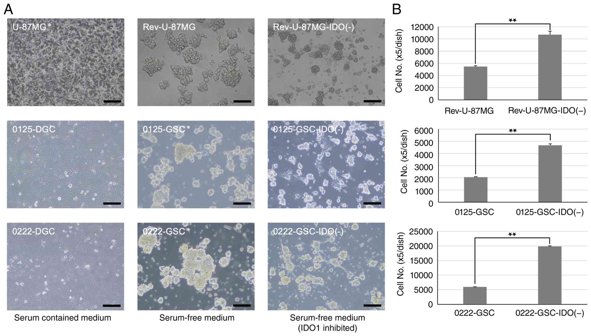 | Figure 1Morphology and cell adherence in each
cell line. (A) Morphology of the cell lines used in this study. Top
row shows the commercially available human glioblastoma cell line
U-87MG (left), and the derived lines Rev-U-87MG (center) and
Rev-U-87MG-IDO(-) (right). Rev-U-87MG was generated by culturing
U-87MG in serum-free medium for 2 weeks and formed spheres.
Rev-U-87MG-IDO(-) was established by culturing Rev-U-87MG with 10
µM 1-methyl-L-tryptophan added at each passage for 2 weeks and
formed smaller spheres. Middle and bottom rows show newly
established glioma stem-like cell lines 0125-GSC/0222-GSC (center),
and their derived lines 0125-DGC/0222-DGC (left) and
0125-GSC-IDO(-)/0222-GSC-IDO(-) (right). 0125-DGC and 0222-DGC were
generated by culturing 0125-GSC and 0222-GSC in serum-containing
medium for 2 weeks, and adhered to the culture dish with no sphere
formation. 0125-GSC-IDO(-) and 0222-GSC-IDO(-) were established by
culturing 0125-GSC and 0222-GSC with 10 µM 1-methyl-L-tryptophan at
each passage for 2 weeks, resulting in reduced sphere formation.
Scale bars=0.1 µm. (B) Number of adherent cells in each cell line.
Compared to Rev-U-87MG (top), 0125-GSC (middle), and 0222-GSC
(bottom), the number of adherent cells was significantly increased
in Rev-U-87MG-IDO(-) (top), 0125-GSC-IDO(-) (middle), and
0222-GSC-IDO(-) (bottom). Cells were cultured in 10-cm dishes.
After removal of floating cells, adherent cells were detached using
trypsin and counted with a Z1 Coulter Counter® (Beckman
Coulter, Brea, CA, USA). **P<0.01. DGC,
differentiated glioblastoma cell; GSC, glioma stem-like cell; IDO1,
indoleamine 2,3-dioxygenase 1. |
Effects of IDO1 inhibition on proteins
expression
Western blot analysis was used to examine the
protein expression of IDO1, stem cell markers (Nestin, Nanog, and
SOX2) and astrocyte marker (GFAP) in each cell line. The
IDO1-inhibited cell lines Rev-U-87MG-IDO(-), 0125-GSC-IDO(-), and
0222-GSC-IDO(-) showed significantly reduced IDO1 expression
compared to Rev-U-87MG, 0125-GSC, and 0222-GSC (Fig. 2A). These IDO1-inhibited cell lines
also showed significantly reduced expression of stem cell markers
(Fig. 2B), along with increased
expression of astrocyte marker (Fig.
2C). Additional experiments using 1-MT at concentrations of 1
and 5 µM further demonstrated that IDO1 suppression and the
subsequent reduction in stem cell marker expression occurred in a
concentration-dependent manner (Fig.
S1). These findings suggest that continuous treatment with 10
µM 1-MT for more than 2 weeks effectively inhibited IDO1 expression
which in turn reduced the expression of stem cell markers along
with increased the expression of astrocyte marker, indicating that
IDO1 inhibition promotes the differentiation of GSCs.
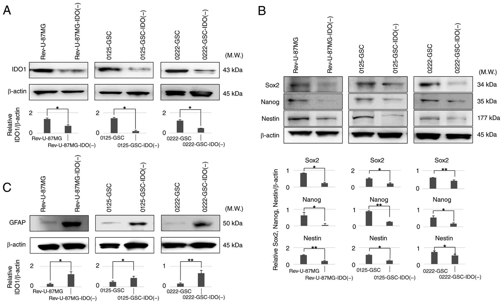 | Figure 2Western blot analysis of protein
expression of (A) IDO1, (B) stem cell markers Sox2 (top), Nanog
(upper middle) and Nestin (lower middle), and (C) astrocyte marker
GFAP in each cell line. (A) Compared to glioma stem-like cell lines
Rev-U-87MG, 0125-GSC, and 0222-GSC, IDO1 expression was
significantly reduced in the IDO1-inhibited cell lines
Rev-U-87MG-IDO(-), 0125-GSC-IDO(-), and 0222-GSC-IDO(-).
*P<0.05. (B) Similarly, expression of the stem cell
markers Sox2, Nanog, and Nestin was significantly decreased in
Rev-U-87MG-IDO(-), 0125-GSC-IDO(-), and 0222-GSC-IDO(-) compared to
their respective stem cell-like glioma cell lines.
*P<0.05; **P<0.01. (C) Conversely,
expression of the astrocyte marker GFAP was significantly increased
in Rev-U-87MG-IDO(-), 0125-GSC-IDO(-), and 0222-GSC-IDO(-) compared
to their respective stem cell-like glioma cell lines.
*P<0.05; **P<0.01. GSC, glioma
stem-like cell; IDO1, indoleamine 2,3-dioxygenase 1. |
Effects of IDO1 inhibition on
temozolomide sensitivity
Based on these results, the drug sensitivity of each
cell line to temozolomide was examined. Rev-U-87MG exhibited
reduced sensitivity (indicating increased resistance) to
temozolomide compared to the parent U-87MG, with significant
differences observed at 1 and 10 µM (both P<0.05) (Fig. 3A, solid and dotted lines). However,
Rev-U-87MG-IDO(-) showed increased sensitivity (indicating reduced
resistance) compared to Rev-U-87MG, with a significant difference
observed at 100 µM (P<0.01) (Fig.
3A, dotted and dashed lines). The half-maximal inhibitory
concentration (IC50) values of temozolomide were 0.5 µM
for U-87MG, 16.0 µM for Rev-U-87MG, and 0.9 µM for
Rev-U-87MG-IDO(-). Similarly, 0125-DGC showed significantly higher
sensitivity to temozolomide compared to the parent 0125-GSC line at
0.1, 1, 10, and 100 µM (all P<0.01) (Fig. 3B, solid and dotted lines).
0125-GSC-IDO(-) also showed enhanced drug sensitivity compared to
the parent 0125-GSC, with significant differences observed at 10
and 100 µM (both P<0.01) (Fig.
3B, dotted and dashed lines). The IC50 values of
temozolomide were 250.6 µM for 0125-GSC, 0.2 µM for 0125-DGC, and
106.7 µM for 0125-GSC-IDO(-). The differentiated line 0222-DGC was
significantly higher sensitive compared to 0222-GSC at 0.1 and 1 µM
(both P<0.01) (Fig. 3C, solid
and dotted lines). 0222-GSC-IDO(-) showed increased sensitivity
compared to 0222-GSC, with significant differences at 0.1, 1, and
100 µM (P<0.05, P<0.01, and P<0.05, respectively)
(Fig. 3C, dotted and dashed
lines). The IC50 values of temozolomide were 155.4 µM
for 0222-GSC, 117.6 µM for 0222-DGC, and 12.2 µM for
0222-GSC-IDO(-). Consistent results were also obtained through
comprehensive quantification using a CCK-8-based metabolic activity
assay to assess the sensitivity of 0125-GSC and 0125-GSC-IDO(-)
cells to temozolomide (Fig. 3D).
0125-GSC-IDO(-) showed enhanced drug sensitivity compared to the
parent 0125-GSC, with significant differences observed at 1, 100,
and 1,000 µM (P<0.05, P<0.01, and P<0.05, respectively)
(Fig. 3D, dotted and dashed
lines). These findings indicate that GSCs exhibit greater
resistance to temozolomide compared to DGCs. However, IDO1
inhibition in GSCs reduces this resistance, supporting the
hypothesis that IDO1 inhibition promotes GSC differentiation and
decreases chemoresistance.
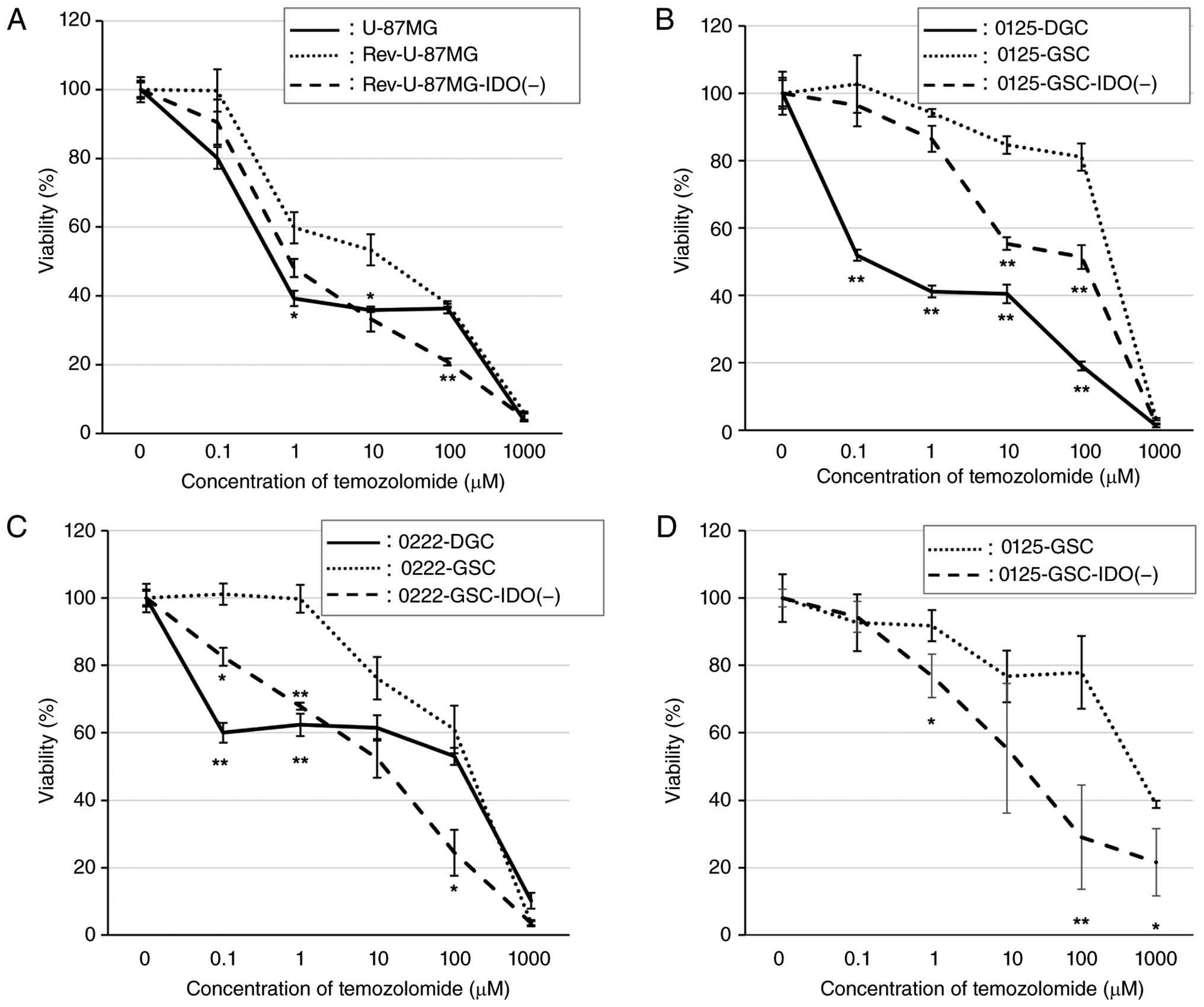 | Figure 3Drug sensitivity to temozolomide was
evaluated in each cell line. A total of 1x10⁴ cells were
seeded into 10-cm dishes, and the medium was replaced with fresh
medium containing varying concentrations of temozolomide 24 h
later. After 72 h, cells were harvested and counted using a (A-C)
Z1 Coulter Counter or analyzed using (D) Cell Counting Kit-8. (A)
Rev-U-87MG exhibited significant reduction of sensitivity for
temozolomide compared to U-87MG at 1 and 10 µM. Rev-U-87MG-IDO(-)
showed significant increase of sensitivity for temozolomide
compared to Rev-U-87MG at 100 µM. *P<0.05;
**P<0.01. (B) 0125-DGC exhibited significant
sensitivity for temozolomide compared to 0125-GSC at 0.1, 1, 10,
and 100 µM. 0125-GSC-IDO(-) showed significant sensitivity for
temozolomide compared to 0125-GSC at 10 and 100 µM.
**P<0.01. (C) 0222-DGC exhibited significant
sensitivity for temozolomide compared to 0222-GSC at 0.1 and 1 µM.
0222-GSC-IDO(-) showed significant sensitivity for temozolomide
compared to 0222-GSC at 0.1, 1, and 100 µM.*P<0.05;
**P<0.01. (D) 0125-GSC-IDO(-) showed significant
sensitivity for temozolomide compared to 0125-GSC at 1, 100, and
1,000 µM. *P<0.05; **P<0.01. GSC,
glioma stem-like cell; IDO1, indoleamine 2,3-dioxygenase 1. |
Discussion
This study demonstrated that inhibition of IDO1
expression in GSCs leads to a loss of stem cell characteristics and
promotes differentiation. Induction of differentiation through IDO1
inhibition also reduced temozolomide resistance in GSCs.
Immunotherapy has recently gained attention as a
novel strategy for the treatment of malignant gliomas. Among the
targets under investigation, IDO1, a tryptophan-metabolizing
enzyme, has drawn particular interest (10,12,15,16,19).
Glioblastoma cells show high levels of expression of IDO1, which
suppresses antitumor immunity by catabolizing tryptophan, an amino
acid critical for immune cell function (12). IDO1 inhibitors enhance the
effectiveness of immunotherapy in a murine glioblastoma model
(19), and combined treatment with
IDO1 inhibitors and temozolomide also achieves synergistic effects
(10). Furthermore, IDO1
expression is particularly high in glioblastoma patients with poor
prognosis (16), and is
significantly higher in World Health Organization grade III and IV
gliomas compared to lower grade I or II gliomas (15). IDO1 expression is clearly
associated with glioblastoma malignancy, but few studies have
specifically addressed the relationship between IDO1 and GSCs,
which are critical drivers of glioblastoma aggressiveness. Our
previous study analyzed IDO1 expression in various cell types,
including DGCs and GSCs, and found that IDO1 expression was
significantly higher in GSC lines than in DGC lines (8).
Recent reports have shown that interferon-beta
(IFN-β) expression is upregulated in IDO1-knockout mice (20). IFN-β is known to promote cell
differentiation and exert antitumor effects (6,7,17).
Therefore, we hypothesized that IDO1 inhibition leads to increased
IFN-β expression, which in turn promotes the differentiation of
GSCs. Differentiation of GSCs or undifferentiated glioblastoma
cells could enhance their susceptibility to chemotherapy and/or
radiotherapy, so overcoming the inherent treatment resistance of
glioblastoma. This concept formed the basis for our present study.
However, the relationship between IDO1 and IFN-β was not verified
in the present study. Beyond IFN-β signaling, the mechanisms by
which IDO1 inhibition promotes differentiation remain to be
elucidated and should be addressed in future studies.
The present study observed that the culture
conditions affected the differentiation status of each cell line.
U-87MG cultured in serum-free medium (Rev-U-87MG) formed spheres,
which is characteristic of neuron and/or glioma stem cells. We
previously reported that this change promotes higher expression of
stem cell markers (8). In
contrast, the present study found continuous treatment with 1-MT
suppressed sphere formation and reduced expression of stem cell
markers [Rev-U-87MG-IDO(-)]. Similarly, 0125-GSC and 0222-GSC
cultured in serum-containing medium (0125-DGC and 0222-DGC) formed
no spheres. We previously reported that this change leads to
reduced expression of stem cell markers (8). Inhibition of IDO1 in 0125-GSC and
0222-GSC led to similar reductions in both sphere formation and
stem cell marker expression [0125-GSC-IDO(-) and 0222-GSC-IDO(-)].
These results clearly indicate that IDO1 inhibition promotes
differentiation of GSCs.
Moreover, the present study demonstrated that IDO1
inhibition-induced differentiation reduces resistance to
temozolomide in GSCs. Rev-U-87MG exhibited increased resistance
compared to U-87MG, but this resistance was attenuated in
Rev-U-87MG-IDO(-). Likewise, 0125-GSC and 0222-GSC showed decreased
resistance after IDO1 expression was inhibited. These findings
suggest that differentiation-inducing therapy via IDO1 inhibition
enhances drug sensitivity in GSCs.
Importantly, the differentiation-inducing approach
examined in this study is not intended to replace temozolomide
therapy but rather to complement and enhance its therapeutic
efficacy. Differentiation therapy has long been explored in
hematological malignancies, but no effective method has yet been
established for glioblastomas. Some molecules, such as bone
morphogenetic proteins or FOXO3, have been studied for their
potential to induce glioblastoma differentiation, clinical
translation remains limited (21-23).
However, IDO1 has not previously been considered a target for
differentiation-inducing therapy. IDO1 has been extensively studied
from the perspective of immunotherapy, but few studies have
investigated its relationship with GSCs or as a target for
differentiation-inducing therapy. The present findings provide the
first evidence supporting the use of IDO1 inhibition as a novel
strategy to induce differentiation and overcome chemotherapy
resistance in glioblastomas.
Various limitations and future prospects for this
research need to be discussed further. In the present study, IDO1
was only inhibited by pharmacological action via continuous
administration of 1-MT. To establish the specificity of IDO1 more
clearly, future studies employing genetic approaches, such as IDO1
knockdown or knockout cell models, are required. Also, the
mechanistic basis by which IDO1 inhibition leads to the loss of
stem cell character was not experimentally validated in the present
study, so further studies will be required to elucidate the
intracellular signaling pathways linking IDO1 inhibition to the
loss of stem cell character. Furthermore, other effects of
inhibiting IDO1 in glioblastoma remain to be investigated. In
particular, whether IDO1 inhibition affects additional malignant
phenotypes, such as migration and invasion, was not evaluated in
the present study and is therefore beyond the scope of the current
dataset; accordingly, our conclusions are limited to
differentiation-related changes and altered drug sensitivity. To
further clarify the mechanism of synergistic effects with
temozolomide, the apoptotic pathway and the mechanisms of cell
death must be elucidated. In addition, proliferation and apoptosis
were not directly evaluated in the present GSCs models. In this
study, IDO1-inhibited cell lines were established by continuous
pre-treatment with 1-MT to minimize the effects of acute
cytotoxicity. Although previous studies have reported that
treatment with 1-MT alone does not significantly affect in vitro
cell viability under standard culture conditions (10), the possibility that cytostatic or
cytotoxic effects may contribute to the observed phenotypic changes
cannot be completely excluded.
Supplementary Material
Western blot analysis of protein
expression of IDO1 (top), and the stem cell markers Sox2 (upper
middle) and Nanog (lower middle) in 0125-GSC cells following
continuous treatment with 1-methyl-L-tryptophan (1-MT) at
concentrations of 1 μM (left), 5 μM (middle), and 10
μM (right) for 2 weeks. Treatment with 1 or 5 μM 1-MT
did not sufficiently suppress the expression of IDO1 or the stem
cell markers, whereas treatment with 10 μM markedly
suppressed their expression. β-actin was used as a loading control.
**P<0.01. 1-MT, 1-methyl-L-tryptophan; GSC, glioma
stem-like cell; IDO1, indoleamine 2,3-dioxygenase 1.
Acknowledgements
Not applicable
Funding
Funding: This study was supported by the Japan Society for the
Promotion of Science (JSPS) KAKENHI (grant no. JP 20K17980).
Availability of data and materials
The data generated in the present study may be
requested from the corresponding author.
Authors' contributions
KN performed the experiments, generated all of the
data presented in the results and figures, prepared the figures and
drafted the original manuscript. SY conceived the study, acquired
funding, developed the study methodology, managed the project, and
reviewed and edited the manuscript. YA performed the experiments
together with KN, and contributed to the collection of all the data
presented in the results and figures. YO conceived the study
together with SY, and contributed to the generation of the data
presented in the results and figures. ES performed the experiments
together with KN, and contributed to the collection of all the data
presented in the results and figures. AY managed and supervised the
project, reviewed and edited the manuscript, and contributed to the
analysis and interpretation of the data. KN and SY confirm the
authenticity of all the raw data. All authors read and approved the
final manuscript.
Ethics approval and consent to
participate
This study was approved by the Institutional Review
Board of Nihon University School of Medicine (approval no. 2026-09;
Tokyo, Japan).
Patient consent for publication
Not applicable.
Competing interests
The authors declare that they have no competing
interests.
References
|
1
|
Stupp R, Hegi ME, Mason WP, van den Bent
MJ, Taphoorn MJB, Janzer RC, Ludwin SK, Allgeier A, Fisher B,
Belanger K, et al: Effects of radiotherapy with concomitant and
adjuvant temozolomide versus radiotherapy alone on survival in
glioblastoma in a randomised phase III study: 5-year analysis of
the EORTC-NCIC trial. Lancet Oncol. 10:459–466. 2009.PubMed/NCBI View Article : Google Scholar
|
|
2
|
Stupp R, Mason WP, van den Bent MJ, Weller
M, Fisher B, Taphoorn MJB, Belanger K, Brandes AA, Marosi C,
Bogdahn U, et al: Radiotherapy plus concomitant and adjuvant
temozolomide for glioblastoma. N Engl J Med. 352:987–996.
2005.PubMed/NCBI View Article : Google Scholar
|
|
3
|
Wakabayashi T, Natsume A, Mizusawa J,
Katayama H, Fukuda H, Sumi M, Nishikawa R, Narita Y, Muragaki Y,
Maruyama T, et al: JCOG0911 INTEGRA study: A randomized screening
phase II trial of interferonβ plus temozolomide in comparison with
temozolomide alone for newly diagnosed glioblastoma. J Neurooncol.
138:627–636. 2018.PubMed/NCBI View Article : Google Scholar
|
|
4
|
Gupta PB, Chaffer CL and Weinberg RA:
Cancer stem cells: Mirage or reality? Nat Med. 15:1010–1012.
2009.PubMed/NCBI View Article : Google Scholar
|
|
5
|
Gupta PB, Fillmore CM, Jiang G, Shapira
SD, Tao K, Kuperwasser C and Lander ES: Stochastic state
transitions give rise to phenotypic equilibrium in populations of
cancer cells. Cell. 146:633–644. 2011.PubMed/NCBI View Article : Google Scholar
|
|
6
|
Natsume A, Ito M, Katsushima K, Ohka F,
Hatanaka A, Shinjo K, Sato S, Takahashi S, Ishikawa Y, Takeuchi I,
et al: Chromatin regulator PRC2 is a key regulator of epigenetic
plasticity in glioblastoma. Cancer Res. 73:4559–4570.
2013.PubMed/NCBI View Article : Google Scholar
|
|
7
|
Yamamuro S, Sano E, Okamoto Y, Ochiai Y,
Ohta T, Ogino A, Natsume A, Wakabayashi T, Ueda T, Hara H, et al:
Antitumorigenic effect of interferon-β by inhibition of
undifferentiated glioblastoma cells. Int. J Oncol. 47:1647–1654.
2015.PubMed/NCBI View Article : Google Scholar
|
|
8
|
Ozawa Y, Yamamuro S, Sano E, Tatsuoka J,
Hanashima Y, Yoshimura S, Sumi K, Hara H, Nakayama T, Suzuki Y and
Yoshino A: Indoleamine 2,3-dioxygenase 1 is highly expressed in
glioma stem cells. Biochem Biophys Res Commun. 524:723–729.
2020.PubMed/NCBI View Article : Google Scholar
|
|
9
|
Yamamuro S, Okamoto Y, Sano E, Ochiai Y,
Ogino A, Ohta T, Hara H, Ueda T, Nakayama T, Yoshino A and Katayama
Y: Characterization of glioma stem-like cells from human
glioblastomas. Int. J Oncol. 47:91–96. 2015.PubMed/NCBI View Article : Google Scholar
|
|
10
|
Hanihara M, Kawataki T, Oh-Oka K, Mitsuka
K, Nakao A and Kinouchi H: Synergistic antitumor effect with
indoleamine 2,3-dioxygenase inhibition and temozolomide in a murine
glioma model. J Neurosurg. 124:1594–1601. 2016.PubMed/NCBI View Article : Google Scholar
|
|
11
|
Hosseinalizadeh H, Mahmoodpour M, Samadani
AA and Roudkenar MH: The immunosuppressive role of indoleamine 2,
3-dioxygenase in glioblastoma: Mechanism of action and
immunotherapeutic strategies. Med Oncol. 39(130)2022.PubMed/NCBI View Article : Google Scholar
|
|
12
|
Uyttenhove C, Pilotte L, Théate I,
Stroobant V, Colau D, Parmentier N, Boon T and Van den Eynde BJ:
Evidence for a tumoral immune resistance mechanism based on
tryptophan degradation by indoleamine 2,3-dioxygenase. Nat. Med.
9:1269–1274. 2003.PubMed/NCBI View
Article : Google Scholar
|
|
13
|
Munn DH and Mellor AL: IDO in the tumor
microenvironment: Inflammation, counter-regulation, and tolerance.
Trends Immunol. 37:193–207. 2016.PubMed/NCBI View Article : Google Scholar
|
|
14
|
Jacquerie A, Hoeben A, Eekers DBP, Postma
AA, Vanmechelen M, de Smet F, Ackermans L, Anten M, Severens K,
Hausen AZ, et al: Prognostic relevance of high expression of
kynurenine pathway markers in glioblastoma. Sci Rep.
14(14975)2024.PubMed/NCBI View Article : Google Scholar
|
|
15
|
Kesarwani P, Prabhu A, Kant S, Kumar P,
Graham SF, Buelow KL, Wilson GD, Miller CR and Chinnaiyan P:
Tryptophan metabolism contributes to radiation-induced immune
checkpoint reactivation in glioblastoma. Clin. Cancer Res.
24:3632–3643. 2018.PubMed/NCBI View Article : Google Scholar
|
|
16
|
Mitsuka K, Kawataki T, Satoh E, Asahara T,
Horikoshi T and Kinouchi H: Expression of indoleamine
2,3-dioxygenase and correlation with pathological malignancy in
gliomas. Neurosurgery. 72:1031–1039. 2013.PubMed/NCBI View Article : Google Scholar
|
|
17
|
Yuki K, Natsume A, Yokoyama H, Kondo Y,
Ohno M, Kato T, Chansakul P, Ito M, Kim SU and Wakabayashi T:
Induction of oligodendrogenesis in glioblastoma-initiating cells by
IFN-mediated activation of STAT3 signaling. Cancer Lett. 284:71–79.
2009.PubMed/NCBI View Article : Google Scholar
|
|
18
|
Nishide T, Yamamuro S, Sano E, Aida Y,
Nara K, Yazawa G, Ozawa Y and Yoshino A: Synergistic effect of
perampanel with temozolomide on glioblastoma cells in vivo.
Heliyon. 11(e43167)2025.
|
|
19
|
Wainwright DA, Chang AL, Dey M,
Balyasnikova IV, Kim CK, Tobias A, Cheng Y, Kim JW, Qiao J, Zhang
L, et al: Durable therapeutic efficacy utilizing combinatorial
blockade against IDO, CTLA-4, and PD-L1 in mice with brain tumors.
Clin Cancer Res. 20:5290–5301. 2014.PubMed/NCBI View Article : Google Scholar
|
|
20
|
Hoshi M, Saito K, Hara A, Taguchi A,
Ohtaki H, Tanaka R, Fujigaki H, Osawa Y, Takemura M, Matsunami H,
et al: The absence of IDO upregulates type I IFN production,
resulting in suppression of viral replication in the
retrovirus-infected mouse. J Immunol. 185:3305–3312.
2010.PubMed/NCBI View Article : Google Scholar
|
|
21
|
Caja L, Tzavlaki K, Dadras MS, Tan EJ,
Hatem G, Maturi NP, Morén A, Wik L, Watanabe Y, Savary K, et al:
Snail regulates BMP and TGFβ pathways to control the
differentiation status of glioma-initiating cells. Oncogene.
37:2515–2531. 2018.PubMed/NCBI View Article : Google Scholar
|
|
22
|
Jin X, Jin X, Kim LY, Dixit D, Jeon HY,
Kim EJ, Kim JK, Lee SY, Yin J, Rich JN and Kim H: Inhibition of
ID1-BMPR2 intrinsic signaling sensitizes glioma stem cells to
differentiation therapy. Clin. Cancer Res. 24:383–394.
2018.PubMed/NCBI View Article : Google Scholar
|
|
23
|
Sato A, Sunayama J, Okada M, Watanabe E,
Seino S, Shibuya K, Suzuki K, Narita Y, Shibui S, Kayama T and
Kitanaka C: Glioma-initiating cell elimination by metformin
activation of FOXO3 via AMPK. Stem Cells Transl Med. 1:811–824.
2012.PubMed/NCBI View Article : Google Scholar
|

















