Introduction
Recently, several studies have reported a low
physical activity among adult women worldwide (1-6).
Lower levels of physical activity have developed into
lifestyle-related diseases, such as obesity and type 2 diabetes.
The reduction in physical activity has become that a social issue
that needs to be solved in middle-aged, as well as older adults,
but also in the younger generation. Some studies have reported that
a loss of skeletal muscle mass and muscle strength was caused by
long duration spent in inactivity, with more prominent effects
observed in the lower extremities than in the upper extremities
(7-9).
Thus, long-term physical inactivity and lower levels of physical
activity significantly affect the loss of skeletal muscle mass and
muscle strength in the lower extremities.
Regular exercise habits and physical activity in
young adults affect the age-related loss of skeletal muscle mass
and muscle strength in middle-aged and older adults (10). Previous studies have suggested that
peak values of skeletal muscle mass and muscle mass achieved in
early adulthood may be an explanatory factor for the effects of
age-related loss of skeletal muscle mass and strength (1). Several studies have shown that the
regular exercise habits and physical activity levels in young
adults determine whether an individual will be healthy during
middle age and old age (10). Thus
far, the rate of skeletal muscle mass loss with aging is greater in
females than in males (1). In
particular, it can be presumed that the formation of regular
exercise habits in young adults to maintain and increase higher
levels of physical activity is particularly critical for women to
stay healthy during middle age and old age.
Ectopic adipose tissue, which accumulates within
skeletal muscle [intramuscular adipose tissue (IntraMAT)
accumulates due to aging (11-13).
IntraMAT has also been implicated in physical impairments and the
risk of falls (11). The factors
affecting the IntraMAT content other than aging include blood
properties, physical activity levels, physical performance and the
dietary intake status (11).
However, the association between these factors remains unclear.
Among recent technological advancements, diagnostic imaging using
ultrasound tomography has gained prominence, enabling the
non-invasive and highly accurate evaluation of muscle mass,
IntraMAT and subcutaneous fat thickness in addition to organs and
blood vessels. It is widely used for various purposes from bedside
use by trained clinicians to applications in sports fields
(10).
However, to date, to the best of our knowledge, only
a limited number of studies have evaluated muscle thickness and
IntraMAT in young women using ultrasound tomography. The authors
deemed that elucidating the association between physical activity
levels and muscle thickness, evaluated using ultrasound tomography,
may contribute to improving the health and increasing the physical
activity of young women. Based on the hypothesis that physical
activity levels in young women may be related to lower limb
skeletal muscle thickness, IntraMAT and muscle strength, the
present study aimed to investigate the association between physical
activity levels and lower limb skeletal muscle thickness, IntraMAT
and muscle strength in healthy young collegiate women.
Subjects and methods
Study participants
The present study included 20 healthy young
collegiate women [mean age, 20.7±1.0 years; mean body mass index
(BMI), 22.8±3.2 kg/m2; mean number of step counts,
7599.6±2338.9 counts]. Among the study participants, 12
participants did not have regular exercise habits and 8
participants had regular exercise habits, including 3 participants
engaged in judo, 1 participant in soccer and 4 participants
attending a sports course lecture. Patients with injuries to the
lower extremities during the study period and those with a history
of surgery, injury, or surgery during the past 6 months were
excluded from the study. Prior to the study, the outline, purpose,
risks, effectiveness associated with the study were explained to
the participants, and written informed consent was obtained. The
present study was approved by the Ethics Committee of Teikyo Heisei
University (no. 2022-022-1) and was conducted in accordance with
the guidelines of the Declaration of Helsinki.
Anthropometry
The height, body weight, waist circumference and hip
circumference of the participants were measured, and their BMI was
calculated as the ratio of body weight (kg) to height squared
(m2). The height and weight of the participants were
measured to the nearest 0.1 cm or 0.1 kg while wearing light
clothing. Height was measured using a metal stadiometer [Yoshida
YS201-S(2M)]. Weight was measured using a scale (BS-302WT; Dretec).
Waist and hip circumference were measured in the standing position
using a tape measure. Waist circumference was measured at the level
of the umbilicus and hip circumference was measured at the widest
part of the hips.
Assessment of physical activity
levels
Physical activity levels were measured using a
uniaxial accelerometer (Lifecorder EX; Suzuken Co. Ltd.). During
the study, the participants were instructed to wear the
accelerometer in a horizontal position for 16 consecutive days.
Physical activity levels were measured for 14 days, excluding the
first and last days. The measurements included the number of step
counts, the total energy expenditure per day, energy expenditure
during exercise and activity time categorized by intensity. Each
participant wore an accelerometer on a belt at the waist level
during the study period. Accelerometers were worn on the lower back
from the time of waking up until bedtime, and removed during sports
activities, bathing and swimming. Participants whose measurements
were obtained for at least 10 out of the 14 days were included in
the analysis, and the data were averaged for the 10-14-day period.
The accelerometer used in the present study could determine the
activity level from 1 to 9. Kumahara et al (14) reported a high correlation between
energy expenditure calculated from this accelerometer and that
measured using a metabolic chamber system, which is widely used in
Japan. These physical activity levels were broadly classified as
light [levels 1-3; ≥1.5 to <3 metabolic equivalents (MET)],
moderate (levels 4-6; ≥3 to <6 MET) and vigorous intensity
(levels 7-9; ≥6 MET), according to a previous study (14).
Ultrasonic measurement of muscle
thickness
Lower limb muscle thickness was measured using
ultrasound tomography. Ultrasound tomography and data analysis were
performed by a single investigator. All the participants refrained
from participating in intense sports for 2 days prior to the
ultrasound tomography. The system-setting parameters for ultrasound
tomography (LOGIQ e V2; Cytiva) were as follows: B-mode; frequency,
8.0 MHz; gain, 80 dB; and depth, 7 cm. Imaging was performed with
the participants placed in the prone position with the legs fully
extended and relaxed. The ultrasound tomography positions were
marked on the body surface in the dorsal recumbent and knee
extension (0˚) positions using a glide meter (Martin Anthropometer;
Takei Scientific Instruments) and measuring tape. The mid-thigh
(midway between the greater trochanter and knee joint cleft) were
measured and marked on the anterior and lateral sides of the
mid-section. Measurements for the rectus femoris and vastus
intermedius were obtained from the anterior part of the thigh and
those for the vastus lateralis and vastus intermedius were obtained
from the lateral part of the thigh. The rectus femoris and vastus
lateralis were defined as the superficial and ventral fascia of
each muscle, respectively, and the vastus intermedius was defined
as the area between the fascia and femur. Subcutaneous tissue
thickness was measured between the uppermost part of the skin and
the superficial fascia of the muscle at the anterior and lateral
sites. A total of five images were scanned for each section of the
thigh and were averaged for future analysis. Ultrasound tomography
and data analysis were performed as previously described (15). To minimize the difference in body
size, muscle thicknesses of the rectus femoris and vastus
intermedius were normalized by thigh length (muscle thickness per
thigh length).
Ultrasonic measurement of
intramuscular fat measurement by echo intensity
IntraMAT was measured using ultrasound tomography.
As the IntraMAT content estimated from echo intensity is consistent
with magnetic resonance imaging and 1H-magnetic resonance
spectroscopy, the intramuscular fat content was measured based on
the ultrasound echo intensity. Echo intensity was analyzed using
ImageJ software (version 1.44; National Institutes of Health). The
region of interest (ROI) was selected to include muscle as much as
possible with reference to a previous study (12), and bone and surrounding fascia were
excluded (12). The mean echo
intensity of the ROI was calculated (8-bit resolution, resulting in
a value between 0 and 255; scale: black=0; white=255). All
ultrasound tomography images of the rectus femoris, vastus
lateralis and vastus intermedius muscles were analyzed using ImageJ
software (version 1.53e; National Institutes Health), as previously
described (12,13,16).
Measurement of muscle strength
The maximal voluntary contraction (MVC) force during
isometric knee extension was measured as the muscle strength of the
right lower limb. Measurements were obtained with the knee joint
flexed at 90˚ (full knee joint extension=0˚), using a measuring
table for knee extension and flexion and a tension meter (T.K.K.
5715a, T.K.K. 5710e; Takei Scientific Instruments). Muscle
contraction was sustained for 3 sec, with a 2-min rest period. The
measurements were performed three times, and the average of the
three measurements was used. Isometric knee extension force is
expressed as an absolute value (Nm). To minimize the difference in
body size, muscle strength was normalized by weight (muscle
strength per weight). Muscle strength measurement and data analysis
were performed according to previous studies (17).
Statistical analysis
All data are expressed as the mean ± standard
deviation. Comparisons of variables between the two groups were
performed using the unpaired t-test, and Pearson's single
correlation was used for associations between continuous variables.
All statistical analyses were performed using SPSS software
(version 23.0 J; SPSS IBM Corp.). Values of P<0.05 were
considered to indicate statistically significant differences.
Results
The physical characteristics of the participants are
presented in Table I. The
accelerometers were worn for an average of 13.3±0.7 days (10-14
days). The mean total energy expenditure, number of step counts,
and time spent in inactivity, light-intensity, and moderate- to
vigorous-intensity activity during the study period were
1791.4±152.7 kcal/day, 7599.6±23 counts, 1363.1±22.9 min/day,
50.3±17.2 min/day and 25.2±11.8 min/day, respectively. The mean
muscle thickness of the vastus lateralis and rectus femoris was
2.11±0.30 and 1.72±0.32 cm, respectively.
 | Table IPhysical characteristics of all of
the study participants. |
Table I
Physical characteristics of all of
the study participants.
| Characteristic | Value |
|---|
| Age (years) | 20.7±1.0 |
| Height (cm) | 158.9±5.1 |
| Weight (kg) | 57.4±7.6 |
| BMI
(kg/m2) | 22.8±3.2 |
| Waist circumference
(cm) | 75.8±7.6 |
| Hip circumference
(cm) | 93.7±6.3 |
| Physical
activity | |
|
Total energy
expenditure (cal/day) | 1791.4±152.7 |
|
Number of
step counts (steps/day) | 7599.6±2338.9 |
|
Time spent
in light intensity (min/day) | 50.3±17.2 |
|
Time spent
in moderate to vigorous intensity (min/day) | 25.2±11.8 |
| Muscle
thickness | |
|
Rectus
femoris (cm) | 1.72±0.32 |
|
Vastus
intermedius of the anterior sites (cm) | 1.47±0.43 |
|
Rectus
femoris + vastus intermedius of the anterior site (cm) | 3.20±0.70 |
|
Echo
intensity of rectus femoris (a.u.) | 64.62±9.83 |
|
Vastus
lateralis (cm) | 2.11±0.30 |
|
Vastus
intermedius of the lateral sites (cm) | 1.80±0.34 |
|
Vastus
lateralis + vastus intermedius of the lateral site (cm) | 3.91±0.49 |
|
Echo
intensity of vastus lateralis (a.u.) | 56.51±8.74 |
|
Rectus
femoris/thigh length | 0.05±0.01 |
|
Vastus
intermedius of the anterior sites/thigh length | 0.04±0.02 |
|
Rectus
femoris + vastus intermedius of the anterior site/thigh length | 0.10±0.03 |
|
Vastus
lateralis/thigh length | 0.06±0.01 |
|
Vastus
intermedius of the lateral sites/thigh length | 0.05±0.01 |
|
Vastus
lateralis + vastus intermedius of the lateral site/thigh
length | 0.12±0.02 |
| Muscle
strength | |
|
MVC force
during isometric knee extension/weight (Nm/kg) | 2.79±0.50 |
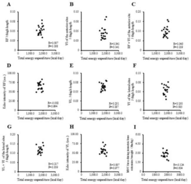
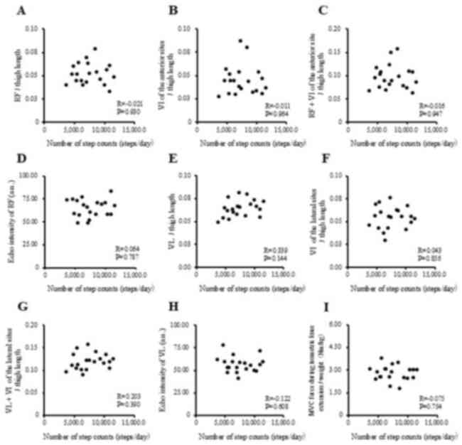
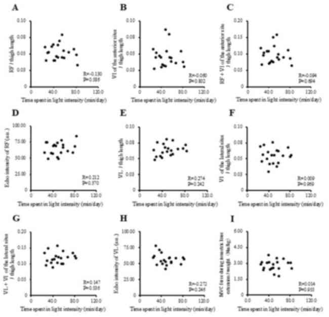
The correlations between physical activity levels
with muscle thickness and muscle strength in the study participants
are presented in Fig. 1, Fig. 2, Fig.
3 and Fig. 4 and Table II. Single correlation analysis did
not reveal any significant correlation between the physical
activity level and muscle thickness or muscle strength (Fig. 1, Fig.
2, Fig. 3 and Fig. 4 and Table
II).
| Table IICorrelation of total energy
expenditure and time spent in physical activity with muscle
thickness and muscle strength in all the study participants. |
Table II
Correlation of total energy
expenditure and time spent in physical activity with muscle
thickness and muscle strength in all the study participants.
| | Total energy
expenditure | Number of step
counts | Time spent in light
intensity | Time spent in
moderate to vigorous intensity |
|---|
| Parameter | R | P-value | R | P-value | R | P-value | R | P-value |
|---|
| Rectus
femoris/thigh length | 0.307 | 0.188 | -0.021 | 0.930 | -0.130 | 0.586 | 0.159 | 0.502 |
| Vastus intermedius
of the anterior sites/thigh length | 0.341 | 0.141 | -0.011 | 0.964 | -0.060 | 0.802 | 0.084 | 0.725 |
| Rectus femoris +
vastus intermedius of the anterior site/thigh length | 0.343 | 0.139 | -0.016 | 0.947 | -0.094 | 0.694 | 0.121 | 0.610 |
| Echo intensity of
rectus femoris (a.u.) | -0.002 | 0.994 | 0.064 | 0.787 | 0.212 | 0.370 | -0.113 | 0.635 |
| Vastus
lateralis/thigh length | 0.251 | 0.287 | 0.339 | 0.144 | 0.274 | 0.242 | 0.204 | 0.387 |
| Vastus intermedius
of the lateral sites/thigh length | 0.130 | 0.585 | 0.043 | 0.856 | 0.009 | 0.969 | 0.145 | 0.543 |
| Vastus lateralis +
vastus intermedius of the lateral site/thigh length | 0.217 | 0.359 | 0.203 | 0.390 | 0.147 | 0.536 | 0.203 | 0.391 |
| Echo intensity of
vastus lateralis (a.u.) | 0.007 | 0.977 | -0.122 | 0.608 | -0.272 | 0.246 | 0.118 | 0.620 |
| MVC force during
isometric knee extension/weight (Nm/kg) | -0.124 | 0.604 | -0.075 | 0.754 | 0.014 | 0.955 | -0.095 | 0.691 |
The differences in the physical characteristics of
participants with or without regular exercise habits are presented
in Table III. The present study
aimed to clarify whether physical activity levels, muscle thickness
and muscle strength were influenced by regular exercise habits. The
group without regular exercise habits had significantly lower
values of rectus femoris muscle thickness per thigh length
(P=0.009), rectus femoris and vastus intermedius muscle thickness
at the anterior site per thigh length (P=0.011) and muscle strength
per weight (P<0.001). This group also trended to have a higher
echo intensity of the rectus femoris (a.u.) (P=0.056) compared with
the group with regular exercise habits (Table III).
| Table IIIDifferences in physical
characteristics between the groups with and without regular
exercise habits. |
Table III
Differences in physical
characteristics between the groups with and without regular
exercise habits.
| Morphometrics | Without habit group
(n=12) | Exercise habit
group (n=8) | P-value |
|---|
|
Age
(years) | 20.9±1.2 | 20.3±0.5 | 0.111 |
|
Height
(cm) | 158.8±5.9 | 159.2±4.2 | 0.858 |
|
Weight
(kg) | 55.6±7.0 | 60.2±8.1 | 0.213 |
|
BMI
(kg/m2) | 22.0±2.3 | 23.9±4.2 | 0.287 |
|
Waist
circumference (cm) | 75.5±7.4 | 76.4±8.3 | 0.797 |
|
Hip
circumference (cm) | 92.6±5.9 | 95.2±6.9 | 0.398 |
| Physical
activity | | | |
|
Total energy
expenditure (kcal/day) | 1759.1±162.5 | 1839.9±131.7 | 0.239 |
|
Number of
step counts (steps/day) | 7542.1±2549.9 | 7685.9±2148.5 | 0.893 |
|
Time spent
in light intensity (min/day) | 50.0±18.2 | 50.9±16.8 | 0.914 |
|
Time spent
in moderate to vigorous intensity (min/day) | 24.6±10.5 | 26.2±14.3 | 0.787 |
| Muscle
thickness | | | |
|
Rectus
femoris/thigh length | 0.05±0.01 | 0.06±0.01 | 0.009 |
|
Vastus
intermedius of the anterior sites/thigh length | 0.04±0.01 | 0.06±0.02 | 0.025 |
|
Rectus
femoris + vastus intermedius of the anterior site/thigh length | 0.08±0.02 | 0.12±0.03 | 0.011 |
|
Echo
intensity of rectus femoris (a.u.) | 67.84±10.16 | 59.78±7.43 | 0.056 |
|
Vastus
lateralis/thigh length | 0.06±0.01 | 0.06±0.01 | 0.620 |
|
Vastus
intermedius of the lateral sites/thigh length | 0.05±0.01 | 0.06±0.01 | 0.126 |
|
Vastus
lateralis + vastus intermedius of the lateral site/thigh
length | 0.11±0.02 | 0.12±0.02 | 0.437 |
|
Echo
intensity of vastus lateralis (a.u.) | 56.65±9.04 | 56.31±8.88 | 0.936 |
| Muscle
strength | | | |
|
MVC force
during isometric knee extension/weight (Nm/kg) | 2.52±0.41 | 3.19±0.32 | <0.001 |
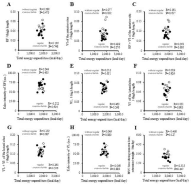
The correlations between physical activity levels
and muscle thickness and muscle strength in the groups with and
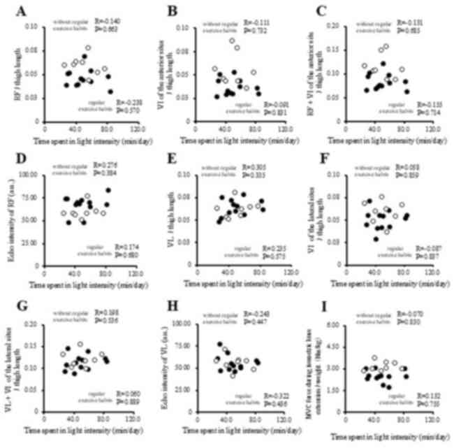
without regular exercise habits are illustrated in Fig. 5, Fig.
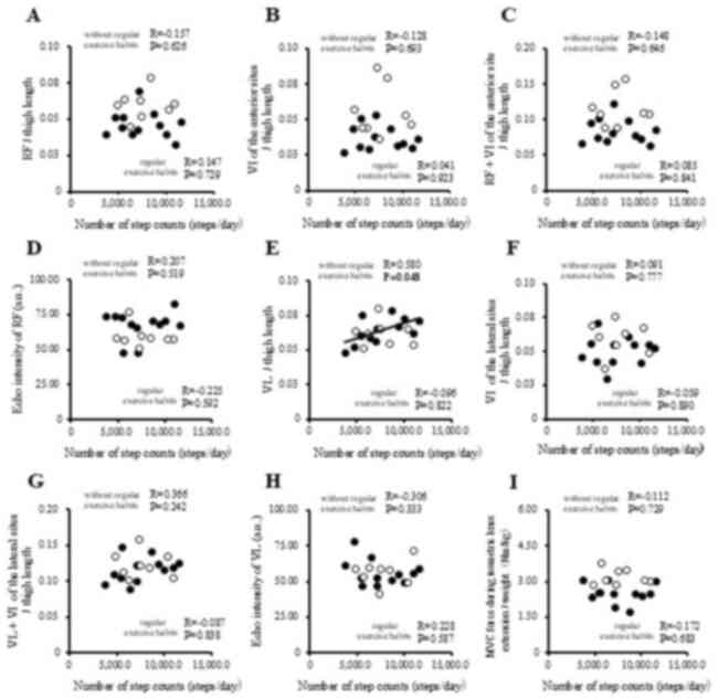
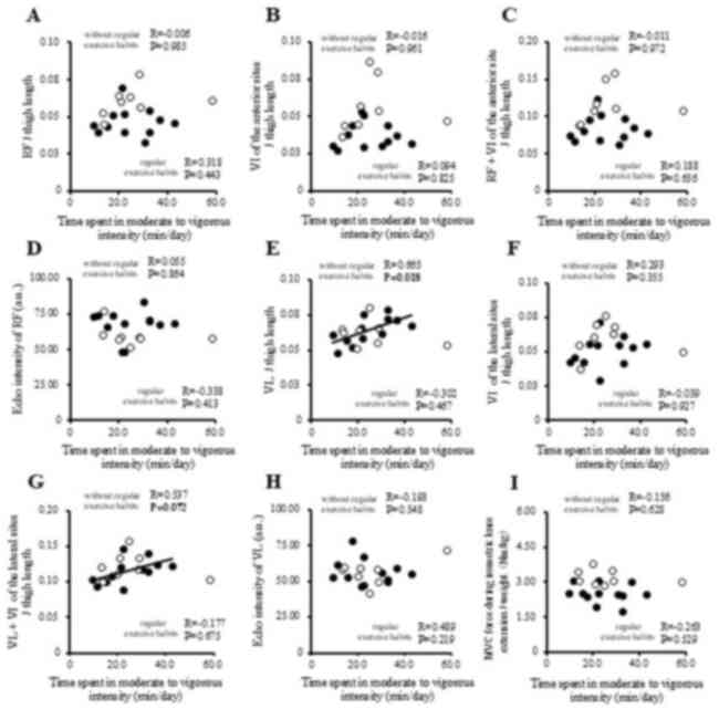
6, Fig. 7 and Fig. 8 and Table
IV. Of note, white circles represent the group with regular
exercise habits, and black circles represent the group without
regular exercise habits. In Fig. 5,
Fig. 6, Fig. 7 and Fig.
8, some similarities may appear between the locations of the
dots with those shown in Fig. 1,
Fig. 2, Fig. 3 and Fig.
4. Fig. 1, Fig. 2, Fig.
3 and Fig. 4 include all the
study participants, and Fig. 5,
Fig. 6, Fig. 7 and Fig.
8 include all the study participants divided into two groups.
In the group with regular exercise habits, no significant
correlation was observed between the physical activity level and
muscle thickness or muscle strength (Fig. 5, Fig.
6, Fig. 7 and Fig. 8). In the group without regular
exercise habits, physical activity levels, particularly the time
spent in moderate- to vigorous-intensity activity, positively
correlated with vastus lateralis muscle thickness per thigh length
(R=0.665, P=0.018; Fig. 8E and
Table IV); a positive correlation
was also found between the time spent in moderate to vigorous
intensity and vastus lateralis and vastus intermedius muscle
thickness at the lateral site per thigh length (R=0.537, P=0.072;
Fig. 8G and Table IV), as well as between the number of
step counts and the vastus lateralis muscle thickness at the
lateral site per thigh length muscle thickness (R=0.580, P=0.048;
Fig. 6E and Table IV). However, ImtraMAT was not
significantly correlated with any of the physical activity levels
and muscle thickness or muscle strength in the study
participants.
| Table IVCorrelation of total energy
expenditure and time spent in physical activity with muscle
thickness, and muscle strength in groups with and without regular
exercise habits. |
Table IV
Correlation of total energy
expenditure and time spent in physical activity with muscle
thickness, and muscle strength in groups with and without regular
exercise habits.
| | Total energy
expenditure | Number of step
counts | Time spent in light
intensity | Time spent in
moderate to vigorous intensity |
|---|
| | Group without
exercise | Group with exercise
habits | Group without
exercise | Group with exercise
habits | Group without
exercise | Group with exercise
habits | Group without
exercise | Group with exercise
habits |
|---|
| Parameter | R | P-value | R | P-value | R | P-value | R | P-value | R | P-value | R | P-value | R | P-value | R | P-value |
|---|
| Rectus
femoris/thigh length | 0.288 | 0.475 | 0.136 | 0.748 | -0.157 | 0.626 | 0.147 | 0.729 | -0.140 | 0.663 | -0.238 | 0.570 | -0.006 | 0.985 | 0.318 | 0.443 |
| Vastus intermedius
of the anterior sites/thigh length | 0.077 | 0.812 | 0.439 | 0.276 | -0.128 | 0.693 | 0.041 | 0.923 | -0.111 | 0.732 | -0.091 | 0.831 | -0.016 | 0.961 | 0.094 | 0.825 |
| Rectus femoris +
vastus intermedius of the anterior site/thigh length | 0.161 | 0.617 | 0.361 | 0.380 | -0.148 | 0.646 | 0.085 | 0.841 | -0.131 | 0.685 | -0.155 | 0.714 | -0.011 | 0.972 | 0.188 | 0.656 |
| Echo intensity of
rectus femoris (a.u.) | 0.249 | 0.435 | -0.212 | 0.614 | 0.207 | 0.519 | -0.225 | 0.592 | 0.276 | 0.384 | 0.174 | 0.680 | 0.055 | 0.864 | -0.338 | 0.413 |
| Vastus
lateralis/thigh length | 0.211 | 0.511 | 0.463 | 0.248 | 0.580 | 0.048 | -0.096 | 0.822 | 0.305 | 0.335 | 0.235 | 0.575 | 0.665 | 0.018 | -0.302 | 0.467 |
| Vastus intermedius
of the lateral sites/thigh length | 0.029 | 0.929 | 0.051 | 0.905 | 0.091 | 0.777 | -0.059 | 0.890 | 0.058 | 0.859 | -0.087 | 0.837 | 0.293 | 0.355 | -0.039 | 0.927 |
| Vastus lateralis +
vastus intermedius of the lateral site/thigh length | 0.130 | 0.687 | 0.265 | 0.526 | 0.366 | 0.242 | -0.087 | 0.838 | 0.198 | 0.536 | 0.060 | 0.889 | 0.537 | 0.072 | -0.177 | 0.675 |
| Echo intensity of
vastus lateralis (a.u.) | 0.049 | 0.880 | -0.060 | 0.888 | -0.306 | 0.333 | 0.228 | 0.587 | -0.243 | 0.447 | -0.322 | 0.436 | -0.193 | 0.548 | 0.489 | 0.219 |
| MVC force during
isometric knee extension/weight (Nm/kg) | -0.466 | 0.127 | -0.315 | 0.447 | -0.112 | 0.729 | -0.712 | 0.683 | -0.070 | 0.830 | 0.132 | 0.755 | -0.156 | 0.628 | -0.263 | 0.529 |
Discussion
The major findings of the present study were that
the number of step counts and time spent performing moderate- to
vigorous-intensity activity were positively associated with vastus
lateralis muscle thickness at the lateral site per thigh length in
the group without regular exercise habits. These results support
the hypothesis that physical activity levels in young women may be
related to lower limb muscle thickness and strength. It has been
found that compared to those with moderate-to-vigorous-intensity
physical activity (MVPA), individuals with lower physical activity
levels exhibited significantly lower skeletal muscle mass index,
and this association was observed in older adults, as well as in
younger women (1,18-20).
Oshita and Myotsuzono (1) reported
that current physical activity among female students particularly
affects the lower limb muscle mass. They demonstrated that physical
inactivity or low physical activity may cause difficulty in
independent mobility in the younger generation, even among those
with regular exercise habits in the past (1). Previous studies have reported that
30-50% of healthy young Japanese women already fall under the
cut-off value for skeletal muscle mass, a precursor to sarcopenia
(1,21,22).
Therefore, to maintain skeletal muscle mass, time spent in MVPA
needs to be continued by the younger generation. Another study
reported that the cross-sectional area (CSA) of the vastus
lateralis of the quadriceps muscles decreased by ~40% in
individuals aged between 20 and 80 years of age (23). The loss in the size of the quadriceps
with age may contribute to low physical activity in habitual sports
and physical activity, which require higher exercise intensities
than most daily activities (24-26).
Thus, based on the results of the present study and previous
studies (1,21,22),
maintaining lower limb muscle thickness and continuous engagement
in MVPA are required to maintain and improve future health, even in
healthy young women.
The results of the present study did not reveal any
significant association between the physical activity level and
muscle thickness or strength in the group with regular exercise
habits. However, in the participants without regular exercise
habits, physical activity levels, particularly the time spent in
moderate- to vigorous intensity activity, were positively
associated with vastus lateralis muscle thickness at the lateral
site per thigh length. In previous research, the relative weight
percentage of lower limb muscle thickness indicated that the thigh
muscle group accounts for approximately half (46.9%) of the total
skeletal muscle mass, with the quadriceps muscle accounting for the
highest percentage (27).
Furthermore, the CSA of the quadriceps muscle is composed of four
muscles, of which the vastus lateralis occupies the largest
physiological area, at ~30% (26,27).
Jacob et al (28) also
reported an association between the functional capability of lower
limb muscle thickness in healthy adults aged 18-70 years during the
course of daily life and the morphology of vastus lateralis muscle
thickness. These findings indicate that vastus lateralis muscle
thickness is negatively associated with age, and this association
may be caused by the intensity and frequency of physical activity
(28). Young adults engage in more
vigorous-intensity weight shifts compared to older adults, while
older adults engage in more light-intensity weight shifts compared
to younger individuals. Physical activity of light- and
vigorous-intensity is inferred from the types of mobilized muscles,
and the difference in energy expenditure. The results of the
present study did not reveal any significant association between
the physical activity level and muscle thickness or strength in the
group with regular exercise habits. The group with regular exercise
habits practiced vigorous-intensity competitive sports in daily
life and maintained vastus lateralis muscle thickness. Therefore,
physical activities other than competitive sports may have been
unaffected.
The results of the present study also revealed no
significant association between the physical activity level and
echo intensity in the study participants. However, in the
participants without regular exercise habits, a higher echo
intensity of the rectus femoris (a.u.) was found compared to the
participants with regular exercise habits. Therefore, it was
suggested that the continuation of exercise affects IntraMAT.
Previous research has suggested that skeletal muscle size is a
critical factor for the regulation of IntraMAT content and that
increases in metabolic capacity of already existing muscles
contribute to decreases in the IntraMAT content (29). Even among young women, continuous
exercise is associated with an decreased IntraMAT, regardless of
the amount of physical activity. These findings suggest that it may
be crucial to establish regular exercise habits to prevent an
increase in IntraMAT in young women.
In a previous study, moderate-intensity exercise in
older adults in the exercise group was associated with
significantly higher vastus lateralis CSA and intracellular
resistance index scores on segmental bioelectrical impedance
spectroscopy exercise than those of the older adults without
regular exercise habits (30).
However, in the present study, physical activity levels,
particularly the time spent performing moderate- to
vigorous-intensity activity by the participants without regular
exercise habits were significantly and positively associated with
vastus lateralis muscle thickness at the lateral site per thigh
length. The group without regular exercise habits may have
exhibited a correlation between the duration of MVPA and lower
extremity muscle thickness as they did not engage in
vigorous-intensity competitive sports and were more susceptible to
the influence of the intensity and frequency of physical activity.
MVPA refers to activities of daily living and exercise at an
intensity of ≥3 MET, such as walking at a moderate speed or jogging
(31). A previous study demonstrated
that the vertical and anterior-posterior center-of-mass
accelerations of the vastus lateralis muscle during gait increase
with increasing gait speed that was the muscle activity of the
vastus lateralis muscle increases with increasing gait speed
(32). Furthermore, previous studies
have reported a significant positive association between maximum
walking speed and lower limb muscle strength (23,33), and
lower limb muscle mass and lower limb muscle strength are closely
related to walking function (34). A
previous study demonstrated the association between running speed
and the electromyography of lower limb muscle mass, reporting that
the activity of the vastus lateralis exhibited the greatest
increase in the pre-contact and braking phases with the increasing
running speed (35). Previous
studies have shown that walking and running movements associated
with MVPA affect vastus lateralis muscle thickness in the lower
extremities (23,31,33,35). The
present study demonstrated that vastus lateralis muscle thickness
was associated with the time spent performing moderate0 to
vigorous-intensity activity in the group without regular exercise
habits. Thus, these results suggest that the walking and running
movements associated with the time spent performing MVPA may have
increased the muscle activity of the vastus lateralis muscle and
thickened in healthy young collegiate women without regular
exercise habits.
The present study has some limitations which should
be mentioned. First, the number of participants in the study was
small and the study was limited to collegiate healthy young women
attending the same university. Therefore, there was potential
selection bias in the present study, and it is unclear whether
these results are applicable to young males and other populations.
Second, it was not possible to determine the causal association
between physical activity levels and lower limb muscle thickness in
collegiate healthy young women as the present study design was a
cross-sectional study. Finally, the physical activity levels during
sports activities were not evaluated using the accelerometer in the
group with regular exercise habits, possibly leading to an
underestimation of the physical activity levels. In addition, the
limitations of the accelerometer include the impossibility of
measuring activity while swimming or bathing, difficulty in
evaluating upper limb movement, and the inability to determine
physical activity levels, while moving such as bicycling.
Therefore, the inability to measure these activities may have
introduced errors when evaluating energy expenditure and time spent
in physical activity.
Another limitation to the present study is the lack
of dietary analyses that may be related to lower limb muscle
thickness and IntraMAT. A previous study compared the association
between IntraMAT and diet in older and younger women and found an
association between macronutrients and alcohol intake (36). Therefore, it is possible that
differences in composition due to diet may influence lower limb
muscle thickness and IntraMAT.
However, few studies have evaluated muscle thickness
in young women using ultrasound tomography (15). The results of the present study
suggest that time spent preforming MVPA may be associated to
lower-limb muscle thickness in female college students without
regular exercise habits. The results presented herein suggest that
continuous physical activity at moderate to vigorous intensity may
contribute to maintaining and improving lower limb muscle thickness
in young women. However, further studies with a greater number of
participants, addressing the current limitations, and conducting
longitudinal analyses are required to clarify the association
between physical activity levels and lower limb muscle thickness
and IntraMAT in healthy young collegiate women, as well as the
influence of diet.
In conclusion, the present study examined the
association of physical activity with lower limb muscle thickness,
IntraMAT and muscle strength. No significant association between
the physical activity level and muscle thickness or strength was
observed in the group with regular exercise habits. However, in
participants without regular exercise habits, physical activity
levels, particularly the time spent performing moderate- to
vigorous-intensity activity, were positively associated with vastus
lateralis muscle thickness per thigh length and also with a higher
echo intensity of the rectus femoris (a.u.). These results suggest
that the presence or absence of an exercise habit and a reduction
in the time spent in MVPA may affect lower limb muscle thickness
and IntraMAT in healthy young collegiate women without regular
exercise habits. Therefore, the lifestyle guidance of a reduction
in the time spent in physical inactivity and continued time spent
in MVPA is necessary to maintain skeletal muscle mass and to
decrease in IntraMAT in young women.
Acknowledgements
Not applicable.
Funding
Funding: No funding was received.
Availability of data and materials
The data generated in the present study may be
requested from the corresponding author.
Authors' contributions
MA and MH designed the study and prepared the
initial drafts of the manuscript. MA, MH, MS and HT collected data.
RM contributed to the data analysis and interpretation, and
assisted in the preparation of the manuscript. All the other
authors contributed to the critical reviewing of the manuscript. MA
and MH confirm the authenticity of all the raw data. All the
authors have read and approved the final version of the manuscript
and agree to be accountable for all aspects of the work, ensuring
that questions related to the accuracy or integrity of any part of
the work were appropriately investigated and resolved.
Ethics approval and consent to
participate
The present study was approved by the Ethics
Committee of Teikyo Heisei University (No. 2022-022-1) and was
conducted in accordance with the guidelines of the Declaration of
Helsinki. All participants provided written informed consent.
Patient consent for publication
Not applicable.
Competing interests
The authors declare that they have no competing
interests.
References
|
1
|
Oshita K and Myotsuzono R: An association
between the physical activity level and skeletal muscle mass index
in female university students with a past exercise habituation.
Osteoporos Sarcopenia. 7:146–152. 2021.PubMed/NCBI View Article : Google Scholar
|
|
2
|
Brown WJ and Trost SG: Life transitions
and changing physical activity patterns in young women. Am J Prev
Med. 25:140–143. 2003.PubMed/NCBI View Article : Google Scholar
|
|
3
|
Engberg E, Alen M, Kukkonen-Harjula K,
Peltonen JE, Tikkanen HO and Pekkarinen H: Life events and change
in leisure time physical activity: A systematic review. Sports Med.
42:433–447. 2012.PubMed/NCBI View Article : Google Scholar
|
|
4
|
Guthold R, Stevens GA, Riley LM and Bull
FC: Worldwide trends in insufficient physical activity from 2001 to
2016: A pooled analysis of 358 population-based surveys with 1·9
million participants. Lancet Glob Health. 6:e1077–e1086.
2018.PubMed/NCBI View Article : Google Scholar
|
|
5
|
Peng B, Ng JYY and Ha AS: Barriers and
facilitators to physical activity for young adult women: A
systematic review and thematic synthesis of qualitative literature.
Int J Behav Nutr Phys Act. 20(23)2023.PubMed/NCBI View Article : Google Scholar
|
|
6
|
Uijtdewilligen L, Peeters GMEE, van
Uffelen JGZ, Twisk JWR, Singh AS and Brown WJ: Determinants of
physical activity in a cohort of young adult women. Who is at risk
of inactive behaviour? J Sci Med Sport. 18:49–55. 2015.PubMed/NCBI View Article : Google Scholar
|
|
7
|
Paddon-Jones D, Sheffield-Moore M, Urban
RJ, Sanford AP, Aarsland A, Wolfe RR and Ferrando AA: Essential
amino acid and carbohydrate supplementation ameliorates muscle
protein loss in humans during 28 days bedrest. J Clin Endocrinol
Metab. 89:4351–4358. 2004.PubMed/NCBI View Article : Google Scholar
|
|
8
|
Akima H, Kubo K, Kanehisa H, Suzuki Y,
Gunji A and Fukunaga T: Leg-press resistance training during 20
days of 6˚ head-down-tilt bed rest prevents muscle deconditioning.
Eur J Appl Physiol. 82:30–38. 2000.PubMed/NCBI View Article : Google Scholar
|
|
9
|
Landers KA, Hunter GR, Wetzstein CJ,
Bamman MM and Weinsier RL: The interrelationship among muscle mass,
strength, and the ability to perform physical tasks of daily living
in younger and older women. J Gerontol A Biol Sci Med Sci.
56:B443–B448. 2001.PubMed/NCBI View Article : Google Scholar
|
|
10
|
Cruz-Jentoft AJ, Bahat G, Bauer J, Boirie
Y, Bruyère O, Cederholm T, Cooper C, Landi F, Rolland Y, Sayer AA,
et al: Sarcopenia: Revised European consensus on definition and
diagnosis. Age Ageing. 48:16–31. 2019.PubMed/NCBI View Article : Google Scholar
|
|
11
|
Kitagawa F, Ogawa M, Yoshiko A, Oshida Y,
Koike T, Akima H and Tanaka NI: Factors related to trunk
intramuscular adipose tissue content-A comparison of younger and
older men. Exp Gerontol. 168(111922)2022.PubMed/NCBI View Article : Google Scholar
|
|
12
|
Hioki M, Kanehira N, Koike T, Saito A,
Shimaoka K, Sakakibara H, Oshida Y and Akima H: Relationship
between adiponectin and intramuscular fat content determined by
ultrasonography in older adults. PLoS One.
17(e0262271)2022.PubMed/NCBI View Article : Google Scholar
|
|
13
|
Akima H, Hioki M, Yoshiko A, Koike T,
Sakakibara H, Takahashi H and Oshida Y: Intramuscular adipose
tissue determined by T1-weighted MRI at 3 T primarily reflects
extramyocellular lipids. Magn Reson Imaging. 34:397–403.
2016.PubMed/NCBI View Article : Google Scholar
|
|
14
|
Kumahara H, Schutz Y, Ayabe M, Yoshioka M,
Yoshitake Y, Shindo M, Ishii K and Tanaka H: The use of uniaxial
accelerometry for the assessment of physical-activity-related
energy expenditure: A validation study against whole-body indirect
calorimetry. Br J Nutr. 91:235–243. 2004.PubMed/NCBI View Article : Google Scholar
|
|
15
|
Hioki M, Kanehira N, Koike T, Saito A,
Shimaoka K, Sakakibara H, Oshida Y and Akima H: Age-related changes
in muscle volume and intramuscular fat content in quadriceps
femoris and hamstrings. Exp Gerontol. 132(110834)2020.PubMed/NCBI View Article : Google Scholar
|
|
16
|
Young HJ, Jenkins NT, Zhao Q and Mccully
KK: Measurement of intramuscular fat by muscle echo intensity.
Muscle Nerve. 52:963–971. 2015.PubMed/NCBI View Article : Google Scholar
|
|
17
|
Akima H, Hioki M and Furukawa T: Effect of
arthroscopic partial meniscectomy on the function of quadriceps
femoris. Knee Surg Sports Traumatol Arthrosc. 16:1017–1025.
2008.PubMed/NCBI View Article : Google Scholar
|
|
18
|
Foong YC, Chherawala N, Aitken D, Scott D,
Winzenberg T and Jones G: Accelerometer-determined physical
activity, muscle mass, and leg strength in community-dwelling older
adults. J Cachexia Sarcopenia Muscle. 7:275–283. 2016.PubMed/NCBI View Article : Google Scholar
|
|
19
|
Rosique-Esteban N, Babio N, Díaz-López A,
Romaguera D, Alfredo Martínez J, Sanchez VM, Schröder H, Estruch R,
Vidal J, Buil-Cosiales P, et al: Leisure-time physical activity at
moderate and high intensity is associated with parameters of body
composition, muscle strength and sarcopenia in aged adults with
obesity and metabolic syndrome from the PREDIMED-Plus study. Clin
Nutr. 38:1324–1331. 2019.PubMed/NCBI View Article : Google Scholar
|
|
20
|
Wu F, Wills K, Laslett LL, Oldenburg B,
Jones G and Winzenberg T: Moderate-to-vigorous physical activity
but not sedentary time is associated with musculoskeletal health
outcomes in a cohort of Australian middle-aged women. J Bone Miner
Res. 32:708–715. 2017.PubMed/NCBI View Article : Google Scholar
|
|
21
|
Miyatani M, Kanehisa H, Azuma K, Kuno S
and Fukunaga T: Site-related differences in muscle loss with aging.
Int J Sport Health Sci. 1:34–40. 2003.
|
|
22
|
Yamada Y, Yamada M, Yoshida T, Miyachi M
and Arai H: Validating muscle mass cutoffs of four International
sarcopenia-working groups in Japanese people using DXA and BIA. J
Cachexia Sarcopenia Muscle. 12:1000–1010. 2021.PubMed/NCBI View Article : Google Scholar
|
|
23
|
Lexell J, Taylor CC and Sjöström M: What
is the cause of the ageing atrophy? J Neurol Sci. 84:275–294.
1988.PubMed/NCBI View Article : Google Scholar
|
|
24
|
Abe T, Sakamaki M, Yasuda T, Bemben MG,
Kondo M, Kawakami Y and Fukunaga T: Age-related, site-specific
muscle loss in 1507 Japanese men and women aged 20 to 95 years. J
Sports Sci Med. 10:145–150. 2011.PubMed/NCBI
|
|
25
|
Ito J: Morphological analysis of the human
lower extremity based on the relative muscle weight. Okajimas Folia
Anat Jpn. 73:247–251. 1996.PubMed/NCBI View Article : Google Scholar
|
|
26
|
Akima H, Foley JM, Prior BM, Dudley GA and
Meyer RA: Vastus lateralis fatigue alters recruitment of musculus
quadriceps femoris in humans. J Appl Physiol (1985). 92:679–684.
2002.PubMed/NCBI View Article : Google Scholar
|
|
27
|
Trappe TA, Lindquist DM and Carrithers JA:
Muscle-specific atrophy of the quadriceps femoris with aging. J
Appl Physiol (1985). 90:2070–2074. 2001.PubMed/NCBI View Article : Google Scholar
|
|
28
|
Jacob I, Johnson MI, Jones G, Jones A and
Francis P: Age-related differences of vastus lateralis muscle
morphology, contractile properties, upper body grip strength and
lower extremity functional capability in healthy adults aged 18 to
70 years. BMC Geriatr. 22(538)2022.PubMed/NCBI View Article : Google Scholar
|
|
29
|
Akima H, Yoshiko A, Hioki M, Kanehira N,
Shimaoka K, Koike T, Sakakibara H and Oshida Y: Skeletal muscle
size is a major predictor of intramuscular fat content regardless
of age. Eur J Appl Physiol. 115:1627–1635. 2015.PubMed/NCBI View Article : Google Scholar
|
|
30
|
Otsuka Y, Yamada Y, Maeda A, Izumo T, Rogi
T, Shibata H, Fukuda M, Arimitsu T, Miyamoto N and Hashimoto T:
Effects of resistance training intensity on muscle quantity/quality
in middle-aged and older people: A randomized controlled trial. J
Cachexia Sarcopenia Muscle. 13:894–908. 2022.PubMed/NCBI View Article : Google Scholar
|
|
31
|
Ainsworth BE, Haskell WL, Herrmann SD,
Meckes N, Bassett DR Jr, Tudor-Locke C, Greer JL, Vezina J,
Whitt-Glover MC and Leon AS: 2011 compendium of physical
activities: A second update of codes and MET values. Med Sci Sports
Exerc. 43:1575–1581. 2011.PubMed/NCBI View Article : Google Scholar
|
|
32
|
Liu MQ, Anderson FC, Schwartz MH and Delp
SL: Muscle contributions to support and progression over a range of
walking speeds. J Biomech. 41:3243–3252. 2008.PubMed/NCBI View Article : Google Scholar
|
|
33
|
Hayashida I, Tanimoto Y, Takahashi Y,
Kusabiraki T and Tamaki J: Correlation between muscle strength and
muscle mass, and their association with walking speed, in
community-dwelling elderly Japanese individuals. PLoS One.
9(e111810)2014.PubMed/NCBI View Article : Google Scholar
|
|
34
|
Uesugi Y, Kanaya S, Nakanishi H and Naito
Y: The relationship between locomotive syndrome risk, gait pattern,
and standing posture in young Japanese women: A cross-sectional
study. Healthcare (Basel). 8(565)2020.PubMed/NCBI View Article : Google Scholar
|
|
35
|
Kyröläinen H, Avela J and Komi PV: Changes
in muscle activity with increasing running speed. J Sports Sci.
23:1101–1109. 2005.PubMed/NCBI View Article : Google Scholar
|
|
36
|
Sjöholm K, Gripeteg L and Larsson I:
Macronutrient and alcohol intake is associated with intermuscular
adipose tissue in a randomly selected group of younger and older
men and women. Clin Nutr ESPEN. 13:e46–e51. 2016.PubMed/NCBI View Article : Google Scholar
|