1. Introduction
Chronic periodontitis, as one of the most prevalent
bacterial infectious oral diseases, causes gingival inflammation,
the loss of periodontal attachment, destruction of alveolar bone,
and ultimately, tooth loss (1).
While stratifying the 2009-2010 National Health and Nutrition
Examination Survey (NHANES) data by patient age, the prevalence of
periodontal disease increases from 24.4% in individuals aged 30-34
years to 70.1% in those aged ≥65 years. Atherosclerosis and
Alzheimer's disease are some of the age-related conditions that
have been connected to periodontitis and have more severe mortality
and morbidity profiles (2). (As
Porphyromonas gingivalis (P. gingivalis) has a
significant impact on the local microbiota, it is regarded as a
‘keystone’ pathogen in periodontal disease. P. gingivalis
disrupts innate immunity, causing dysbiosis, in which symbiotic
commensals turn into accessory pathogens via unknown methods
(2). Although current periodontal
therapy is targeted at the reduction or elimination of disease
progression, the regeneration of the lost periodontal structures is
however, unsuccessful or compromised (3). During the past decades, regenerative
periodontal therapy used guided procedures; however, the fact of
predictable regenerative outcomes remains questionable (4).
Almost 30 years have passed since the inception of
tissue engineering and regenerative medicine. Studies are being
carried out to promote predictable and successful regeneration
(5); yet, the complexity of the oral
environment local inflammatory milieu and the tissue damage brought
on by periodontitis may be the limiting factors for successful
therapeutic outcomes. To achieve the predictable and successful
outcomes, further studies on newer agents that may enhance
regeneration and pave way to overcome these challenges are
required. Multipotent stem cells, or mesenchymal stem cells (MSCs)
can self-renew and develop in multiple ways. MSC-derived
extracellular vesicles (EVs) are deemed to be the most efficient
substitutes for MSCs due to their low immunogenicity, high safety
and variety of bioactivities; as with their parent MSCs, these EVs
can exert immunomodulatory effects and stimulate osteogenesis,
cementogenesis and angiogenesis (6).
The MSC secretory profile is composed of exosomes
that play a pivotal role in MSC cytotherapy. Exosomes are composed
of various membrane proteins, lipoproteins, heat shock proteins and
transport-related proteins. Recent research has demonstrated that
exosomes secreted by MSCs can initiate tissue regeneration and can
potentially be used as therapeutic agents for various diseases
(7). Specifically, the importance of
exosomes in the development and treatment of periodontal disease is
addressed in the present review, and recent breakthroughs in
exosome therapy and regeneration are also mentioned (1).
2. Exosomes
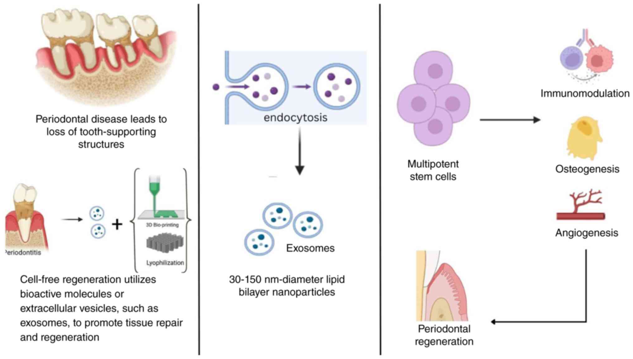
Exosomes are endocytic EVs and can be secreted by
various cell types. Three distinct stages are recognizable in
exosomal biogenesis: i) Endocytic vesicles are formed by the plasma
membrane. Early endosomes are created when endocytic vesicles fuse,
and these endosomes eventually develop into late endosomes. ii)
Intraluminal vesicles (ILVs) are created when the late endosome
membranes budding inward. The term multivesicular bodies (MVBs)
refers to the accumulation of ILVs in late endosomes. iii) ILVs,
often referred to as exosomes, are released when MVBs fuse with the
plasma membrane (8). Exosomes are
lipid bilayer nanoparticles 30-150 nm-diameter (9). There are numerous factors outside the
cell, including cell type, serum conditions, cytokines and growth
hormones, that affect the production of exosomes. Exosomes are
heterogeneous based on the particular shape, content and function
(10). Exosomes are also
membrane-bound carriers. They are composed of metabolites, proteins
and nucleic acids that reflect the physiological state and
constitution of the donor cell (11). Exosomes tend to be highly
proteinaceous; exosomal proteins play various roles, ranging from
tetraspanins (CD9, CD63, CD81 and CD82) involved in cell
penetration, invasion and fusion processes to heat shock proteins
(HSPs; HSP70 and HSP90), which are implicated in antigen
presentation and binding as components of the stress response; some
proteins implicated in exosome release (ALIX-TSG101) (12). These molecules not only aid in the
identification of exosomes, but also enable them to specifically
target recipient cells. Exosomes can reach the distant tissues or
organs via the circulatory system after being taken up by recipient
cells through autocrine or paracrine signalling; thus, they
contribute to multiple physiological and pathological processes
involved in tissue repair. Previous studies have also revealed that
miRNAs carried within exosomes can induce specific biological
effects in recipient cells (5,10).
3. Functions of exosomes
Exosomes are extracellular vesicles released by
cells that facilitate the transfer of proteins and RNAs across
cells (13). Through their paracrine
action, exosomes can be employed in tissue regeneration therapies,
specifically via influencing cellular activities associated with
inflammation, healing, matrix remodelling, differentiation and
death (14). Three mechanisms can
facilitate the transmission of intercellular signals: Direct
membrane fusion, receptor-ligand interaction, or
endocytosis/internalization followed by fusing with the endosomal
limiting membrane of the receiving cells (15). Cell division, apoptosis and
proliferation are all directly related to healthy tissue growth and
regeneration. Various growth factors, such as epithelial growth
factor, vascular endothelial growth factor (VEGF) and fibroblast
growth factor (FGF) are present in exosomes and play a role in cell
division and proliferation. As demonstrated in the study by Liang
et al (16), exosomes
produced from MSCs activated the AKT/mTOR pathway, which in turn
promoted angiogenesis and bone repair. Through the
hypoxia-inducible factor 1α pathway, exosomes have been shown to
accelerate fracture healing, stimulate angiogenesis and activate
VEGF expression (17).
4. Exosomes in periodontal tissue
repair
Numerous studies have highlighted that, due to their
potent repair-promoting properties, MSC derivatives including EVs,
exosomes, matrix vesicles and apoptotic bodies can be utilized as
substitutes for MSCs in order to support periodontal tissue
regeneration. MSC-derived exosomes may improve cell viability and
alter gene expression by triggering pro-survival signalling in
periodontal ligament cells. The pro-survival AKT and ERK signalling
pathways may be activated by adenosine receptors, and related genes
may be expressed to promote the migration and proliferation of
periodontal ligament cells. MSC exosomes contain
CD73/ecto5'-nucleotidase, which hydrolyses extracellular adenosine
monophosphate to produce extracellular adenosine (14). When exosomes are released by cells,
they function as messengers and are absorbed by recipient cells
through membrane fusion, endocytosis/phagocytosis, or binding to
target cell receptors. This allows them to transfer different
bioactive substances and contributes to intercellular communication
(18).
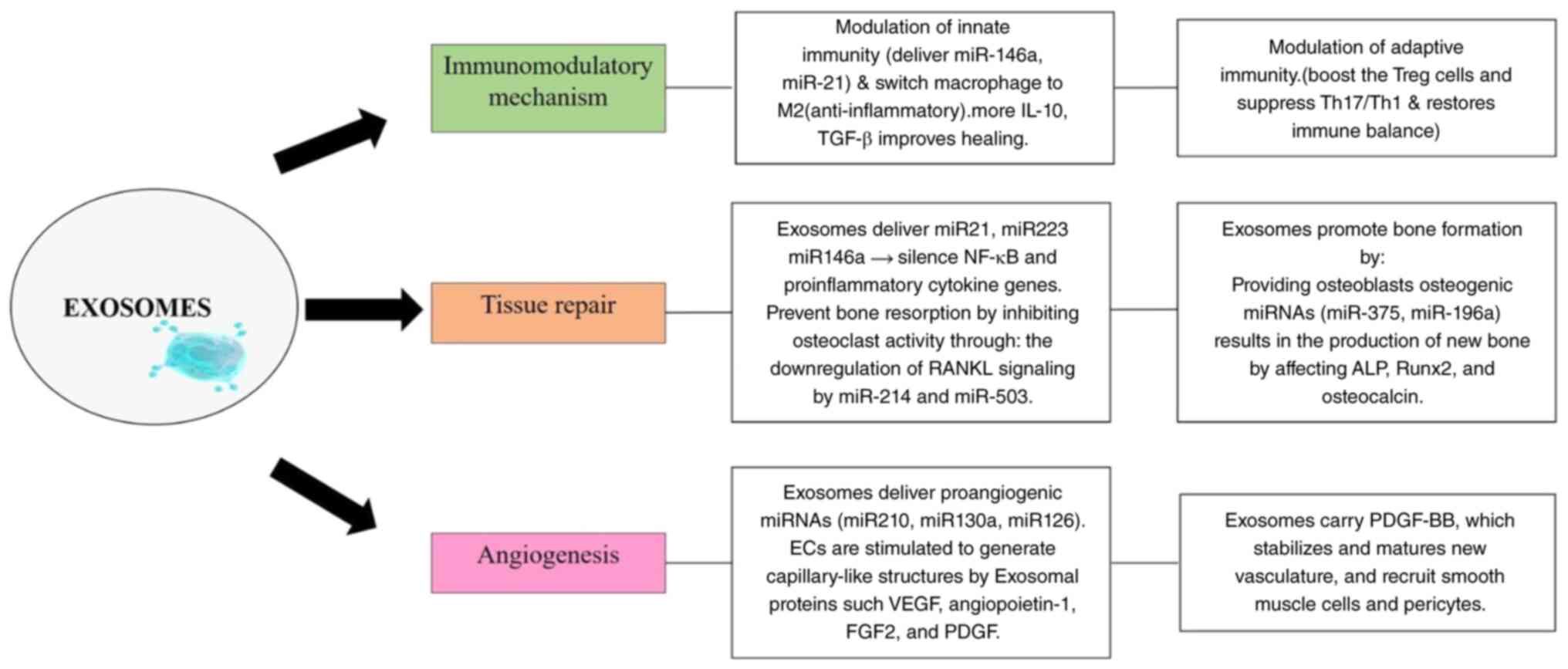
Both innate and adaptive immune responses are
regulated by exosomes (11). By
promoting macrophage polarization from the M1 to the M2 phenotype,
the delivery of miR-146a and miR-21 increases anti-inflammatory
activity through the release of TGF-β and IL-10(19). They support tissue repair and
immunological homeostasis by increasing Treg cell activity and
inhibiting Th17/Th1 responses (13).
Exosomal miR-21, miR-223 and miR-146a decrease osteoclast-mediated
bone resorption by downregulating RANKL and inhibiting NF-κB
signalling and pro-inflammatory cytokine production (20,21).
Osteogenic miRNAs, such as miR-196a, increase the production of
ALP, Runx2 and osteocalcin in osteoblasts, which promotes the
development of new bone (20).
Through the VEGF, angiopoietin-1, FGF2 and platelet-derived growth
factor (PDGF) pathways, pro-angiogenic miRNAs (miR-210, miR-130a
and miR-126) that are delivered by exosomes stimulate endothelial
cells to produce capillary-like structures (18,20,22).
Additionally, they carry PDGF-BB, which recruits smooth muscle
cells and pericytes to sustain vascular integrity by stabilizing
and maturing new vasculature (18,23)
(Figs. 1 and 2).
Exosomes have been shown to regulate the expression
of pro-inflammatory cytokines, such as interleukin (IL)-6 IL-1β and
tumour necrosis factor α (TNF-α) by suppressing pathways, such as
the Th17/Treg/miR- 155-5p/SIRT1 and IL-6/JAK2/STAT3 pathways
(20). Alternatively, M2 macrophages
secrete anti- inflammatory cytokines. This alteration provides an
environment that supports tissue regeneration (24). Alveolar bone resorption is
facilitated by the production of several pro-inflammatory factors
(IL1β, TNF-α and IL-6) due to an imbalance favouring M1 macrophages
in periodontitis. Additionally, M1 macrophages stimulate the RANKL
pathway, which increases bone resorption by stimulating osteoclast
differentiation in periodontal tissues. On the other hand, M2
macrophages release anti-inflammatory cytokines (VEGF, TGF-β and
IL-10), which support bone healing and immune homeostasis (20). EVs from polarized macrophages may
also have an impact on osteogenesis. Kang et al (25) found the comparatively high expression
of miR-155 in M1 EVs and miR-378a in M2 EVs based on miRNA-sequence
analysis of EV cargo. The BMP2 pathway is the specific target of
miR-378a, a positive regulator of osteogenesis, and miR-155 is
known to play an essential role in inflammatory signalling
(25). In order to identify specific
molecular mechanisms, Nakao et al (19) investigated the therapeutic effect of
TNF-α preconditioned- exosomes from gingival tissue-derived MSCs on
ligature-induced periodontitis in mice. Exosomal miR-1260b was a
crucial regulator for preventing inflammatory bone loss, and
TNF-α-induced exosomal CD73 expression helped to polarize M2
macrophages (19). A numb er of
previous studies have focused on role of exosomes in periodontal
regeneration and these are summarized in Table I (26-32).
 | Table ISummary of studies on the role of
exosomes in periodontal tissue regeneration. |
Table I
Summary of studies on the role of
exosomes in periodontal tissue regeneration.
| Authors | Year of
publication | Aim/objective | Type of study | Mode/system | Key
findings/results | (Refs.) |
|---|
| Zhang et
al | 2021 | Compare 3D vs. 2D
MSC exosome effects on periodontal healing | Experimental | Rat periodontitis;
3D MSC culture | 3D culture boosted
exosome yield and regenerative efficacy | (13) |
| Sun et
al | 2022 | Test gingival MSC
exosomes on inflammation & repair | Experimental | LPS-stimulated
PDLSCs; rat periodontitis | Suppressed
IL-6/TNF-α; Improved periodontal regeneration | (26) |
| Zhao et
al | 2022 | Evaluate PDLSC-Exos
in Gel-Alg hydrogel for bone repair | Experimental | Rat alveolar
defect; BMSC culture | Hydrogel-Exos
improved bone fill and osteogenesis | (27) |
| Lin et
al | 2022 | Summarize
exosome-base d therapies | Systematic
review | Preclinical/early
clinical | Strong preclinical
support; need human trials | (10) |
| Huang et
al | 2023 | Study inflammatory
preconditioning on exosome function | Experimental | LPS-periodontitis
model | TNF-α priming
enhanced M2 polarization; prevented bone loss | (21) |
| Mousavi et
al | 2023 | Review MSC-exosome
roles across tissues | Review | Multisystem | Highlighted
paracrine signalling; translational potential | (28) |
| Albougha et
al | 2024 | Test osteogenic
ability of PDLSC-exos | Experimental | Osteoblast-like
cells; rat calvarial defect | Stimulated
mineralization and defect healing | (29) |
| Peng et
al | 2020 | Review exosome
effects on relapse and remodelling | Review | Bone remodelling
models | Balanced RANKL/OPG
axis; promoted bone regeneration | (15) |
| Wang et al
(PMC) | 2024 | Review composite
scaffolds with exosomes | Review | Biomaterial
studies | Scaffold-exosome
synergy - improved retention and osteogenesis | (30) |
| Deng et
al | 2024 | To outline the role
of exosomes and exosome loaded scaffolds in bone regeneration | Review | Based on recent
in vitro, in vivo and preclinical studies | Exosome enhance
osteogenesis, angiogenesis, and immune modulation | (18) |
| Zhou et
al | 2025 | Create
metal-polyphenol-network exosome delivery system | Experimental | PDLSC + rat
defect | Prolonged
release-superior periodontal regeneration | (31) |
| Chen et
al | 2025 | Examine how LPS
preconditioning alters exosomal miRNA cargo | Experimental | PDLSC/macrophage +
rat periodontitis | miR-21/miR-146a-M2
polarization and bone healing | (32) |
5. Applications of exosomes in periodontal
regeneration
A previous study examined the impact of periodontal
ligament stem cell (PDLSC)-derived exosomes (PDLSCs-Exos) on the
proliferation and osteogenic differentiation of bone marrow-derived
MSCs (BMSCs) using CCK-8 assay and alkaline phosphatase staining
(27). Alveolar bone defects were
surgically induced mesial to the bilateral maxillary first molars
of rats in vivo. Bone regeneration was detected by
haematoxylin and eosin staining, Masson's trichrome staining and
micro-computed tomography. The results indicated that PDLSCs-Exos
significantly accelerated BMSC proliferation and osteogenic
differentiation. In addition, rats receiving the exosome-hydrogel
composite exhibited significantly greater new bone formation at the
site of defects when compared to the control and hydrogel groups
(27).
The effectiveness of human bone marrow MSC-derived
exosomes (HBMSC-Exos) in modulating periodontitis was explored in
another study (33). Exosomes were
harvested from culture supernatants using a sequence of
centrifugations, namely 500 x g for 10 min, 2,000 x g for 10 min
and 10,000 x g for 30 min, before being subjected to
ultracentrifugation at 100,000 x g for 70 min For the removal of
further protein contaminants, pellets were washed and again
centrifuged at 100,000 x g for 70 min in 0.9% NaCl, and then
resuspended and stored at -80˚C. In vitro, HBMSC-Exos
inhibited macrophage inflammatory response induced by P.
gingivalis. In vivo, treatment with HBMSC-Exos relieved
periodontal inflammation and inhibited alveolar bone loss in
experimental periodontitis, indicating their therapeutic potential
as a host modulating strategy for periodontitis (33).
Another study also demonstrated that exosomes
derived from dental pulp stem cells (DPSC-Exos) proved to have
therapeutic potential in regulating immune responses and
periodontal repair (34).
Periodontitis, which is induced by host immuno-inflammatory
responses to infection, results in an imbalance between anti- and
pro-inflammatory macrophages. DPSC-Exo-loaded chitosan hydrogel
(DPSC-Exo/CS) successfully promoted the repair of alveolar bone and
periodontal epithelium in murine models. Gene Ontology term
enrichment analysis demonstrated that this effect was achieved
through the regulation of immune and inflammatory pathways. miRNA
sequencing identified miR-1246 as the most prevalent miRNA in
DPSC-Exos, driving macrophage polarization from pro-inflammatory to
anti-inflammatory phenotype. The knockdown of miR-1246 using
antagomir markedly reduced the therapeutic benefit of DPSC-Exo/CS,
validating its pivotal regulatory function in periodontal
regeneration (34).
The modification of the surface of exosomes is a
vital function in the activity and biological activity, which can
compensate for the limitation of the natural exosomes. A critical
technological skill for expanding the applications of exosomes is
their chemical modification, which may increase the stability of
loading, prevent immune detection and target exosomes (35). Furthermore, non-covalent methods such
as binding of ligands and receptors, as well as hydrophobic and
electrostatic insertion, may be applied for designing, modifying
and remodelling the exosome membrane. For the integrity of the
exosome membrane, as well as for enhancing their applications in
fluorescence, drug delivery and targeting, exosomes modified by
exosome engineering techniques are highly efficient and have no
adverse effects. Only protein and peptide components synthesized by
genetic material may be encapsulated by designed exosomes, and this
method has a number of drawbacks (35).
In recent times, drug delivery and regenerative
medicine have also exhibited interest in using exosome mimetics,
which are prepared using unconventional techniques, such as
extrusion. Among various techniques, metal-phenolic network
technology was previously found to have immense potential compared
to other techniques. As previously demonstrated, tannic acid
(TA)-Fe3+-coated exosome mimetics led to the increased
resistance and improved preservation of particles and proteins
(31). EM@[TA-Fe3+] also
led to a significant reduction in the proliferation of P.
gingivalis. Pro-inflammatory cytokines, including IL-6, IL-1B,
and IL-8, were found to be reduced in BMSCs by
EM@[TA-Fe3+] (31).
Exosomes, as opposed to other nanocarriers, such as
liposomes and polymeric nanoparticles, are naturally occurring
carriers for genetic material transfer between cells and mediators
of gene expression in recipient cells. They are found in numerous
bodily fluids and have several advantages over conventional
synthetic delivery vectors, such as direct drug administration to
cells, improved biocompatibility, increased stability, less
immunogenicity and high blood stability (36).
6. Standardization and comparative analysis
of exosome isolation methods
It is possible to separate large numbers of EVs from
serum or plasma (37). One of the
most common isolation methods is ultracentrifugation, which is a
method of applying a high centrifugal force to a fluid to allow the
deposition of particles according to their size. First, a low-speed
centrifugation step is carried out to remove dead cells and other
cell debris, while a high-speed step at increasing speeds (~10,000
x g) is required to deposit larger EVs as micro vesicles, followed
by a high-speed ultracentrifugation step at 100,000 x g to pellet
smaller EVs such as exosomes (38).
However, this method is time-consuming (4-5 h), requires an
ultracentrifuge and results in a comparatively poor recovery of
EVs, ranging from 5-25% (39).
Alternatively, exosomes are isolated from biological fluids by
altering the solubility or dispersibility of the exosomes. For this
purpose, water-excluding polymers, such as polyethylene glycol
(PEG) are used. For the incubation of the sample, a precipitating
solution with PEG and a molecular weight of 8000 Da is used. The
precipitated exosomes are isolated by filtering or low-speed
centrifugation following an overnight incubation period at 4˚C.
Exosome precipitation is easy and does not require special
equipment. It allows for the easy integration of this technique
with clinical practice by using existing technology. The major
drawback of exosome precipitation by polymers is the
co-precipitation of other impurities such as proteins and polymeric
materials (40).
It has been demonstrated that EVs may be effectively
separated from contaminated plasma proteins and high-density
lipoproteins (HDLs) using size exclusion chromatography (SEC)
(41). SEC has a number of practical
and technological drawbacks, although it is the preferred technique
for separating relatively uncontaminated EVs from plasma. Only EVs
larger than the pore size of the matrix, 70 nm for CL-2B Sepharose,
can be effectively isolated with SEC. EV-rich fractions may still
contain trace amounts of other lipoproteins, such as chylomicrons
(100-600 nm) and very low-density lipoproteins (30-80 nm), even
though they are devoid of HDLs. The relatively low yield of
vesicles and the dilution of the purified sample, which
necessitates additional steps and could result in the decline of
the yield of the sample, are two more limitations of SEC procedure
(41).
In general, various isolation methods have different
challenges in terms of particle sizes, the concentration of the
particles and purity of the isolated particles, etc. Therefore,
obtaining pure exosomes fraction remains challenging. Researchers
have to find a middle ground between specificity and yield in
selecting an appropriate exosome isolation method (37). The short half-life of exosomes
renders it difficult to both effectively deliver them to sites of
injury and maintain their activity at the desired location
(42).
7. Current challenges and limitations
Although the most common isolation methods,
including ultracentrifugation, SEC and precipitation have been
investigated, standardization will be necessary to improve the
yield and purity of the exosomes. Ultracentrifugation is the
standard method used for the isolation of exosomes; yet, it is a
time-consuming process that requires expensive equipment
(ultracentrifuge), apart from being associated with a lower exosome
recovery rate. Viscous biological fluids may require extended
periods of centrifugation and even multiple ultracentrifugation
steps, which may damage the exosomes (38). Furthermore, the centrifugal force can
readily mechanically damage exosomes, rendering it challenging to
preserve their bioactivity and morphological integrity (43). SEC is most well suited for further
studies due to high levels of purity and the conservation of
exosome structure, although its low yields and dilution render it
unsuitable for scaling up (41). On
the other hand, precipitation techniques are simple, rapid and do
not require any special equipment; thus. They are appropriate for
routine and clinical applications; however, their major
disadvantage is low specificity caused by the co-precipitation of
non exosomal material (40).
The purification of exosomes with high purity, yield
and recovery remains challenging owing to the heterogeneity of
exosomes and the complexity of the matrix. The currently available
methods for separating exosomes are mainly dependent on their size
and surface-specific proteins. Although size-based isolation does
not involve labelling, their purity is low. Specifically, the
ensuing tests will be impacted by co-isolated non-exosomal
functional vesicles. The purification and isolation of high-quality
exosomes are currently being achieved using different methods, such
as ultracentrifugation along with SEC. The entire approach performs
exceptionally well in enhancing the label-free recovery of highly
pure exosomes (43). To overcome
these challenges, adherence to established guidelines, such as the
Minimal Information for Studies of Extracellular Vesicles (MISEV)
is strongly recommended (44).
8. Recent advancements in surface
engineering of exosomes
The surface modifications of exosomes can be
classified as genetic, chemical, or physical modifications. Genetic
modifications comprise the genetic manipulation of donor cells to
generate specific ligands or proteins to coat the exosome surfaces
to achieve targeted delivery to the desired tissues. On the other
hand, chemical modifications are based on the conjugation process
via click or covalent modifications that allow the attachment of
drugs or peptides to enhance therapeutic capabilities (35,36).
Physically driven modifications, such as
electroporation and sonication increase the loading capacity and
are commonly used for cargo enrichment. However, these
modifications may lead to exosomal damage due to mechanical
disruption and aggregation of the exosome particles. Non-covalent
approaches, such as hydrophobic insertions and ligand-receptor
binding allow for functionalization without compromising the
integrity of the lipid bilayer (35).
Moreover, advancements in material science
technologies have allowed for more sophisticated exosome
modification strategies to enhance therapeutic functions.
Specifically, the coating of exosomes using metal-polyphenol
networks improves their stability, protects exosomal cargos and
increases effectiveness (31).
Furthermore, hydrogel-mediated exosome delivery
platforms have been developed, which offer controlled and localized
release of exosomes, resulting in the improved localization of the
exosomes to the target organ and enhanced regenerative capacity
(18,30). Moreover, exosome mimetics prepared
using extrusion techniques have also been explored due to their
scalability and suitability for mass production (36).
9. Conclusion
The various functions of exosomes in osteogenesis,
angiogenesis, immunomodulation and repair of soft tissues have
positioned them as an emerging cell-free therapeutic treatment for
periodontal regeneration. Preclinical and in vitro studies
(5,20) have revealed that they possess the
ability to regenerate periodontal tissues, with outcomes comparable
to or even superior to those from conventional regenerative
treatments. Exosomes possess special advantages over stem cell
therapy, such as lower immunogenicity, easier storage and higher
safety for use in the clinic. Nevertheless, there are several
issues that restrict their use, including the lack of
well-established protocols for isolation and characterization,
unclear dosage optimization, and challenges to large-scale
production and regulatory approval in routine clinical practice.
These limitations may be overcome and their therapeutic
effectiveness enhanced through the assistance of advancements in
bioengineering, exosome manipulation and biomaterial-mediated
delivery systems. In periodontal regenerative treatment, exosomes
are a novel and highly effective tool as a whole; however, further
well-designed clinical trials are warranted to validate their
safety, efficacy and long-term outcomes.
Acknowledgements
Not applicable.
Funding
Funding: Not funding was received.
Availability of data and materials
Not applicable.
Authors' contributions
The manuscript was prepared by GKa and GKu. GKa, GKu
and PLR contributed to the conception and oversight of the study.
GS, KV and VE were involved in the manuscript review and editing.
The final text has been reviewed and approved by all the authors.
Data authentication is not applicable.
Ethics approved and consent to
participate
Not applicable.
Patient consent for publication
Not applicable.
Competing interests
The authors declare that they have no competing
interests.
Use of artificial intelligence tools
During the preparation of this work, AI tools
(ChatGPT) were used to improve the readability and language of the
manuscript or to generate images, and subsequently, the authors
revised and edited the content produced by the AI tools as
necessary, taking full responsibility for the ultimate content of
the present manuscript.
References
|
1
|
Koca-Ünsal RB and Chaurasia A: Roles of
exosomes in regenerative periodontology: A narrative review. Mol
Biol Rep. 49:12219–12225. 2022.PubMed/NCBI View Article : Google Scholar
|
|
2
|
Elsayed R, Elashiry M, Liu Y, El-Awady A,
Hamrick M and Cutler CW: Porphyromonas gingivalis provokes exosome
secretion and paracrine immune senescence in bystander dendritic
cells. Front Cell Infect Microbiol. 11(669989)2021.PubMed/NCBI View Article : Google Scholar
|
|
3
|
Chew JRJ, Chuah SJ, Teo KYW, Zhang S, Lai
RC, Fu JH, Lim LP, Lim SK and Toh WS: Mesenchymal stem cell
exosomes enhance periodontal ligament cell functions and promote
periodontal regeneration. Acta Biomater. 89:252–264.
2019.PubMed/NCBI View Article : Google Scholar
|
|
4
|
Wang X, Chen J and Tian W: Strategies of
cell and cell-free therapies for periodontal regeneration: The
state of the art. Stem Cell Res Ther. 13(536)2022.PubMed/NCBI View Article : Google Scholar
|
|
5
|
Chen L, Zhu S, Guo S and Tian W:
Mechanisms and clinical application potential of mesenchymal stem
cells-derived extracellular vesicles in periodontal regeneration.
Stem Cell Res Ther. 14(26)2023.PubMed/NCBI View Article : Google Scholar
|
|
6
|
Jing L, Wang HY, Zhang N, Zhang WJ, Chen
Y, Deng DK, Li X, Chen FM and He XT: Critical roles of
extracellular vesicles in periodontal disease and regeneration.
Stem Cells Transl Med. 14(szae092)2025.PubMed/NCBI View Article : Google Scholar
|
|
7
|
Lei F, Li M, Lin T, Zhou H, Wang F and Su
X: Treatment of inflammatory bone loss in periodontitis by stem
cell-derived exosomes. Acta Biomater. 141:333–343. 2022.PubMed/NCBI View Article : Google Scholar
|
|
8
|
Wang X, He L, Huang X, Zhang S, Cao W, Che
F, Zhu Y and Dai J: Recent progress of exosomes in multiple
myeloma: Pathogenesis, diagnosis, prognosis and therapeutic
strategies. Cancers (Basel). 13(1635)2021.PubMed/NCBI View Article : Google Scholar
|
|
9
|
Kong Q, Wang Y, Jiang N, Wang Y, Wang R,
Hu X, Mao J and Shi X: Exosomes as promising therapeutic tools for
regenerative endodontic therapy. Biomolecules.
14(330)2024.PubMed/NCBI View Article : Google Scholar
|
|
10
|
Lin H, Chen H, Zhao X, Ding T, Wang Y,
Chen Z, Tian Y, Zhang P and Shen Y: Advances of exosomes in
periodontitis treatment. J Transl Med. 20(279)2022.PubMed/NCBI View Article : Google Scholar
|
|
11
|
Gurung S, Perocheau D, Touramanidou L and
Baruteau J: The exosome journey: From biogenesis to uptake and
intracellular signalling. Cell Commun Signal. 19(47)2021.PubMed/NCBI View Article : Google Scholar
|
|
12
|
Zhang Y, Liu Y, Liu H and Tang WH:
Exosomes: Biogenesis, biologic function and clinical potential.
Cell Biosci. 9(19)2019.PubMed/NCBI View Article : Google Scholar
|
|
13
|
Zhang Y, Chen J, Fu H, Kuang S, He F,
Zhang M, Shen Z, Qin W, Lin Z and Huang S: Exosomes derived from
3D-cultured MSCs improve therapeutic effects in periodontitis and
experimental colitis and restore the Th17 cell/Treg balance in
inflamed periodontium. Int J Oral Sci. 13(43)2021.PubMed/NCBI View Article : Google Scholar
|
|
14
|
Novello S, Pellen-Mussi P and Jeanne S:
Mesenchymal stem cell-derived small extracellular vesicles as
cell-free therapy: Perspectives in periodontal regeneration. J
Periodontal Res. 56:433–442. 2021.PubMed/NCBI View Article : Google Scholar
|
|
15
|
Peng Q, Yang JY and Zhou G: Emerging
functions and clinical applications of exosomes in human oral
diseases. Cell Biosci. 10(68)2020.PubMed/NCBI View Article : Google Scholar
|
|
16
|
Liang B, Liang JM, Ding JN, Xu J, Xu JG
and Chai YM: Dimethyloxaloylglycine-stimulated human bone marrow
mesenchymal stem cell-derived exosomes enhance bone regeneration
through angiogenesis by targeting the AKT/mTOR pathway. Stem Cell
Res Ther. 10(335)2019.PubMed/NCBI View Article : Google Scholar
|
|
17
|
Liu C, Xiao K and Xie L: Advances in the
use of exosomes for the treatment of ALI/ARDS. Front Immunol.
13(971189)2022.PubMed/NCBI View Article : Google Scholar
|
|
18
|
Deng L, Liu Y, Wu Q, Lai S, Yang Q, Mu Y
and Dong M: Exosomes to exosome-functionalized scaffolds: A novel
approach to stimulate bone regeneration. Stem Cell Res Ther.
15(407)2024.PubMed/NCBI View Article : Google Scholar
|
|
19
|
Nakao Y, Fukuda T, Zhang Q, Sanui T,
Shinjo T, Kou X, Chen C, Liu D, Watanabe Y, Hayashi C, et al:
Exosomes from TNF-α-treated human gingiva-derived MSCs enhance M2
macrophage polarization and inhibit periodontal bone loss. Acta
Biomater. 122:306–324. 2021.PubMed/NCBI View Article : Google Scholar
|
|
20
|
Chen M, Huang B and Su X: Mesenchymal stem
cell-derived extracellular vesicles in periodontal bone repair. J
Mol Med (Berl). 103:137–156. 2025.PubMed/NCBI View Article : Google Scholar
|
|
21
|
Huang X, Li Y, Liao H, Luo X, Zhao Y,
Huang Y, Zhou Z and Xiang Q: Research advances on stem cell-derived
extracellular vesicles promoting the reconstruction of alveolar
bone through RANKL/RANK/OPG pathway. J Funct Biomater.
14(193)2023.PubMed/NCBI View Article : Google Scholar
|
|
22
|
Bian X, Ma K, Zhang C and Fu X:
Therapeutic angiogenesis using stem cell-derived extracellular
vesicles: An emerging approach for treatment of ischemic diseases.
Stem Cell Res Ther. 10(158)2019.PubMed/NCBI View Article : Google Scholar
|
|
23
|
Hellström M, Kalén M, Lindahl P, Abramsson
A and Betsholtz C: Role of PDGF-B and PDGFR-beta in recruitment of
vascular smooth muscle cells and pericytes during embryonic blood
vessel formation in the mouse. Development. 126:3047–3055.
1999.PubMed/NCBI View Article : Google Scholar
|
|
24
|
Ahmad P, Estrin N, Farshidfar N, Zhang Y
and Miron RJ: Mechanistic insights into periodontal ligament stem
cell-derived exosomes in tissue regeneration. Clin Oral Investig.
29(357)2025.PubMed/NCBI View Article : Google Scholar
|
|
25
|
Kang M, Huang CC, Lu Y, Shirazi S,
Gajendrareddy P, Ravindran S and Cooper LF: Bone regeneration is
mediated by macrophage extracellular vesicles. Bone.
141(115627)2020.PubMed/NCBI View Article : Google Scholar
|
|
26
|
Sun J, Wang Z, Liu P, Hu Y, Li T, Yang J,
Gao P and Xu Q: Exosomes derived from human gingival mesenchymal
stem cells attenuate the inflammatory response in periodontal
ligament stem cells. Front Chem. 10(863364)2022.PubMed/NCBI View Article : Google Scholar
|
|
27
|
Zhao Y, Gong Y, Liu X, He J, Zheng B and
Liu Y: The experimental study of periodontal ligament stem cells
derived exosomes with hydrogel accelerating bone regeneration on
alveolar bone defect. Pharmaceutics. 14(2189)2022.PubMed/NCBI View Article : Google Scholar
|
|
28
|
Mousavi E, Khosravi A, Sedigh SS, Mayanei
SAT, Banakar M, Karimzadeh M and Fathi A: Exosomes derived from
mesenchymal stem cells: Heralding a new treatment for
periodontitis? Tissue Cell. 82(102070)2023.PubMed/NCBI View Article : Google Scholar
|
|
29
|
Albougha MS, Sugii H, Adachi O, Mardini B,
Soeno S, Hamano S, Hasegawa D, Yoshida S, Itoyama T, Obata J and
Maeda H: Exosomes from human periodontal ligament stem cells
promote differentiation of osteoblast-like cells and bone healing
in rat calvarial bone. Biomolecules. 14(1455)2024.PubMed/NCBI View Article : Google Scholar
|
|
30
|
Wang T, Zhou Y, Zhang W, Xue Y, Xiao Z,
Zhou Y and Peng X: Exosomes and exosome composite scaffolds in
periodontal tissue engineering. Front Bioeng Biotechnol.
11(1287714)2024.PubMed/NCBI View Article : Google Scholar
|
|
31
|
Zhou ZY, Li ZB, Shi NS, Feng SS, Han YR,
Liu MT, Chen H, Li JH, Ge SH and Yu Y: Metal-polyphenol
network-engineered mesenchymal stem cell-derived exosome mimetics
mediate inflammatory/immune regulation for enhanced periodontal
tissue regeneration. Biomaterials. 327(123696)2026.PubMed/NCBI View Article : Google Scholar
|
|
32
|
Chen L, Zhang J, Yu J, Guo S and Tian W:
LPS pretreated dental follicle stem cell derived exosomes promote
periodontal tissue regeneration via miR-184 and PPARα-Akt-JNK
signaling pathway. Stem Cell Res Ther. 16(347)2025.PubMed/NCBI View Article : Google Scholar
|
|
33
|
Yue C, Cao J, Wong A, Kim JH, Alam S,
Luong G, Talegaonkar S, Schwartz Z, Boyan BD, Giannobile WV, et al:
Human bone marrow stromal cell exosomes ameliorate periodontitis. J
Dent Res. 101:1110–1118. 2022.PubMed/NCBI View Article : Google Scholar
|
|
34
|
Shen Z, Kuang S, Zhang Y, Yang M, Qin W,
Shi X and Lin Z: Chitosan hydrogel incorporated with dental pulp
stem cell-derived exosomes alleviates periodontitis in mice via a
macrophage-dependent mechanism. Bioact Mater. 5:1113–1126.
2020.PubMed/NCBI View Article : Google Scholar
|
|
35
|
Chao T, Zhao J, Gao R, Wang H, Guo J, Gao
Z and Wang Y: Exosome surface modification and functionalization: A
narrative review of emerging technologies and their application
potential in precision medicine. Adv Technol Neurosci. 2:27–33.
2025.
|
|
36
|
Zeng H, Guo S, Ren X, Wu Z, Liu S and Yao
X: Current strategies for exosome cargo loading and targeting
delivery. Cells. 12(1416)2023.PubMed/NCBI View Article : Google Scholar
|
|
37
|
Skuratovskaia D, Vulf M, Khaziakhmatova O,
Malashchenko V, Komar A, Shunkin E, Gazatova N and Litvinova L:
Exosome limitations in the treatment of inflammatory diseases. Curr
Pharm Des. 27:3105–3121. 2021.PubMed/NCBI View Article : Google Scholar
|
|
38
|
Martins TS, Vaz M and Henriques AG: A
review on comparative studies addressing exosome isolation methods
from body fluids. Anal Bioanal Chem. 415:1239–1263. 2023.PubMed/NCBI View Article : Google Scholar
|
|
39
|
Momen-Heravi F, Balaj L, Alian S, Mantel
PY, Halleck AE, Trachtenberg AJ, Soria CE, Oquin S, Bonebreak CM,
Saracoglu E, et al: Current methods for the isolation of
extracellular vesicles. Biol Chem. 394:1253–1262. 2013.PubMed/NCBI View Article : Google Scholar
|
|
40
|
Li P, Kaslan M, Lee SH, Yao J and Gao Z:
Progress in exosome isolation techniques. Theranostics. 7:789–804.
2017.PubMed/NCBI View Article : Google Scholar
|
|
41
|
Stranska R, Gysbrechts L, Wouters J,
Vermeersch P, Bloch K, Dierickx D, Andrei G and Snoeck R:
Comparison of membrane affinity-based method with size-exclusion
chromatography for isolation of exosome-like vesicles from human
plasma. J Transl Med. 16(1)2018.PubMed/NCBI View Article : Google Scholar
|
|
42
|
Tzng E, Bayardo N and Yang PC: Current
challenges surrounding exosome treatments. Extracell Vesicle.
2(100023)2023.PubMed/NCBI View Article : Google Scholar
|
|
43
|
Gao J, Li A, Hu J, Feng L, Liu L and Shen
Z: Recent developments in isolating methods for exosomes. Front.
Bioeng. Biotechnol. 10(1100892)2023.PubMed/NCBI View Article : Google Scholar
|
|
44
|
Théry C, Witwer KW, Aikawa E, Alcaraz MJ,
Anderson JD, Andriantsitohaina R, Antoniou A, Arab T, Archer F,
Atkin-Smith GK, et al: Minimal information for studies of
extracellular vesicles 2018 (MISEV2018): A position statement of
the International Society for Extracellular Vesicles and update of
the MISEV2014 guidelines. J Extracell Vesicles.
7(1535750)2018.PubMed/NCBI View Article : Google Scholar
|