Introduction
The treatment of cancer includes surgery, radiation,
chemotherapy and immunotherapy, and the efficacy of these
treatments continues to improve. However, cancer is the second
leading cause of mortality, and numerous patients with cancer
succumb to the disease due to metastasis, recurrence, and
resistance to drugs and radiation (1,2). Cancer
characteristics, such as metastasis, recurrence and acquired
resistance are considered to be caused by cancer stem cells (CSCs)
within tumors (3). Shibue and
Weinberg (4) indicated that
conventional treatments are not sufficiently effective against CSCs
and that recurrence due to CSCs is a serious issue.
Currently, patients with cancer receive external
X-ray beam radiotherapy during the course of their treatment
(5). Despite the implementation of
novel treatment strategies, resistance to radiotherapy and disease
recurrence remain the major limitations in radiation oncology. CSC
radioresistance is a major cause of the failure of radiotherapy and
tumor recurrence, even following radiotherapy (6). Oral cancer is one of the most prevalent
types of cancer and causes of cancer-related mortality worldwide.
Oral squamous cell carcinoma (OSCC) is the most frequently
diagnosed form of oral cancer (7).
Despite advances in radiotherapy, the prognosis of
patients with OSCC has not improved over the past 30 years, and the
acquisition of radioresistance during fractionated irradiation is
considered to be one of the reasons for the poor prognosis
(8). In a previous study, the
prognostic analysis of patients with advanced-stage OSCC treated
with intra-arterial chemoradiotherapy revealed that the 5-year
survival, disease-free survival and local control rates were 64.5,
59.9 and 85.5%, respectively (9).
These results suggested that metastasis by CSCs reduced the
survival rate, although a high control rate was achieved (9).
Salinomycin (Sal) has been widely used as an
antibiotic in livestock and has been shown to exert anticancer
effects. Its effectiveness against CSCs and anticancer
drug-resistant cells has particularly attracted attention (10). Gupta et al (11) were the first to demonstrate its
efficacy against CSCs. They screened chemicals against breast CSCs
and concluded that Sal was the most effective (11). It has been demonstrated that Sal
suppresses CSCs in a number of types of cancer, including breast
cancer, neuroblastoma, glioblastoma, medulloblastoma, pancreatic
cancer, colon cancer, prostate cancer, melanoma and lung cancer
(12).
However, studies on radiotherapy combined with Sal
have not yet been conducted for OSCC, at least to the best of our
knowledge. Radioresistant cell lines, such as oral squamous
carcinoma SAS-R cells, can be experimentally established following
fractionated X-ray irradiation at 2 Gy/day for >30 days, which
is often performed as a standard radiotherapy plan (13). The present study examined the effects
of Sal to sort these radioresistant cells.
Materials and methods
Reagents and antibodies
Sal from Streptomyces albus was purchased
from MilliporeSigma (cat. no. S4526), and dissolved in dimethyl
sulfoxide (DMSO, Wako Pure Chemical Industries, Ltd.).
Phycoerythrin (PE)-conjugated monoclonal mouse anti-human CD44
(cat. no. 338803) and mouse IgG1, κ isotype control (cat. no.
400114), as well as fluorescein isothiocyanate (FITC)-conjugated
monoclonal mouse anti-human CD24 (cat. no. 311104) and mouse IgG2a,
isotype control (cat. no. 400208) were purchased from BioLegend,
Inc.
Cells and cell culture
The human oral squamous carcinoma cell line, SAS,
and its radioresistant counterpart, SAS-R, were used. These cell
lines were obtained from the Cell Resource Center for Biomedical
Research, Institute of Development, Aging and Cancer, Tohoku
University. It should be noted that the SAS-R cells acquired
radioresistance following exposure to 2 Gy of X-rays daily for
>30 days in vitro, as previously described (13). These cell lines were maintained at
37˚C in a 5% CO2 environment in Roswell Park Memorial
Institute (RPMI)-1640 medium (Thermo Fisher Scientific, Inc.)
supplemented with 10% heat-inactivated fetal bovine serum (FBS;
Japan Bio Serum) and 1% penicillin/streptomycin (Life Technologies;
Thermo Fisher Scientific, Inc.).
Irradiation conditions
In a single-dose experiment, the cultured cells were
irradiated with kilovoltage X-rays (150 kVp, 1.0 Gy/min) through an
additional 0.5 mm aluminum and 0.3 mm copper filter using an X-ray
generator (MBR-1520R-3; Hitachi Medical Co., Ltd.). The
concertation in the air was monitored using a thimble ionization
chamber placed next to the sample during irradiation. The
uncertainty of the absorbed dose measured by the thimble ionization
chamber was ±1%.
Colony formation assay
The clonogenic potency was evaluated using a colony
formation assay. Seeded cells were allowed to attach to the bottom
of the culture dish for 2 h, and then Sal was administered at
concentrations of 0, 0.01, 0.03, 0.1, 0.3, 0.6 and 1 µM 1 h prior
to irradiation. For the experiment involving combined treatment
with 2 Gy, the 0.01 µM concentration was excluded as it was not
expected to affect the surviving fraction. The cells were fixed
with methanol at room temperature for 1 min (Wako Pure Chemical
Industries, Ltd.) 7-10 days following irradiation and stained with
Giemsa staining solution at room temperature for 1 h (Wako Pure
Chemical Industries, Ltd.). Colonies containing >50 cells were
counted under a microscope (IX71N-22PH, Olympus Corporation). The
surviving fraction for each cell line was calculated as the ratio
of the plating efficiency of irradiated cells to that of
non-irradiated cells.
Flow cytometric analysis for detecting
CSC marker-positive fractions
The CSC marker-positive fractions of SAS and SAS-R
cells were measured by flow cytometry using a FACS Aria flow
cytometer (BD Biosciences). The present study used CD24 and CD44,
which are CSC markers for head and neck cancer. Trypsinized cells
were adjusted to a density of 1x10⁶ cells/ml and washed
twice with phosphate-buffered saline without calcium and magnesium
chloride [PBS(-), Takara Bio, Inc.]. The cells were then incubated
with FITC-CD24 (3 µl/106 cells) and PE-CD44 (3
µl/106 cells) or respective isotype control antibodies
(3 µl/106 cells) for 15 min at 4˚C in the dark. The
cells were then washed twice with PBS(-) containing 5% FBS and
analyzed. The CD24+/CD44+ and
CD24-/CD44+ fractions were sorted based on
the isotype control measurement.
Spheroid assays
Cells (1.0x10³ cells/dish) were plated on
a φ35 low-adhesive dish (PrimeSurface®; Sumitomo
Bakelite Co., Ltd.) in serum-free RPMI-1640 supplemented with
epidermal growth factor (10 ng/ml; Wako Pure Chemical Industries,
Ltd.) and basic fibroblast growth factor (10 ng/ml; Wako Pure
Chemical Industries, Ltd.). The cells were treated with Sal (0.3
µM). Spheroid formation was observed over a 1-week period. Spheroid
formation was assessed in the microscopic field of view using an
Olympus microscope (Olympus Corporation) equipped with a TH4-100
light source and 4X objective. Spheroids >50 µm in length (n=3
dishes) were counted for each of the treatment groups.
Apoptotic cell detection
Apoptotic cells were detected using FITC-conjugated
Annexin V (cat. no. 640906, BioLegend, Inc.) and 7-AAD (cat. no.
420404, BioLegend, Inc.). Trypsinized cells were adjusted to a
density of 1x10⁶ cells/ml and washed with PBS(-); the
cells were then incubated for 20 min at 4˚C in the dark following
the addition of FITC-Annexin V (5 µl/106 cells) and
7-AAD (10 µl/106 cells) and analyzed using a FACS Aria
flow cytometer (BD Biosciences). The percentage of FITC-Annexin
V-positive cells was defined as the percentage of apoptotic cells,
i.e., FITC-Annexin V(+)/7-AAD(-) fraction and FITC-Annexin
V(+)/7-AAD(+) fraction.
Statistical analysis
The experiment was repeated three times. The value
of 50% inhibition concentration (IC50) was calculated by fitting
the results of colony formation assay using log-logistic regression
model. The significance of the differences between the
non-treatment and treatment (i.e., irradiation and/or Sal) groups
was evaluated using the Tukey-Kramer post-hoc test following
two-way analysis of variance. These analyses were performed using
Microsoft Excel 2010 (Microsoft Corporation). A value of P<0.05
was considered to indicate a statistically significant
difference.
Results
The CSC-like fractions of the SAS and SAS-R cells
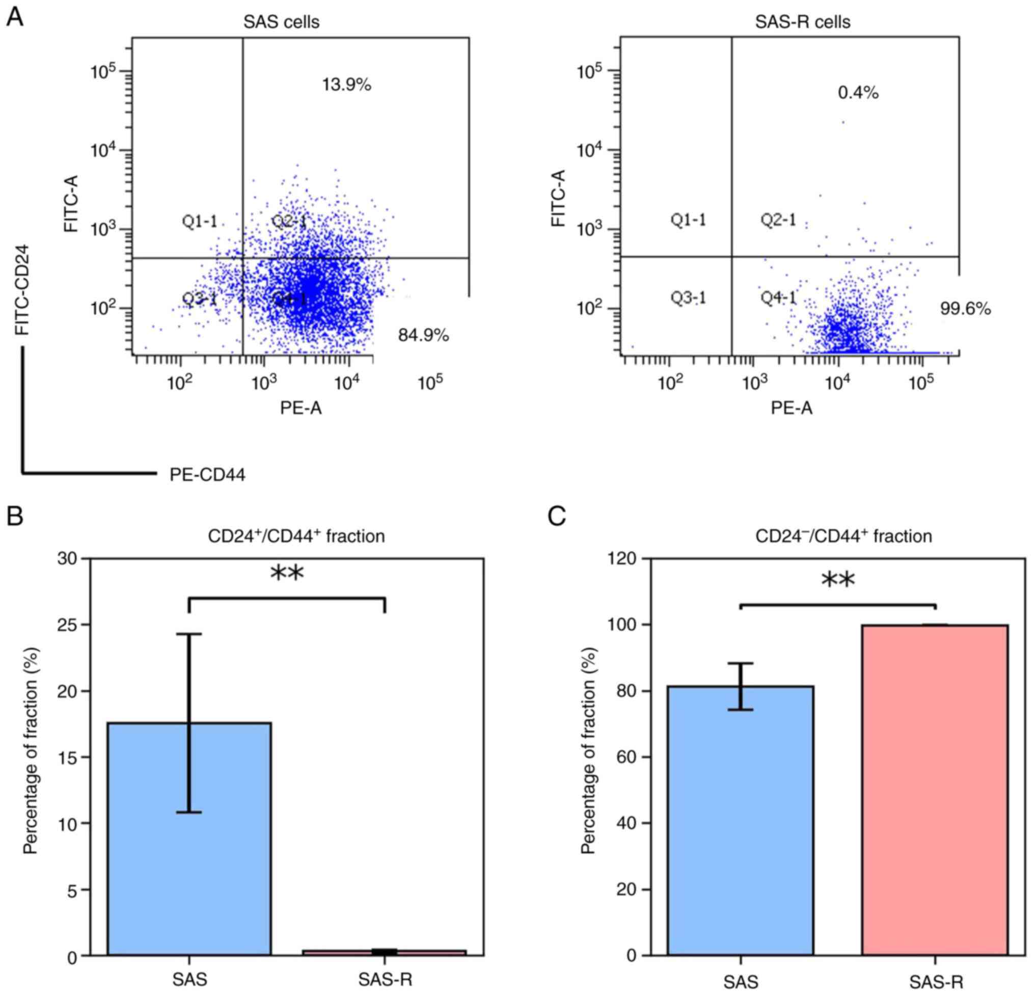
analyzed for CD24 and CD44 are presented in Fig. 1. The percentage of the
CD24+/CD44+ fraction in the SAS-R cells was
significantly lower than that in the SAS cells (0.30±0.10% vs.
17.53±6.73%, P<0.01). The percentage of the
CD24-/CD44+ fraction was higher in the SAS-R
cells compared with that in the SAS cells (99.70±0.1 vs.
81.17±6.99, P<0.01), indicating an expanded population of the
CSC-like fraction in the SAS-R cells compared with that in the SAS
cells.
The results of colony formation and spheroid assays
following treatment with Sal at the concentration of 0, 0.01, 0.03,
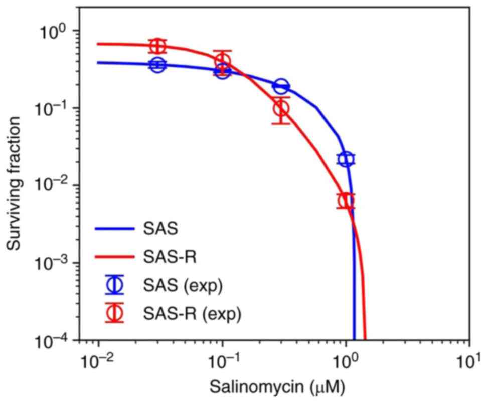
0.1, 0.3, 0.6 and 1 µM are illustrated in Fig. 2. The surviving fraction of SAS and
SAS-R cells decreased in a concentration-dependent manner following
treatment with Sal (Fig. 2A). The
IC50 value calculated from the log-logistic model of the SAS and
SAS-R cells was 0.332 and 0.250 µM, respectively. The addition of
0.3 µM Sal, which was a concentration near IC50 for both cell
lines, resulted in a significant decrease in the sphere formation
of SAS-R cells compared with that of the controls (Fig. 2B and C, P<0.01).
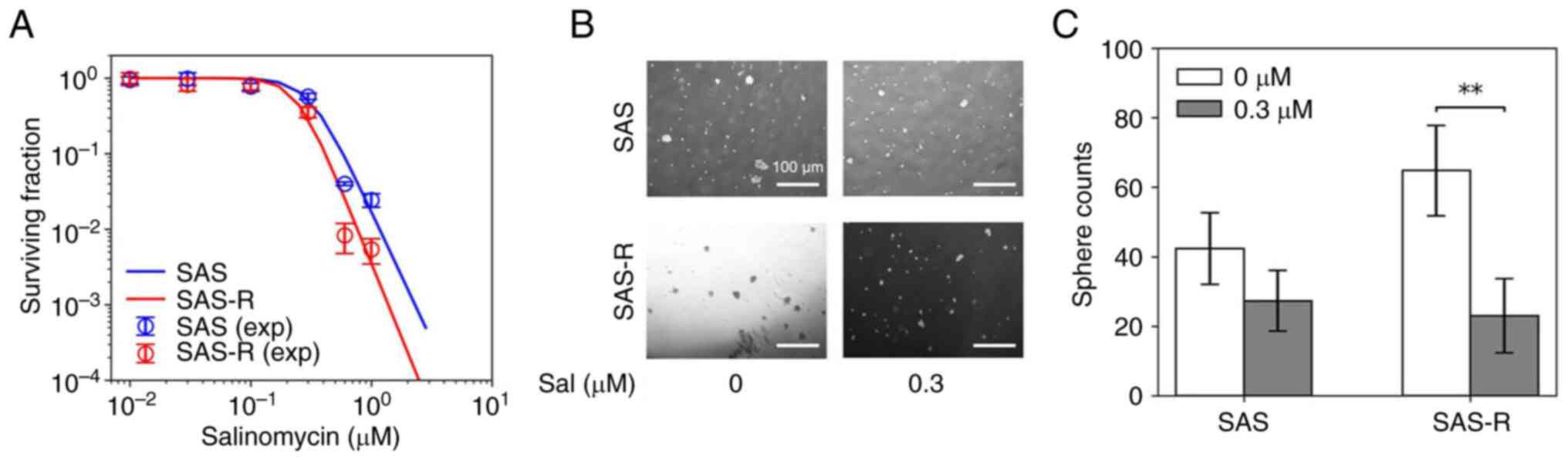 | Figure 2Cell surviving fraction and sphere
formation following treatment with Sal. (A) The cell surviving
fraction of the SAS and SAS-R cells, Sal concentration (i.e., 0,
0.01, 0.03, 0.1, 0.3, 0.6 and 1 µM) gradient. Exp represents
experimental data. Blue and Red solid lines indicate log-logistic
sigmoidal curve fitting SAS and SAS-R cell surviving fraction,
respectively. (B) Representative images are shown. White scale
bars, 100 µm. (C) Sphere counts following treatment with 0.3 µM
Sal. Values are presented as the mean ± standard deviation.
**P<0.01, significant differences. Sal,
salinomycin. |
The cell surviving fraction following 2 Gy
irradiation was evaluated under the Sal concentrations 0, 0.03,
0.1, 0.3, 0.6, and 1 µM (Fig. 3).
The surviving fraction was significantly lower in the SAS-R cells
than in the SAS cells (Fig. 3).
These results indicate that Sal can efficiently kill SAS-R cells in
which the CSC-like population is expanded.
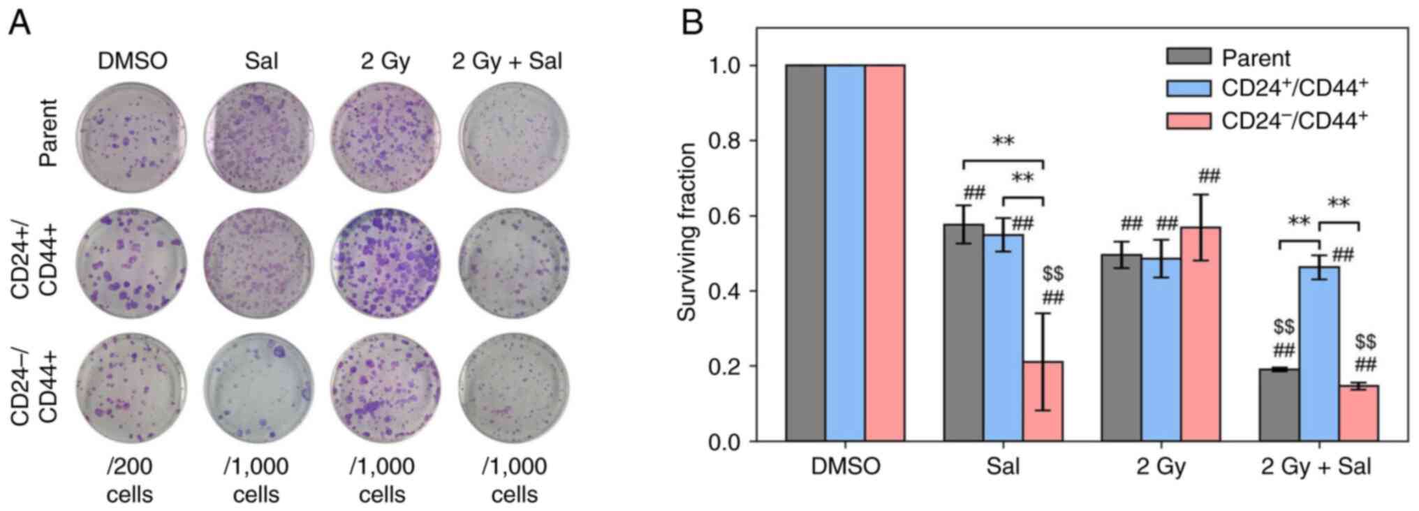
To demonstrate that Sal was effective against
CSC-like fractions, the CD24-/CD44+ and
CD24+/CD44+ fractions were sorted from the
parental SAS cells, and the sensitivity of Sal was measured using a
colony formation assay (Fig. 4).
Two-way ANOVA revealed a significant interaction between Sal
treatment and irradiation in all three cell populations (parent,
P<0.05; CD24-/CD44+, P<0.01; and
CD24+/CD44+, P<0.01). The
CD24-/CD44+ cells tended to exhibit a higher
surviving fraction following irradiation alone than the parental
and CD24+/CD44+ cells. By contrast, treatment
with Sal significantly decreased the surviving fraction of the
CD24-/CD44+ cells compared with the other
fractions. Taken together, these results suggest that the CSC-like
CD24-/CD44+ fraction is highly sensitive to
Sal treatment and exhibits a more pronounced reduction in surviving
fraction following the combined treatment with irradiation and Sal
than the other cell fractions.
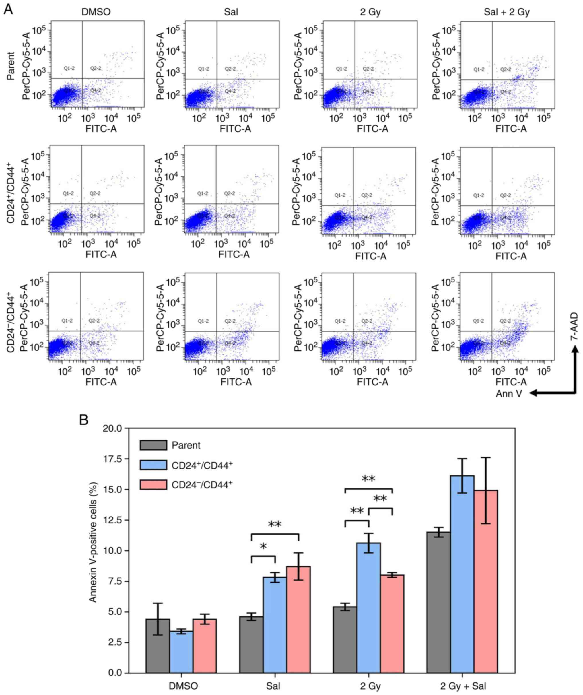
The results of apoptosis at 24 h following
experimental treatment in the fraction of the
CD24+/CD44+ and
CD24-/CD44+ cells sorted from parental SAS
cells are presented in Fig. 5. The
addition of Sal significantly increased apoptosis in both fractions
of the cells compared with the parental SAS cells (Fig. 5B; P<0.05 vs.
CD24+/CD44+ and P<0.01 vs.
CD24-/CD44+). Following treatment with 2 Gy
irradiation alone, the apoptosis of the
CD24+/CD44+ cells was significantly increased
compared with the other two groups (P<0.01). These results were
consistent with those of the colony formation assay; however, there
was no significant difference in the 2 Gy + Sal cell group, and the
apoptosis of the CD24-/CD44+ fraction was
significantly increased compared with the parental SAS cells
treated with 2 Gy irradiation alone (P<0.01). It should be noted
that the apoptosis assay and the clonogenic assay evaluate
different biological endpoints. Whereas the clonogenic assay
measures the long-term reproductive capacity of clonogenic cells,
apoptosis was assessed only 24 h following treatment and may not
fully reflect reproductive cell death. Therefore, early apoptosis
is not necessarily associated with clonogenic survival.
Discussion
Several studies have been conducted to specifically
target the DNA damage response to achieve selective cancer cell
radiosensitization (14).
Nevertheless, despite efforts to overcome radioresistance, the
underlying mechanisms behind its development are not yet fully
understood. CSCs with intrinsic or acquired radioresistance have
higher DNA damage repair capabilities than non-CSCs. Considering
these biological characteristics, the possibility of CSC recovery
between dose fractionations cannot be ignored (15). Therefore, the mechanisms underlying
radioresistance in stem cell-like cells must be elucidated.
The results of the present study demonstrated that
there was a high number of CD24-/CD44+ cells
among the SAS-R cells. CD24 and CD44 are widely used as markers for
CSCs (8). Even in OSCC cell lines,
the CD24-/CD44+ fraction has been reported in
CSC-like cells (16). CD24 is a cell
surface protein expressed on epithelial cells, and its expression
has been confirmed in a variety of cancer types (17). One characteristic of cancer cell
populations as they transition to CSCs is epithelial-mesenchymal
transition. Therefore, when CSCs increase, the epithelial marker
CD24+ cells decrease and CD24- cells increase
(18). Previous research has
demonstrated that the expression of CD44 is associated with
prognosis. For example, Kawano et al (19) demonstrated that CD44+
cells with CSC features were associated with a poor prognosis of
patients with head and neck carcinomas. A high expression of CD44
reflects the aggressiveness of tumor cells and the malignancy of
head and neck carcinomas (20).
Furthermore, CD44+ cells are more likely to form lung
metastases in mice, likely due to their increased migratory and
invasive abilities (21).
Various studies have investigated the mechanisms by
which CSCs exhibit resistance to radiation (22). In photon radiotherapy, the majority
of radiation-induced DNA damage in cancer cells is caused by free
radicals, such as reactive oxygen species (ROS) generated by the
ionization of water molecules, that is, the indirect action of
radiation. There are a multitude of publications showing that ROS
scavengers are upregulated and highly efficient in CSCs of various
tumors (22). Tumor cells reduce
levels of ROS and protected CSCs from radiotherapy-induced cell
death (23). In addition to these
mechanisms, some studies have demonstrated that the activation of
signaling pathways related to survival, such as Bcl-2 and
PI3K/Akt/mTOR, also contributes to CSC radioresistance (24). The results of a previous study by the
authors indicated that the double-strand break repair mechanism in
radioresistant cells strongly depends on the NHEJ repair function
(25).
The mechanism of action of Sal in cancer cells has
been studied from various perspectives (26). In a previous study on breast cancer
cells (MDA-MB-231), Sal induced apoptosis and autophagy (27). The Sal-mediated ROS production led to
mitochondrial dysfunction in MDA-MB-231 cells. Notably, treatment
with N-acetyl-L-cysteine, a ROS scavenger, attenuated Sal-induced
apoptosis and autophagy (27). In
another study, Sal induced mitochondrial dysfunction in
cisplatin-resistant breast cancer cells. The analysis of nuclear
translocation of pro-survival transcription factors by western
blotting revealed a distinct role of p65 (NF-κB) in
cisplatin-mediated resistance in breast cancer (28). In chemoresistant populations, Sal
significantly induced programmed cell death by inducing G2/M cell
cycle arrest and inhibiting the MAPK/PI3K pathways (29). That study demonstrated that Sal
induced toxic DNA lesions and prevented subsequent recovery by
targeting homologous recombination repair. In the present study,
the results of the colony formation assay revealed that the number
of SAS-R cells decreased when Sal was added at concentrations of
0.3 µM or higher without radiation. This suggested that Sal was
effective against SAS-R cells as they included a high proportion of
CSCs.
To date, to the best of our knowledge, there have
been no studies on the effects of Sal on radioresistant oral cancer
cells. However, Lim et al (30) investigated the potential of Sal to
enhance radiotherapy and uncovered novel dual functions of this
ionophore inducing DNA damage and preventing repair using
glioblastoma cells. On the other hand, Zhang et al (31) demonstrated that Sal induced apoptosis
and G2/M arrest, increased Bax and cleaved caspase-3, decreased
Bcl-2 expression, and increased the formation of γ-H2AX nuclear
foci in nasopharyngeal carcinoma cells. In addition, a potential
mechanism underlying the radioresistance of SAS-R cells has been
reported (32). SAS-R cells are more
resistant to ferroptosis, an iron-dependent form of cell death,
than the parental SAS cells due to lower intracellular
Fe2+ levels resulting from the higher expression of
miR-7-5p. Moreover, Sal has been reported to eliminate CSCs through
iron-mediated production of ROS (33) and to overcome the radioresistant of
nasopharyngeal carcinoma cells by promoting ROS generation via the
inhibition of Nrf2(34). These
findings may explain the colony formation assay results obtained
herein, in which Sal treatment significantly reduced the survival
of SAS-R cells compared with that of SAS cells.
The present study has several limitations. First,
all experiments were conducted exclusively in vitro, without
in vivo validation using animal models. Second, the
molecular pathways discussed above were not experimentally
validated. Finaly, although the cell-sorting experiment provides
supportive evidence for the involvement of the
CD24-/CD44+ population in the enhanced
response to Sal, it does not directly demonstrate the molecular
mechanism responsible for Sal-mediated radiosensitization. Further
studies are warranted to elucidate the detailed molecular
mechanisms underlying the radiosensitizing effects of Sal and to
evaluate its potential as a novel CSC-targeting
radiosensitizer.
In conclusion, the present study demonstrates that
Sal is effective in reducing the survival of radioresistant OSCC
cells, which contain a high proportion of CSC-like fraction. As
modern radiotherapy mainly relies on fractionated irradiation,
radioresistant cells are considered to contribute to poor clinical
outcomes. The findings presented herein suggest that Sal may serve
as a potential radiosensitizer by reducing the survival of
radioresistant OSCC cells and the CD24-/CD44+
cell fraction in combination with irradiation.
Acknowledgements
Not applicable.
Funding
Funding: The present study was supported by the Japan Society
for the Promotion of Science (JSPS) KAKENHI grant no. 22K07789.
Availability of data and materials
The data generated in the present study may be
requested from the corresponding author.
Authors' contributions
RS, YH and MF designed the study. RS performed the
experiments. RS and YH performed the statistical analysis. RS and
YH wrote the manuscript. MF supervised the study. RS and YH confirm
the authenticity of all the raw data. All authors reviewed the
manuscript, and all authors have read and approved the final
manuscript.
Ethics approval and consent to
participate
Not applicable.
Patient consent for publication
Not applicable.
Competing interests
The authors declare that they have no competing
interests.
References
|
1
|
Sung H, Ferlay J, Siegel RL, Laversanne M,
Soerjomataram I, Jemal A and Bray F: Global cancer statistics 2020:
GLOBOCAN estimates of incidence and mortality worldwide for 36
cancers in 185 countries. CA Cancer J Clin. 71:209–249.
2021.PubMed/NCBI View Article : Google Scholar
|
|
2
|
Ganesh K and Massagué J: Targeting
metastatic cancer. Nat Med. 27:34–44. 2021.PubMed/NCBI View Article : Google Scholar
|
|
3
|
Yin W, Wang J, Jiang L and James Kang Y:
Cancer and stem cells. Exp Biol Med (Maywood). 246:1791–1801.
2021.PubMed/NCBI View Article : Google Scholar
|
|
4
|
Shibue T and Weinberg RA: EMT, CSCs, and
drug resistance: The mechanistic link and clinical implications.
Nat Rev Clin Oncol. 14:611–629. 2017.PubMed/NCBI View Article : Google Scholar
|
|
5
|
Olivares-Urbano MA, Griñán-Lisón C,
Marchal JA and Núñez MI: CSC radioresistance: A therapeutic
challenge to improve radiotherapy effectiveness in cancer. Cancers
(Basel). 9(1651)2020.PubMed/NCBI View Article : Google Scholar
|
|
6
|
Shimura T, Nara H, Yamazaki M, Sasatani M
and Ushiyama A: Suppression of cancer stem-like cell
radioresistance by inhibiting AMPK signaling. J Radiat Res.
66:227–233. 2025.PubMed/NCBI View Article : Google Scholar
|
|
7
|
Yuan SF, Wang YM, Chan LP, Hung AC, Nguyen
HDH, Chen YK, Hu SC, Lo S and Wang YY: IL-1RA promotes oral
squamous cell carcinoma malignancy through mitochondrial
metabolism-mediated EGFR/JNK/SOX2 pathway. J Transl Med.
21(473)2023.PubMed/NCBI View Article : Google Scholar
|
|
8
|
Hasegawa K, Saga R, Takahashi R, Fukui R,
Chiba M, Okumura K, Tsuruga E and Hosokawa Y: 4-Methylumbelliferone
inhibits clonogenic potency by suppressing high molecular
weight-hyaluronan in fibrosarcoma cells. Oncol Lett. 19:2801–2808.
2020.PubMed/NCBI View Article : Google Scholar
|
|
9
|
Oyama T, Hosokawa Y, Abe K, Hasegawa K,
Fukui R, Aoki M and Kobayashi W: Prognostic value of quantitative
FDG-PET in the prediction of survival and local recurrence for
patients with advanced oral cancer treated with superselective
intra-arterial chemoradiotherapy. Oncol Lett. 19:3775–3780.
2020.PubMed/NCBI View Article : Google Scholar
|
|
10
|
Fuchs D, Heinold A, Opelz G, Daniel V and
Naujokat C: Salinomycin induces apoptosis and overcomes apoptosis
resistance in human cancer cells. Biochem Biophys Res Commun.
390:743–749. 2009.PubMed/NCBI View Article : Google Scholar
|
|
11
|
Gupta PB, Onder TT, Jiang G, Tao K,
Kuperwasser C, Weinberg RA and Lander ES: Identification of
selective inhibitors of cancer stem cells by high-throughput
screening. Cell. 138:645–659. 2009.PubMed/NCBI View Article : Google Scholar
|
|
12
|
Qi D, Liu Y, Li J, Huang JH, Hu X and Wu
E: Salinomycin as a potent anticancer stem cell agent: State of the
art and future directions. Med Res Rev. 42:1037–1063.
2022.PubMed/NCBI View Article : Google Scholar
|
|
13
|
Kuwahara Y, Mori M, Kitahara S and
Fukumoto M, Ezaki T, Mori S, Echigo S, Ohkubo Y and Fukumoto M:
Targeting of tumor endothelial cells combining 2 Gy/day of X-ray
with everolimus is the effective modality for overcoming clinically
relevant radioresistant tumors. Cancer Med. 3:310–321.
2014.PubMed/NCBI View
Article : Google Scholar
|
|
14
|
Begg AC, Stewart FA and Vens C: Strategies
to improve radiotherapy with targeted drugs. Nat Rev Cancer.
11:239–253. 2011.PubMed/NCBI View
Article : Google Scholar
|
|
15
|
Fukui R, Saga R, Matsuya Y, Tomita K,
Kuwahara Y, Ohuchi K, Sato T, Okumura K, Date H, Fukumoto M and
Hosokawa Y: Tumor radioresistance caused by radiation-induced
changes of stem-like cell content and sub-lethal damage repair
capability. Sci Rep. 12(1056)2022.PubMed/NCBI View Article : Google Scholar
|
|
16
|
Todoroki K, Ogasawara S, Akiba J, Nakayama
M, Naito Y, Seki N, Kusukawa J and Yano H: CD44v3+/CD24- cells
possess cancer stem cell-like properties in human oral squamous
cell carcinoma. Int J Oncol. 48:99–109. 2016.PubMed/NCBI View Article : Google Scholar
|
|
17
|
Panagiotou E, Syrigos NK, Charpidou A,
Kotteas E and Vathiotis IA: CD24: A novel target for cancer
immunotherapy. J Pers Med. 12(1235)2022.PubMed/NCBI View Article : Google Scholar
|
|
18
|
Blick T, Hugo H, Widodo E, Waltham M,
Pinto C, Mani SA, Weinberg RA, Neve RM, Lenburg ME and Thompson EW:
Epithelial mesenchymal transition traits in human breast cancer
cell lines parallel the CD44(hi)/CD24(lo/-) stem cell phenotype in
human breast cancer. J Mammary Gland Biol Neoplasia. 15:235–252.
2010.PubMed/NCBI View Article : Google Scholar
|
|
19
|
Kawano T, Yanoma S, Nakamura Y, Ozeki A,
Kokatsu T, Kubota A, Furukawa M and Tsukuda M: Soluble CD44
standard, CD44 variant 5 and CD44 variant 6 and their relation to
staging in head and neck cancer. Acta Otolaryngol. 125:392–397.
2005.PubMed/NCBI View Article : Google Scholar
|
|
20
|
Joshua B, Kaplan MJ, Doweck I, Pai R,
Weissman IL, Prince ME and Ailles LE: Frequency of cells expressing
CD44, a head and neck cancer stem cell marker: Correlation with
tumor aggressiveness. Head Neck. 34:42–49. 2012.PubMed/NCBI View Article : Google Scholar
|
|
21
|
Davis SJ, Divi V, Owen JH, Bradford CR,
Carey TE, Papagerakis S and Prince ME: Metastatic potential of
cancer stem cells in head and neck squamous cell carcinoma. Arch
Otolaryngol Head Neck Surg. 136:1260–1266. 2010.PubMed/NCBI View Article : Google Scholar
|
|
22
|
Arnold CR, Mangesius J, Skvortsova II and
Ganswindt U: The role of cancer stem cells in radiation resistance.
Front Oncol. 10(164)2020.PubMed/NCBI View Article : Google Scholar
|
|
23
|
Szumiel I: Ionizing radiation-induced
oxidative stress, epigenetic changes and genomic instability: The
pivotal role of mitochondria. Int J Radiat Biol. 91:1–12.
2015.PubMed/NCBI View Article : Google Scholar
|
|
24
|
Xia P and Xu XY: PI3K/Akt/mTOR signaling
pathway in cancer stem cells: From basic research to clinical
application. Am J Cancer Res. 5:1602–1609. 2015.PubMed/NCBI
|
|
25
|
Ohuchi K, Saga R, Hasegawa K, Tsuruga E,
Hosokawa Y, Fukumoto M and Okumura K: DNA-PKcs phosphorylation
specific inhibitor NU7441 enhances the radiosensitivity of
clinically relevant radioresistant oral squamous cell carcinoma
cells. Biomed Rep. 18(28)2023.PubMed/NCBI View Article : Google Scholar
|
|
26
|
Dewangan J, Srivastava S and Rath SK:
Salinomycin: A new paradigm in cancer therapy. Tumour Biol.
39(1010428317695035)2017.PubMed/NCBI View Article : Google Scholar
|
|
27
|
Kim KY, Park KI, Kim SH, Yu SN, Lee D, Kim
YW, Noh KT, Ma JY, Seo YK and Ahn SC: Salinomycin induces reactive
oxygen species and apoptosis in aggressive breast cancer cells as
mediated with regulation of autophagy. Anticancer Res.
37:1747–1758. 2017.PubMed/NCBI View Article : Google Scholar
|
|
28
|
Tyagi M and Patro BS: Salinomycin reduces
growth, proliferation and metastasis of cisplatin resistant breast
cancer cells via NF-κB deregulation. Toxicol In Vitro. 60:125–131.
2019.PubMed/NCBI View Article : Google Scholar
|
|
29
|
Patel S, Patel A, Nair A, Shah K, Shah K,
Tanavde V and Rawal R: Salinomycin mediated therapeutic targeting
of circulating stem like cell population in oral cancer. J Biomol
Struct Dyn. 40:11141–11153. 2022.PubMed/NCBI View Article : Google Scholar
|
|
30
|
Lim YC, Ensbey KS, Offenhäuser C, D'souza
RCJ, Cullen JK, Stringer BW, Quek H, Bruce ZC, Kijas A, Cianfanelli
V, et al: Simultaneous targeting of DNA replication and homologous
recombination in glioblastoma with a polyether ionophore. Neuro
Oncol. 22:216–228. 2020.PubMed/NCBI View Article : Google Scholar
|
|
31
|
Zhang Y, Zuo Y, Guan Z, Lu W, Xu Z, Zhang
H, Yang Y, Yang M, Zhu H and Chen X: Salinomycin radiosensitizes
human nasopharyngeal carcinoma cell line CNE-2 to radiation. Tumour
Biol. 37:305–311. 2016.PubMed/NCBI View Article : Google Scholar
|
|
32
|
Tomita K, Fukumoto M, Itoh K, Kuwahara Y,
Igarashi K, Nagasawa T, Suzuki M, Kurimasa A and Sato T: MiR-7-5p
is a key factor that controls radioresistance via intracellular
Fe2+ content in clinically relevant radioresistant cells. Biochem
Biophys Res Commun. 518:712–718. 2019.PubMed/NCBI View Article : Google Scholar
|
|
33
|
Mai TT, Hamaï A, Hienzsch A, Cañeque T,
Müller S, Wicinski J, Cabaud O, Leroy C, David A, Acevedo V, et al:
Salinomycin kills cancer stem cells by sequestering iron in
lysosomes. Nat Chem. 9:1025–1033. 2017.PubMed/NCBI View Article : Google Scholar
|
|
34
|
Zhang G, Wang W, Yao C, Ren J, Zhang S and
Han M: Salinomycin overcomes radioresistance in nasopharyngeal
carcinoma cells by inhibiting Nrf2 level and promoting ROS
generation. Biomed Pharmacother. 91:147–154. 2017.PubMed/NCBI View Article : Google Scholar
|