Introduction
Salivary adenoid cystic carcinoma (SACC) is one of
the most frequent malignant salivary gland tumors, constituting
approximately 8% of all salivary gland neoplasms (1). SACC is known for its prolonged
clinical course and delayed onset of distant metastases (2). Perineural invasion (PNI), a frequent
occurrence in SACC, is difficult to identify clinically, which may
prevent a complete surgical resection (3). Vrielinck et al reported a
correlation between PNI and poor prognosis (4).
Spindle cell melanoma is a melanoma that has a
notable propensity for PNI. Iwamoto et al (5,6)
demonstrated that Schwann cell markers, including the p75
neurotrophin receptor (p75NTR) and glial fibrillary acidic protein
(GFAP), were overexpressed in spindled melanoma compared with the
epithelioid melanoma which does not have the characteristics of
PNI. In addition, the authors hypothesized that Schwann cell
differentiation may be involved in the process of PNI. Leu-7 (human
natural killer-1, HNK-1) belongs to the CD57 antibody group. Leu-7
may react with antigens in the medullary sheath of the central and
peripheral nervous systems and is regarded as a new Schwann cell
marker (7). α-smooth muscle actin
(α-SMA) is regarded as a useful probe in the study of smooth muscle
differentiation in normal and pathological conditions (8). The expression of α-SMA has been
observed in human myoepithelial cells of salivary (9), mammary (10), sweat (11) and bronchial mucous glands (12).
The aim of the current study was to investigate the
expression pattern of a new Schwann cell marker (Leu-7) and α-SMA
in SACC and acinic cell carcinoma (ACA) and to confirm the
correlation between Schwann-like cell differentiation and PNI in
SACC.
Patients and methods
Patients and samples
The present study comprised 28 SACC patients (16
males and 12 females; mean age, 50 years; range, 26–67) and 10 ACA
patients (6 males and 4 females; mean age, 51.4 years; range,
31–65) treated at the Department of Oral and Maxillofacial Surgery,
School of Stomatology, Fourth Military Medical University between
2004 and 2008. The present study was approved by the Ethics
Committee of the Fourth Military Medical University. Informed
consent was obtained from all subjects prior to obtaining the
tissue samples during surgery.
Samples were divided into two sections. For
immunohistochemical staining and hematoxylin and eosin (H&E)
staining, one section was fixed immediately in 10% formalin for 24
h and was embedded in paraffin. Subsequently, 4-μm serial
sections were prepared with a microtome. For pre-embedding
immunogold-silver cytochemistry, the tissue was incised into a 2×2
mm slice and fixed into a mixture of 4% paraformaldehyde, 0.05%
glutaraldehyde and 15% saturated picric acid in 0.1 M phosphate
buffer (pH 7.4) at room temperature.
Assessment of PNI
Slides were prepared from each sample using
4-μm H&E-stained serial sections and reviewed by two
independent oral pathologists who were blind to the clinical
findings. The presence or absence of microscopic PNI was
recorded.
Immunohistochemical staining
Mouse anti-human Leu-7 was used as the primary
antibody and biotinylated goat anti-mouse IgG was used as the
secondary antibody. The two antibodies were reacted with a
streptavidin-biotin-peroxidase complex. Briefly, sections were
mounted on glass slides and air-dried. Dewaxing and quenching of
endogenous peroxidase was carried out using 3%
H2O2/methanol and subsequently protease
digestion was carried out for antigen retrieval. The samples were
then incubated in goat serum (Invitrogen, Carlsbad, CA, USA) at
1:10 dilution for 20 min to block non-specific binding. Mouse
anti-human monoclonal antibody Leu-7 (Abcam, Cambridge, MA, USA)
was diluted in phosphate-buffered saline (PBS)/0.3% bovine serum
albumin (Sigma-Aldrich, St. Louis, MO, USA) and used at a
predetermined optimal dilution of 1:25. Following overnight
incubation at 4°C, the sections were incubated for 30 min at 37°C
in goat anti-mouse IgG (Invitrogen). The peroxidase-labeled
streptavidin (Invitrogen) was then added for 30 min at 37°C. The
substrate color reaction was developed by incubation in liquid
diaminobenzamine (DAB; Dako, Glostrup, Denmark). For the negative
control sections, the primary antibody was replaced by non-immune
normal goat serum. The results of immunohistochemical staining were
evaluated by two independent pathologists blind to the clinical
findings.
Immunofluorescence double-staining and
confocal laser scanning microscopy (CLSM) study
A total of 22 samples that expressed Leu-7 were
included in immunofluorescence double-staining carried out.
Briefly, sections were stored and then exposed to antigen
retrieval. Mouse anti-human monoclonal antibody Leu-7 (Abcam, 1:50)
and rabbit anti-human polyclonal antibody α-SMA (Abcam, 1:200) were
used. Sections were incubated in a mixture of the two primary
antibodies. Subsequent to overnight incubation at 4°C, the sections
were rinsed three times for 15 min in PBS (0.01 mol/l, pH 7.4) and
incubated for 60 min at 37°C in a biotinylation goat anti-mouse IgG
(Invitrogen, 1:8,000). Sections were then incubated for 3 h at room
temperature in a mixture of goat anti-rabbit FITC-conjugated IgG
(Invitrogen, 1:30) and avidin-conjugated Cy3 (Sigma, 1:100).
The labeled tissues were then rinsed in PBS (0.01
mol/l, pH 7.4), mounted on gelatin-coated slides and coverslipped.
The distribution and intensity of labeling were assessed using a
Zeiss epi-illumination fluorescence microscope and a Zeiss LSM 510
confocal laser scanning microscopy system. The software of the CLSM
exhibited images acquired with 495-nm laser illumination (FITC)
color-coded in green and those acquired at 543 nm (Cy3) color-coded
in red. In each immunostaining run, the two primary antibodies were
replaced by non-immune normal goat serum as negative controls.
Pre-embedding immunogold-silver
cytochemistry
The expression of Leu-7/α-SMA was explored using
pre-embedding immunogold-silver cytochemistry. Sections of
50-μm were cut with a vibratome (VT 1000S; Leica, Mannheim,
Germany) and placed in PBS containing 25% sucrose and 10% glycerol
for 1 h for cryo-protection. Following a freeze-thaw treatment,
sections were blocked with 5% BSA and 5% NGS for 4 h to block
non-specific immunoreactivity. Subsequent immunohistochemical
procedures were performed at room temperature.
For Leu-7/α-SMA double labeling, Leu-7 was detected
by immunoperoxidase and α-SMA by an immunogold-silver staining
method. The sections were incubated overnight in primary antibodies
against Leu-7 with those against α-SMA, diluted to working
concentrations as described above, in PBS containing 1% BSA and 1%
NGS. Following rinsing in PBS, sections were incubated overnight in
a mixture of secondary antibodies of biotinylated goat anti-mouse
IgG and goat anti-rabbit IgG conjugated to 1.4-nm gold particles
(1:100, Nanoprobes, Yaphank, NY, USA). In addition, following
rinsing in PBS, sections were post-fixed in 2% glutaraldehyde in
PBS for 45 min. Silver enhancement was carried out in the dark with
an HQ Silver kit (Nanoprobes). Prior to and following the silver
enhancement, sections were rinsed several times with deionized
water. The sections were then incubated in ABC solution (Sigma,
1:300) for 4 h and visualized using the glucose
oxidase-3,3′-diaminobenzidine method. Immuno-labeled sections were
fixed with 0.5% osmium tetroxide in 0.1 M phosphate buffer for 1 h,
dehydrated in graded ethanol series, then in propylene oxide and
finally flat-embedded in Epon 812. Following polymerization, the
sections were examined under the light microscope. Sections
containing Leu-7/α-SMA immunoreactivity were selected, trimmed
under a stereomicroscope and mounted onto blank resin stubs.
Ultra-thin sections were cut and mounted on mesh grids. The
sections were then counterstained with Uranyl Acetate and lead
citrate and observed under a JEM-1230 electron microscope (JEOL
Ltd., Tokyo, Japan). Electron micrographs were developed in the
dark room.
Statistical analysis
Statistical analysis was performed using SPSS 18.0
software (Chicago, IL, USA). To determine the difference of the
ability of the levels of PNI and Leu-7 expression between SACC and
ACA, statistical analysis was performed using Fisher’s exact test.
To find the correlation between PNI and Leu-7 expression, κ
analysis was used. P<0.05 was considered to indicate a
statistically significant difference.
Results
PNI and the expression of Leu-7 in SACC
and ACA
PNI appeared in 16 out of 28 SACCs (57.1%). In
addition, 1 case of PNI was observed microscopically out of the 10
specimens of ACA (10%). The H&E section findings of PNI are
summarized in Table I. The
differences in the rate of PNI between SACC and ACA were
significant (Fisher’s exact test, P=0.009; Table II).
 | Table IClinical data and Leu-7 expression in
38 salivary malignancies. |
Table I
Clinical data and Leu-7 expression in
38 salivary malignancies.
| Number | Diagnosis | Gender | Age (years) | Tumor site | PNI | Leu-7 |
|---|
| 1 | SACC | M | 52 | Left parotid
gland | − | + |
| 2 | SACC | M | 57 | Left parotid
gland | + | + |
| 3 | SACC | F | 67 | Right submaxillary
gland | + | + |
| 4 | SACC | M | 61 | Right parotid
gland | + | + |
| 5 | SACC | F | 46 | Left submaxillary
gland | − | + |
| 6 | SACC | M | 34 | Right parotid
gland | + | + |
| 7 | SACC | F | 55 | Soft palate | + | + |
| 8 | SACC | M | 54 | Right submaxillary
gland | − | − |
| 9 | SACC | F | 52 | Right parotid
gland | − | − |
| 10 | SACC | M | 62 | Right palate | + | + |
| 11 | SACC | F | 66 | Left submaxillary
gland | + | + |
| 12 | SACC | M | 31 | Left parotid
gland | − | − |
| 13 | SACC | M | 26 | Right palate | − | − |
| 14 | SACC | M | 47 | Right submaxillary
gland | − | + |
| 15 | SACC | M | 49 | Left submaxillary
gland | + | + |
| 16 | SACC | F | 41 | Right sublingual
gland | + | + |
| 17 | SACC | M | 37 | Left parotid
gland | + | + |
| 18 | SACC | F | 45 | Right parotid
gland | + | + |
| 19 | SACC | M | 61 | Left parotid
gland | − | + |
| 20 | SACC | F | 42 | Right submaxillary
gland | + | + |
| 21 | SACC | M | 47 | Right submaxillary
gland | + | + |
| 22 | SACC | F | 60 | Right sublingual
gland | + | + |
| 23 | SACC | F | 62 | Right parotid
gland | + | + |
| 24 | SACC | M | 67 | Left sublingual
gland | − | − |
| 25 | SACC | M | 58 | Right parotid
gland | + | + |
| 26 | SACC | F | 35 | Left sublingual
gland | − | − |
| 27 | SACC | M | 39 | Right submaxillary
gland | − | + |
| 28 | SACC | F | 46 | Right parotid
gland | − | + |
| 29 | ACA | M | 59 | Right parotid
gland | + | − |
| 30 | ACA | M | 39 | Left sublingual
gland | − | − |
| 31 | ACA | F | 61 | Right palate | − | − |
| 32 | ACA | M | 54 | Left parotid
gland | − | − |
| 33 | ACA | F | 43 | Left parotid
gland | − | − |
| 34 | ACA | M | 57 | Right parotid
gland | − | − |
| 35 | ACA | M | 65 | Left palate | − | − |
| 36 | ACA | M | 31 | Left sublingual
gland | − | − |
| 37 | ACA | F | 52 | Right sublingual
gland | − | − |
| 38 | ACA | F | 53 | Right parotid
gland | − | − |
| Table IIDifferences of ability of PNI and
Leu-7 expression between SACC and ACA. |
Table II
Differences of ability of PNI and
Leu-7 expression between SACC and ACA.
| PNIa | Leu-7
expressionb |
|---|
|
|
|
|---|
| (+) | (−) | Total | (+) | (−) | Total |
|---|
| SACC | 16 | 12 | 28 | 22 | 6 | 28 |
| ACA | 1 | 9 | 10 | 0 | 10 | 10 |
| Total | 17 | 21 | 38 | 22 | 16 | 38 |
Positive staining for Leu-7 was revealed in 22 out
of 28 SACCs (78.6%; Fig. 1a),
while none of the 10 ACAs were positive for Leu-7 (Fig. 1b). The differences between SACC and
ACA in the expression of Leu-7 were significant (Fisher’s exact
test, P=0.000; Table II).
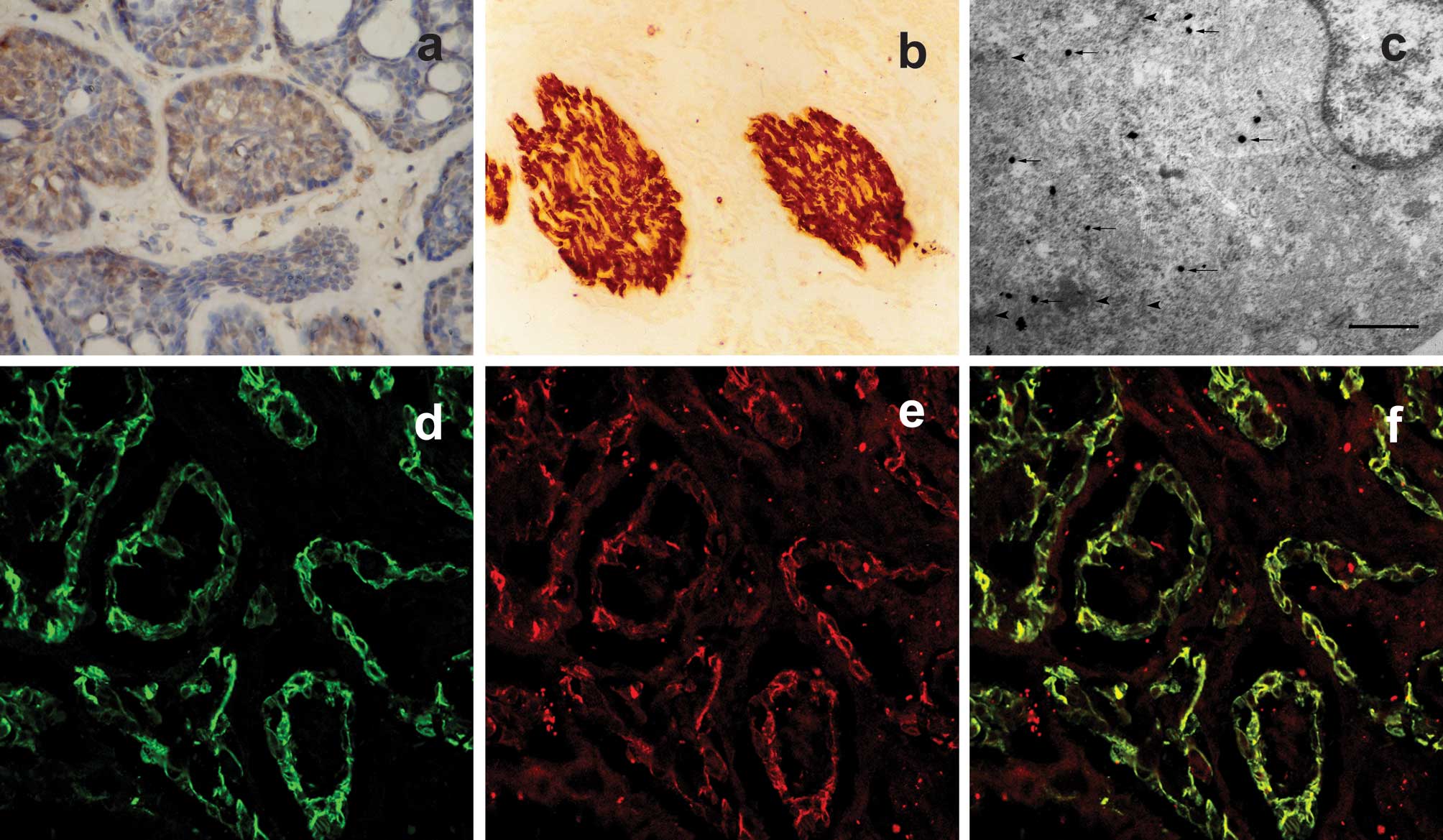 | Figure 1(a) Representative expression of Leu-7
in human SACC which was present diffusely in the cytoplasm of
carcinoma cells (original magnification, ×400). (b) No Leu-7
expression was detected in human ACA while peripheral nerve tracts
revealed strong intensity of staining (original magnification,
×200). (c) Electron micrographs showing double labeling of Leu-7
and α-SMA in the myoepithelial cell in SACC.
Immunogold-silver-enhanced particles, indicative of α-SMA
immunoreactivity, are revealed diffusely in cytoplasm (arrows).
Immunoperoxidase reaction product, indicative of Leu-7
immunoreactivity, is also distributed in cytoplasm (arrowheads).
The particles are located in the same myoepithelial cell of the
SACC tissue. Scale bars=0.5 μm. (d-f) Leu-7/α-SMA expressed
in human SACC tissues by double-label immunofluorescence (original
magnification, ×600). (d) Green fluorescence demonstrated positive
staining of α-SMA. (e) Red fluorescence demonstrated positive
staining of Leu-7. (f) Yellow fluorescence demonstrated positive
staining of Leu-7/α-SMA, which was co-expressed in the same
location. SACC, salivary adenoid cystic carcinoma; ACA, acinic cell
carcinoma; α-SMA, smooth muscle actin. |
Correlation between Leu-7 expression and
PNI in SACC
SACCs (16/28) were observed with PNI and these were
all positive for Leu-7. The κ test revealed that there was a
significant correlation between Leu-7 expression and PNI (κ=0.533,
P=0.01; Table III).
| Table IIICorrelation between the Leu-7
expression and PNI in SACC. |
Table III
Correlation between the Leu-7
expression and PNI in SACC.
| PNI |
|---|
|
|
|---|
| (+) | (−) | Total |
|---|
| Leu-7 (+) | 16 | 6 | 22 |
| Leu-7 (−) | 0 | 6 | 6 |
| Total | 16 | 12 | 28 |
Double-label immunofluorescence and CLSM
for Leu-7/α-SMA
In the SACC, myoepithelial cells were detectable by
α-SMA staining. In the CLSM images, green fluorescence revealed
positive staining of α-SMA, red fluorescence revealed positive
staining of Leu-7 and yellow fluorescence revealed green and red
fluorescence in the same location (Fig. 1d-f). All 22 SACC cases were
double-stained for Leu-7/α-SMA, which were generally localized
diffusely in the cytoplasm of myoepithelial cells.
Electron microscopic examination for
Leu-7/α-SMA
Under the electron microscope, myoepithelial cells
were recognizable by α-SMA staining. Immunogold-silver-enhanced
particles indicative of α-SMA immunoreactivity were present
diffusely in the cytoplasm (arrows, Fig. 1c) in SACC. The immunoperoxidase
reaction product indicative of Leu-7 immunoreactivity was localized
mainly in the cytoplasm (arrowheads, Fig. 1c). Co-expression of Leu-7 and α-SMA
was identified in the same myoepithelial cells (Fig. 1c).
Discussion
SACC has a well-documented propensity for PNI. It is
recognized as one of the most significant prognostic factors.
Previous studies (13,14) have reported the correlation between
PNI and poor prognosis. Identification of the potential mechanisms
or the influential factors of PNI in SACC is likely to aid in its
prevention and treatment. There are several hypotheses for PNI in
SACC. In their study, Cheng et al indicated that perineural
space invasion was thought to occur by infiltration through the
perineural and intraneural lymphatic vessels and/or small veins, or
direct invasion of cancer cells along the perineural space, the
least resistant tissue (15).
Bockman et al suggested that the perineural space provided a
suitable microenvironment for the growth of cancer cells (16). Previous studies have shown that
Schwann cell markers (p75NTR, GFAP) were overexpressed in spindled
melanoma in which PNI was observed. Thus, it was speculated that
Schwann-like cell differentiation was involved in the process of
PNI (5,6).
Leu-7 (HNK-1) belongs to the CD57 antibody group and
has been defined as a new Schwann cell marker (7). Overexpression of Leu-7 has been
identified in human prostate adenocarcinoma in which PNI was
observed (17). The present study
has demonstrated that the Schwann cell marker, Leu-7, is expressed
in the majority of SACCs. Conversely, all 10 specimens of ACA,
which did not have the tendency of PNI, demonstrated negative
staining for Leu-7. Furthermore, statistical analysis revealed that
the ratio of samples with PNI in positive expression of Leu-7
(72.7%) was higher than that of negative expression of Leu-7 (0%).
In addition, the correlation between PNI and Leu-7 expression was
shown to be significant through the κ analysis (κ=0.533, P=0.01).
These results were in accordance with the study of Iwamoto et
al on spindled melanoma (5,6).
Schwann cells are the principal glial cells of the vertebrate
peripheral nervous system. Although these cells serve various
functions in the peripheral nerves and ganglia, they are best known
for their ability to elaborate the myelin sheath (18). Therefore, we suggest that certain
types of cells in SACC differentiate into Schwann-like cells and
this is one of the potential mechanisms of PNI in SACC.
SACC tissue contains myoepithelial and canula
endothelial cells, while no myoepithelial cells exist in ACA
(19). The cells of SACC and ACA
were thought to originate from the reserve and multipotent cells of
the intercalated duct, respectively (20). Therefore, the tendency of
Schwann-like cell differentiation in myoepithelial cells by
immunofluorescence staining and electron microscopic examination
was investigated. α-SMA was used as a myoepithelial marker and
Leu-7 as a Schwann cell marker. Therefore, those markers which were
double-marked with α-SMA and Leu-7 under immunofluorescence
staining and CLSM were considered to be transition state cells
within the process of differentiation from myoepithelial to Schwann
cells. Certain cells were identified with the co-expression of
Leu-7/α-SMA in SACC (Fig. 1c-f).
The electron microscopic examination also confirmed that
Leu-7/α-SMA were co-localized in the cytoplasm of the transition
state cells in the SACC sample. These results indicate that
Schwann-like cell differentiation occurs in myoepithelial cells of
SACC.
Although this study has identified the phenomenon of
Schwann-like cell differentiation of myoepithelial cells in SACCs,
the exact mechanism and possible influential factors remain to be
elucidated. In addition, the morphology and function of the
transition state cell require further analysis.
Acknowledgements
This study was supported by grants from the National
Natural Science Foundation of China (nos. 30271424, 30772428 and
81072230) and Natural Science Foundation of Shannxi Province of
China (no. 2002C2-06).
References
|
1
|
Yang X, Dai J, Li T, et al: Expression of
EMMPRIN in adenoid cystic carcinoma of salivary glands: correlation
with tumor progression and patients’ prognosis. Oral Oncol.
46:755–760. 2010.PubMed/NCBI
|
|
2
|
Bhattacharyya N: Survival and prognosis
for cancer of the submandibular gland. J Oral Maxillofac Surg.
62:427–430. 2004. View Article : Google Scholar : PubMed/NCBI
|
|
3
|
van der Wal JE, Snow GB and van der Waal
I: Intraoral adenoid cystic carcinoma. The presence of perineural
spread in relation to site, size, local extension and metastatic
spread in 22 cases. Cancer. 66:2031–2033. 1990.PubMed/NCBI
|
|
4
|
Vrielinck LJ, Ostyn F, van Damme B, van
den Bogaert W and Fossion E: The significance of perineural spread
in adenoid cystic carcinoma of the major and minor salivary glands.
Int J Oral Maxillofac Surg. 17:190–193. 1988. View Article : Google Scholar : PubMed/NCBI
|
|
5
|
Iwamoto S, Odland PB, Piepkorn M and
Bothwell M: Evidence that the p75 neurotrophin receptor mediates
perineural spread of desmoplastic melanoma. J Am Acad Dermatol.
35:725–731. 1996. View Article : Google Scholar : PubMed/NCBI
|
|
6
|
Iwamoto S, Burrows RC, Agoff SN, Piepkorn
M, Bothwell M and Schmidt R: The p75 neurotrophin receptor,
relative to other Schwann cell and melanoma markers, is abundantly
expressed in spindled melanomas. Am J Dermatopathol. 23:288–294.
2001. View Article : Google Scholar : PubMed/NCBI
|
|
7
|
Nagasaka T, Lai R, Sone M, Nakashima T and
Nakashima N: Glandular malignant peripheral nerve sheath tumor: an
unusual case showing histologically malignant glands. Arch Pathol
Lab Med. 124:1364–1368. 2000.PubMed/NCBI
|
|
8
|
Skalli O, Ropraz P, Trzeciak A, Benzonana
G, Gillessen D and Gabbiani G: A monoclonal antibody against
α-smooth muscle actin: a new probe for smooth muscle
differentiation. J Cell Biol. 103:2787–2796. 1986.
|
|
9
|
Prasad AR, Savera AT, Gown AM and Zarbo
RJ: The myoepithelial immunophenotype in 135 benign and malignant
salivary gland tumors other than pleomorphic adenoma. Arch Pathol
Lab Med. 123:801–806. 1999.PubMed/NCBI
|
|
10
|
Lazard D, Sastre X, Frid MG, Glukhova MA,
Thiery JP and Koteliansky VE: Expression of smooth muscle-specific
proteins in myoepithelium and stromal myofibroblasts of normal and
malignant human breast tissue. Proc Natl Acad Sci USA. 90:999–1003.
1993. View Article : Google Scholar : PubMed/NCBI
|
|
11
|
Schön M, Benwood J, O’Connell-Willstaedt T
and Rheinwald JG: Human sweat gland myoepithelial cells express a
unique set of cytokeratins and reveal the potential for alternative
epithelial and mesenchymal differentiation states in culture. J
Cell Sci. 112:1925–1936. 1999.
|
|
12
|
Tillie-Leblond I, de Blic J, Jaubert F,
Wallaert B, Scheinmann P and Gosset P: Airway remodeling is
correlated with obstruction in children with severe asthma.
Allergy. 63:533–541. 2008. View Article : Google Scholar : PubMed/NCBI
|
|
13
|
da Cruz Perez DE, de Abreu Alves F, Nobuko
Nishimoto I, de Almeida OP and Kowalski LP: Prognostic factors in
head and neck adenoid cystic carcinoma. Oral Oncol. 42:139–146.
2006.PubMed/NCBI
|
|
14
|
Ko YH, Lee MA, Hong YS, et al: Prognostic
factors affecting the clinical outcome of adenoid cystic carcinoma
of the head and neck. Jpn J Clin Oncol. 37:805–811. 2007.
View Article : Google Scholar : PubMed/NCBI
|
|
15
|
Cheng J, Saku T, Okabe H and Furthmayr H:
Basement membranes in adenoid cystic carcinoma. An
immunohistochemical study. Cancer. 69:2631–2640. 1992. View Article : Google Scholar : PubMed/NCBI
|
|
16
|
Bockman DE, Büchler M and Beger HG:
Interaction of pancreatic ductal carcinoma with nerves leads to
nerve damage. Gastroenterology. 107:219–230. 1994.PubMed/NCBI
|
|
17
|
Genega EM, Hutchinson B, Reuter VE and
Gaudin PB: Immunophenotype of high-grade prostatic adenocarcinoma
and urothelial carcinoma. Mod Pathol. 13:1186–1191. 2000.
View Article : Google Scholar : PubMed/NCBI
|
|
18
|
Zorick TS and Lemke G: Schwann cell
differentiation. Curr Opin Cell Biol. 8:870–876. 1996. View Article : Google Scholar
|
|
19
|
Garrett J and Emmelin N: Activities of
salivary myoepithelial cells: a review. Med Biol. 57:1–28.
1979.
|
|
20
|
Kashani IR, Golipoor Z, Akbari M, Mahmoudi
R, Azari S, Shirazi R, Bayat M and Ghasemi S: Schwann-like cell
differentiation from rat bone marrow stem cells. Arch Med Sci.
7:45–52. 2011. View Article : Google Scholar : PubMed/NCBI
|


