Introduction
The preceding article in this series (1) concerned post-mortem measurements of
several plasma variables, percentage of glycated hemoglobin, liver
glucokinase activity and hepatic content of cholesterol,
triglycerides and phospholipids found in control female rats
exposed from the 8th week after birth and for the ensuing 8 weeks
to a diet containing 64% (w/w) starch and 5% (w/w) sunflower oil
(Ssun rats) or to diets containing 64% D-fructose and 5% sunflower
oil (Fsun rats) or 3.4% sunflower oil mixed with 1.6% salmon oil
(Fsal rats) or safflower oil (Fsaf rats). Generally, the
differences between the Ssun and Fsun rats were attenuated or even
eliminated in the Fsaf and the Fsal rats, particularly in the
latter. The present report extends comparable observations to a
further set of post-mortem investigations dealing with systolic
arterial blood pressure, plasma leptin concentrations, the
activities of glutathione reductase, superoxide dismutase and
catalase in liver, heart, kidney, soleus muscle and visceral
adipose tissue, and the kidney proliferating cell nuclear antigen
index.
Materials and methods
The four groups of rats (Ssun, Fsun, Fsal and Fsaf)
examined in the present study were the same as those defined in the
first report in this series (2).
The systolic arterial blood pressure was measured by
a plethysmographic procedure at the tail level, the individual
results representing the mean of four measurements. The plasma
leptin concentration was assessed using a SPI Bio kit (Bertin
Pharma, Montigny-le-Bretonneux, France). The kidney proliferating
cell nuclear antigen index was determined as previously described
(3). The activity of glutathione
reductase was measured by the procedure recommended by Goldberg and
Spooner (4), the results being
expressed as μmol NADPH consumed per min and per mg tissue wet
weight. The activity of superoxide dismutase was assayed according
to the method described by Elstner et al(5) and expressed as units per g protein.
The activity of catalase was measured by the method proposed by
Aebi (6), the results being
indicated as mmol H2O2 consumed per min and
per g protein.
All results are presented as mean values (± SEM)
together with the number of individual observations (n). The
statistical significance of differences between mean values was
assessed using the Student’s t-test.
Results
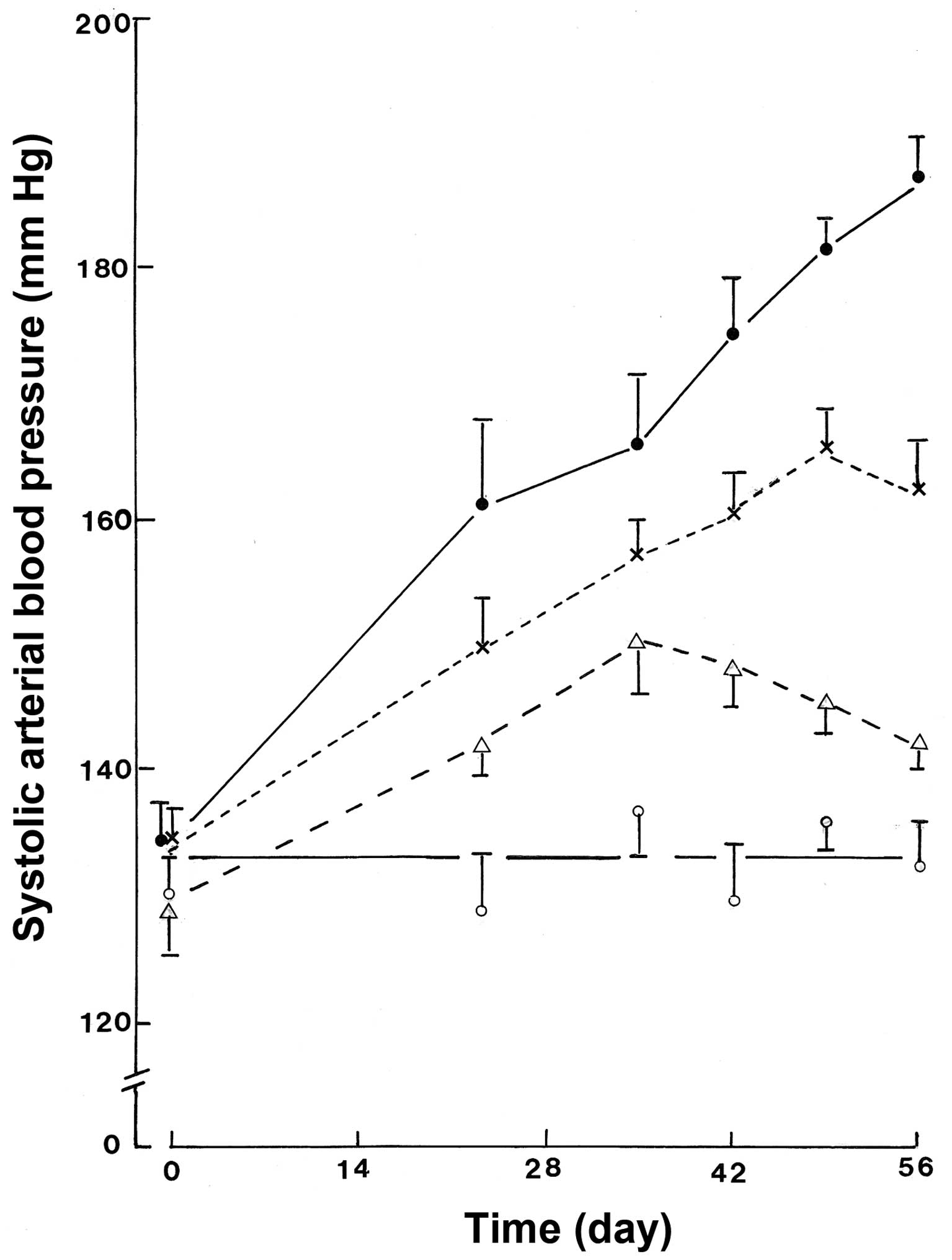
Systolic arterial blood pressure
As illustrated in Fig.
1, the systolic arterial blood pressure remained fairly stable
in the Ssun rats, there being no significant correlation between
the mean measurements above basal value and the length of the
experimental period (r=+0.435; n=6; P>0.1). In the Fsun rats,
however, the blood pressure progressively increased during the
experimental period (r=+0.989; n=6; P<0.001). This was also the
case in the Fsal rats (r=+0.971; n=6; P<0.002). However,
covariance analysis indicated that the slope of the regression line
was significantly lower (F=15.56; f=1, 8; P<0.005) in the Fsal
rats (0.537) than in the Fsun rats (0.919). The increase in blood
pressure was even less pronounced in the Fsaf rats. Indeed, in the
latter rats, the coefficient of correlation between the mean blood
pressure above basal value and the length of the experimental
period did not achieve statistical significance (r=+0.722; n=6;
P<0.11). Moreover, the slope of the regression line was not
significantly different (F=1.54; f=1, 8; P<0.25) in the Fsaf
(0.271) and Ssun rats (0.080). Only the elevation of the regression
line remained significantly higher in the Fsaf rats than in the
Ssun rats (F=16.42; f=1, 9; P<0.005).
The incremental area under the curve, expressed as
mmHg.day, provided comparable information, averaging 89.6±2.7,
1617.0±41.7, 997.2±23.5 and 756.4±16.2 in the Ssun, Fsun, Fsal and
Fsaf rats, respectively (n=6 in each case), as illustrated in
Fig. 2A. The four mean values were
all significantly different from one another (P<0.001).
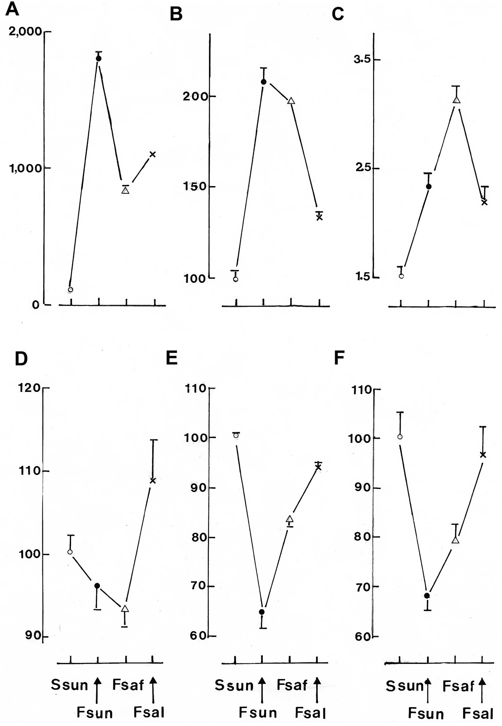 | Figure 2Comparison between Ssun rats (○), Fsun
rats (●), Fsaf rats (△) and Fsal rats (x), in terms of systolic
arterial blood pressure, plasma leptin concentration, kidney
proliferating cell nuclear antigen index, glutathione reductase
activity, superoxide dismutase activity and catalase activity. (A)
The incremental area in blood pressure over the experimental period
expressed as mmHg.day; mean values (± SEM) of 5–6 individual
observations. (B) The plasma leptin concentration expressed as a
percentage of the mean value in Ssun rats; mean value (± SEM) of
5–6 individual measurements. (C) The mean values (± SEM) for the
kidney proliferating cell nuclear antigen index, expressed as
percentages, of 100–120 separate determinations. (D) The activity
of glutathione reductase in liver, heart, kidney and soleus muscle
expressed as percentages of the mean corresponding values in Ssun
rats; mean values (± SEM) of 20–24 individual measurements. The
activity of superoxide dismutase (E) and catalase (F) in liver,
heart, kidney, soleus muscle and visceral adipose tissue expressed
as percentages of the mean corresponding values in Ssun rats; mean
values (± SEM) of 25–30 individual measurements. |
Plasma leptin concentration
The plasma leptin concentration in the Fsun rats
(12.85±0.44 ng/ml; n=6) was double that (P<0.001) in the Ssun
rats (6.18±0.29 ng/ml; n=6). It did not differ significantly
(P>0.18) between the Fsun and Fsaf rats (12.20±0.07 ng/ml; n=6).
In the Fsal rats, however, it averaged 8.24±0.20 ng/ml (n=5) and
was much lower (P<0.001) than that in the Fsun rats and somewhat
higher (P<0.001) than that in the Ssun rats (Fig. 2B).
Enzymatic activities
The results of the two separate assays of
glutathione reductase activity performed in this study yielded
comparable results. In the heart, which yielded one of the lowest
mean values, the coefficient of correlation between the individual
measurements made in these two separate assays was 0.880 (n=23;
P<0.001). In general, only minor differences of glutathione
reductase activity in the liver, heart, kidney, soleus muscle and
visceral adipose tissue homogenates were observed among the four
groups of rats (Table I).
Nevertheless, the trend was, in most cases, towards lower values in
the Fsun and Fsaf rats than in the Ssun or Fsal rats. Indeed, when
ignoring the measurements made in adipose tissue, which yielded the
mean lowest values among the five sampled organs in each group of
rats, the activity of glutathione reductase found in the liver,
heart, kidney and soleus muscle, expressed relative to the mean
reference value found in the same organ in Fsun rats, averaged in
Fsun and Fsal rats 104.0±2.7% (n=44), as distinct (P<0.005) from
only 94.6±1.8% (n=45) in the Fsun and Fsaf rats.
 | Table IGlutathione reductase activity. |
Table I
Glutathione reductase activity.
| Ssun | Fsun | Fsal | Fsaf |
|---|
| Liver | 8.39±0.16a | 8.01±0.14a | 9.68±0.60b | 8.42±0.20a |
| Heart | 1.96±0.10a | 2.00±0.09a | 2.26±0.36b | 1.60±0.03a |
| Kidney | 13.44±0.48a | 12.25±0.22a | 14.16±0.69b | 13.32±0.32a |
| Soleus muscle | 1.81±0.14a | 1.73±0.27c | 1.78±0.09b | 1.65±0.10b |
| Visceral adipose
tissue | 0.75±0.03a | 0.83±0.12a | 0.70±0.06b | 0.89±0.14a |
The activity of superoxide dismutase was, in all
five organs, significantly lower in the Fsun rats than in the Ssun
rats, averaging in the Fsun rats 64.4±3.0% (n=30) of the mean
corresponding reference values in the Ssun rats (100.0±0.8%; n=30).
In the fructose-fed rats, however, it increased (P<0.001) to
83.8±1.8% (n=30) of the same reference value upon partial
substitution of sunflower oil by safflower oil, and further
increased (P<0.001) to 93.7±1.5% (n=25) upon partial
substitution of sunflower oil by salmon oil. The latter percentage
was somewhat lower (P<0.001) than the reference value recorded
in the Ssun rats (Table II).
| Table IISuperoxide dismutase activity. |
Table II
Superoxide dismutase activity.
| Ssun (n=6) | Fsun (n=6) | Fsal (n=5) | Fsaf (n=6) |
|---|
| Liver | 264±1 | 131±2 | 281±5 | 225±6 |
| Heart | 84±1 | 75±1 | 79±1 | 77±1 |
| Kidney | 146±6 | 112±3 | 135±2 | 133±2 |
| Soleus muscle | 245±1 | 146±1 | 216±1 | 203±1 |
| Visceral adipose
tissue | 265±2 | 124±1 | 230±2 | 179±1 |
The activity of catalase in the liver, heart,
kidney, soleus muscle and visceral adipose tissue was also much
lower (P<0.001) in the Fsun rats than in the Ssun rats, the
former averaging 68.0±2.6% (n=30) of the mean corresponding
reference values found in the latter (100.0±5.4%; n=30). As
indicated in Table III, in the
fructose-fed rats catalase activity was increased (P<0.025) to
78.8±3.8% (n=30) upon partial substitution of sunflower oil by
safflower oil and to 96.5±6.0% (n=25) of the mean corresponding
reference values found in Ssun rats upon partial substitution of
sunflower oil by salmon oil. The latter percentage was not
significantly different (P>0.07) from that in the Ssun rats.
| Table IIICatalase activity. |
Table III
Catalase activity.
| Ssun (n=6) | Fsun (n=6) | Fsal (n=5) | Fsaf (n=6) |
|---|
| Liver | 113±15 | 82±9 | 116±13 | 88±2 |
| Heart | 118±14 | 82±5 | 115±12 | 104±8 |
| Kidney | 194±23 | 130±10 | 217±40 | 162±23 |
| Soleus muscle | 115±15 | 80±8 | 98±16 | 83±10 |
| Visceral adipose
tissue | 63±9 | 39±4 | 54±6 | 46±5 |
The correlations between the diet-induced changes
and the glutathione reductase, superoxide dismutase and catalase
activity in the organs under consideration are illustrated in the
lower panels of Fig. 2.
Significant or close-to-significant correlations
were observed between the individual values of superoxide dismutase
and catalase activity in liver (r=+0.504; n=23; P<0.02), heart
(r=+0.493; n=23; P<0.03), kidney (r=+0.474; n=23; P<0.03),
soleus muscle (r=+0.386; n=23; P<0.07) and visceral adipose
tissue (r=+0.558; n=23; P<0.008). Moreover, when the individual
measurements of superoxide dismutase and catalase activity recorded
in each group of rats (Ssun, Fsun, Fsal and Fsaf) and in each organ
(liver, heart, kidney, soleus muscle and visceral adipose tissue)
were expressed relative to the corresponding mean values in order
to eliminate any group effect, a close-to-significant positive
correlation was still observed (r=+0.181; n=115; P<0.06),
indicating parallel changes in these two enzymatic activities at
the individual level. Furthermore, such a correlation achieved
statistical significance (r=+0.236; n=92; P<0.03) when the
results recorded in visceral adipose tissue were excluded. An even
higher significance (r=+0.311; n=69; P<0.01) was achieved when
considering the data collected in the liver, heart and kidney.
Statistical significance (r=+0.416; n=23; P<0.001) was achieved
when only the measurements made in the liver were considered.
Kidney proliferating cell nuclear antigen
index
A total of 20 kidney fields were examined in each
rat. The total number of nuclei examined in each of these fields
ranged between 123 in a Fsal rat and 389 in a Ssun rat. Even so,
the mean number of nuclei under consideration in the twenty fields
did not differ significantly between these two animals (P>0.36),
averaging 283±13 (n=20) in the Fsal rat and 300±13 (n=20) in the
Ssun rat.
In the Ssun rats, the kidney proliferating cell
nuclear antigen index (expressed as a percentage) ranged between
1.35±0.14 (n=20) and 1.87±0.22 (n=20); the difference between the
two mean values failing to achieve statistical significance
(P>0.05). Pooling together all available measurements, such an
index averaged 1.53±0.08 (n=120) in the Ssun rats. A similar SEM
was reached when considering only the mean values, each derived
from 20 determinations; in this case the results from the Ssun rats
averaged 1.53±0.07 (n=6).
In the Fsun rats, only one animal yielded a mean
index within the range of individual values recorded in the Ssun
rats (1.31±0.14; n=20). All other Fsun rats yielded an index
>2.00. The difference between the Ssun and Fsun rats achieved
statistical significance when judged from the mean individual
values recorded in each rat (t=2.90; df=10; P<0.02) or from all
120 measurements (t=6.06; df=238; P<0.001).
The situation found in the Fsal rats was comparable
to that in the Fsun rats (Table
IV). Thus, two Fsal rats yielded mean indices of 1.68±0.21 and
1.69±0.20 which were within the range of values found in the Ssun
rats, whilst the highest individual values in Fsal (3.22±0.49;
n=20) and Fsun rats (3.24±0.25; n=20) were almost identical. The
overall mean value derived from the 20 measurements made in each
animal also failed to differ significantly (P>0.38) in Fsal
(2.20±0.14; n=100) and Fsun rats (2.35±0.11; n=120). Moreover, the
values in the Fsal rats exceeded those in the Ssun rats
(P<0.04), even when the difference was judged from the mean
individual values obtained in the Fsal (2.20±0.28; n=5) and Ssun
(1.53±0.07; n=6) rats.
| Table IVKidney proliferating cell nuclear
antigen index (%). |
Table IV
Kidney proliferating cell nuclear
antigen index (%).
| Ssun | Fsun | Fsal | Fsaf |
|---|
| 1 | 1.41±0.18a | 2.18±0.27a | 2.32±0.20a | 2.91±0.26a |
| 2 | 1.54±0.15a | 2.82±0.31a | 2.07±0.28a | 3.86±0.20a |
| 3 | 1.53±0.16a | 1.31±0.14a | 3.22±0.49a | 4.05±0.33a |
| 4 | 1.35±0.14a | 3.24±0.25a | 1.68±0.21a | 2.74±0.27a |
| 5 | 1.87±0.22a | 2.03±0.21a | 1.69±0.20a | 2.10±0.19a |
| 6 | 1.44±0.24a | 2.54±0.25a | | 3.18±0.29a |
| Overall mean
value | 1.53±0.08b | 2.35±0.11b | 2.20±0.14c | 3.14±0.12b |
| Mean individual
value | 1.53±0.07d | 2.35±0.27d | 2.20±0.28e | 3.14±0.30d |
Finally, in the Fsaf rats, the index was even higher
than in the Fsun rats, such a difference achieving statistical
significance (P<0.001) when judged from the 120 measurements
made in each of these two groups of rats. Furthermore, in the Fsaf
rats, all mean individual values were >2.00 and the highest
individual value was 4.05±0.33 (n=20).
Discussion
The present results reveal that the fructose-induced
metabolic syndrome involves increases in the systolic arterial
blood pressure, plasma leptin concentration and kidney
proliferating cell nuclear antigen index, and decreases in the
activities of glutathione reductase, superoxide dismutase and
catalase in liver, heart, kidney, soleus muscle and visceral
adipose tissue homogenates. In most of these cases, the partial
substitution of sunflower oil in the diet of the fructose-fed rats
by either safflower or salmon oil provoked a partial or complete
restoration of these functional, hormonal and enzymatic variables
towards the reference values found in control rats exposed to a
diet containing starch instead of D-fructose as carbohydrate. As
recently indicated in an extensive review of the relevant
literature (7) presented as the
background information to the present study, the present findings
reinforce the view that the dietary supply of long-chain
polyunsaturated ω6 fatty acids, especially C18:2 ω6, and long-chain
polyunsaturated ω3 fatty acids may exert favourable effects in
terms of correcting the undesirable features otherwise prevailing
in the fructose-induced and possibly other models of metabolic
syndrome, with long-chain polyunsaturated ω3 fatty acids appearing
to be particularly effective.
Acknowledgements
We are grateful to C. Demesmaeker for secretarial
help.
References
|
1
|
Mellouk Z, Louchami K, Hupkens E, Sener A,
Ait Yahia D and Malaisse WJ: The metabolic syndrome of fructose-fed
rats: Effects of long-chain polyunsaturated ω3 and ω6 fatty acids V
Post-mortem findings. Mol Med Reports. (In Press).
|
|
2
|
Mellouk Z, Hachimi Idrissi T, Louchami K,
Hupkens E, Malaisse WJ, Ait Yahia D and Sener A: The metabolic
syndrome of fructose-fed rats: effects of long-chain
polyunsaturated ω3 and ω6 fatty acids. I Intraperitoneal glucose
tolerance test. Int J Mol Med. 28:1087–092. 2011.
|
|
3
|
Belkacemi L, Selselet-Attou G, Giroix MH,
Nortier J, Nguidjoe E, Hupkens E, Sener A and Malaisse WJ:
Intermittent fasting modulation of the diabetic syndrome in
streptozotocin-injected rats: post-mortem investigations. Submitted
for publication.
|
|
4
|
Goldberg DM and Spooner RJ: Glutathione
reductase. Methods of Enzymatic Analysis. Bergmeyer HB: 3. 3rd
edition. Verlag Chemie; Weinheim: pp. 258–265. 1992
|
|
5
|
Elstner EF, Youngman RJ and Obwald W:
Superoxide dismutase. Methods of Enzymatic Analysis. Bergmeyer HB:
3. 3rd edition. Verlag Chemie; Weinheim: pp. 293–302. 1983
|
|
6
|
Aebi H: Catalase. Methods of enzymatic
analysis. Bergmeyer HU: 3. 2nd edition. Verlag Chemie; Weinheim:
pp. 673–684. 1974, View Article : Google Scholar
|
|
7
|
Boukortt FO, Madrani Z, Mellouk Z,
Louchami K, Sener A and Ait Yahia D: Nutritional factors and
fructose-induced metabolic syndrome. Metab Funct Res Diab. 4:18–34.
2011.
|