Introduction
Substantial evidence implicates the Epstein-Barr
virus (EBV) in the pathogenesis of Hodgkin’s lymphoma (HL)
(1–3). EBV detection in HL may be used to
risk-stratify patients and derive optimum treatment strategies.
Investigation into the presence of EBV nucleic acids in affected
tissues in EBV-associated diseases is performed by a variety of
different techniques, including spot hybridization, in situ
hybridization (ISH) and the polymerase chain reaction (PCR)
(2,3). EBV-related proteins, including EBV
nuclear antigen 1 (EBNA1) and the latent membrane proteins (LMP1,
LMP2a and LMP2b) have also been examined by performing
immunohistochemical assays. As previously stated, the percentage of
EBV-positive cases of HL varied among studies, ranging between 20
and 70% (1), and one of the most
significant causes for this wide range may be the sensitivity of
the method employed. To obtain an accurate percentage for the EBV
infection rate in China, three different EBV detecting methods were
employed to analyse 59 paraffin-embedded tissue samples from
national cases of HL.
Materials and methods
Materials and samples
In total, 59 formalin-fixed and paraffin-embedded
archival blocks, obtained between 1997 and 2009, were retrieved
from the pathology departments of four hospitals: Nanfang Hospital
affiliated to the Nanfang Medical University, Guangzhou General
Hospital of the People’s Liberation Army, Guangzhou Children’s
Hospital and Shaanxi Provincial People’s Hospital. All sections had
previously been diagnosed as positive for HL and were re-identified
by two of our pathologists. The diagnosis of HL was established by
finding H/RS cells within an appropriate background of reactive
cells, according to the criteria of the latest WHO classification
(4) and also based on
morphological (H&E section and immunophenotypic criteria
(expression of CD20, CD43 and CD45RO antigen). The study was
approved by the ethics committee of Shaanxi Provincial People’s
Hospital and written informed consent was obtained from the
patients.
Immunohistochemistry (IHC)
Paraffin sections were stained with MAbs (Dako,
Carpinteria, CA, USA) against CD45RO antigen, CD20 antigen (L26),
CD45RO antigen (UCHL1), CD15 antigen and CD30 antigen using a
standard SP immunohistochemistry kit supplied by Beijing Zhongshan
Biological Company (Beijing, China). LMP-1 was detected using a
commercial cocktail of MAb against LMP1 (CS1–4, Dako), diluted at
1:200. The IHC procedure was performed as described previously
(5). Diaminobenzidine (DAB) was
used as a chromogen. Known EBV-positive HL cases were used as
positive controls. Each case was tested a minimum of two or three
times.
EBV-encoded RNA (EBER)1 ISH
EBER expression was detected using 20 bp of doubled
digoxigenin-labeled (5′ end) oligonucleotide probes (antisense),
5′-ctacagccacacacgtctcc-3′, designed by Primer 5.0 software, as the
EBER1 gene fragment (GeneBank gi|16326314|AB065135.1, human
herpesvirus 4 gene for EBER1 small RNA). The probe was synthesized
and labeled by Takara Biotech Co. (Dalian, China). The ISH
procedure used was described in the protocol of Boster
Biotechnology Co. (Wuhan, China).
Briefly, the paraffin sections from each case were
mounted on APES-treated glass slides, dewaxed and hydrated,
predigested with pepsin (3%) for 5–10 min and hybridized for 14–16
h with a probe concentration of 2 ng/μl. The slides were washed
with 2X SSC, 0.5X SSC and 0.2X SSC for 15 min at 37°C, blocked with
BSA at 37°C for 30 min, treated with biotinylated-rabbit antibodies
against digoxin at 37°C for 60 min and washed with 0.5 M PBS for 5
min four times. SABC was added at 37°C for 20 min and
biotin-peroxidase at 37°C for 20 min, then the sections were washed
with 0.5 M PBS for 5 min four times, dyed with DAB for 10 min and
counter-stained with hematoxylin for 8 min. Two known EBV-positive
cases were routinely used as positive controls. Two slides treated
without the probe were used as negative controls.
PCR techniques
DNA preparation
DNA was extracted from the formalin-fixed
paraffin-embedded tissues. Sections (7 μm) were cut from each block
and deparaffinized by three changes of xylene followed by ethanol
washing. The samples were suspended in 50 μl TE buffer, containing
10 mM Tris-hydrochloric acid (pH 8.0) and 1 mM EDTA (pH 8.0). The
DNA was purified using Qiagen columns commercial kit (QIAamp DNA
Mini kit; Qiagen, Shanghai, China), and when negative amplification
for β-globin (housekeeping gene) was encountered, DNA was
re-extracted.
PCR procedure
The first primer pair was a housekeeping gene
β-globin: (PC04, 5′-caacttcatccacgttcacc-3′; GH20,
5′-gaagagccaaggacaggtac-3′; expected size 267 bp). The second was
designed covering 253 bp of the EBV BamHI-W fragment, based
on the DNA sequences of GenBank (forward, 5′-aatgggcgccattttgt-3′
and reverse, 5′-tccctagaactgacaatt-3′). The PCR mixture contained 2
μl template DNA, 2.0 μl 10X PCR buffer [containing 100 mM Tris-HCl
pH 9.0, 100 mM KCl, 80 mM
(NH4)2SO4 and 0.1% NP40], 2.0 mm
MgCl2, 400 μM dNTP mixture, 10 pmol of each primer and
1.5 units Taq Polymerase (Takara Bio, Inc.) in a final
volume of 20 μl. The PCR procedure consisted of initial incubation
for 5 min at 94°C, 30 cycles of 94°C for 30 sec, then 56°C for 30
sec, 72°C for 30 sec and a final extension at 72°C for 5 min. PCR
products were visualized under short-wavelength ultraviolet light
following ethidium bromide staining of the agarose gels.
Results
Clinical data
There were 59 cases in total, 42 males and 17
females with a gender ratio of 2.5:1. The average age was 24.7
(range, 3–57) years old. The number of adolescent patients with HL
was 27 (45.8%), with 18 males and 9 females. Out of these cases, 30
were the lymphocyte predominance subtype (LR), 18 cases had mixed
cellularity (MC), 8 cases had nodular sclerosis (NS) and 3 cases
had the lymphocyte depletion (LD) subtype. These results are
summarized in Table I.
 | Table IComparison of the EBV-positive rate
observed using different detection methods in 59 cases of HL. |
Table I
Comparison of the EBV-positive rate
observed using different detection methods in 59 cases of HL.
| | Positive cases
(%) |
|---|
| |
|
|---|
| Type of HL | Cases (%) | LMP1 | EBER1 | PCR
(BamHI-W) |
|---|
| LR | 30 (50.8) | 21 (70.0) | 22 (73.3) | 24 (80.0) |
| MC | 18 (30.5) | 12 (66.7) | 13 (72.2) | 13 (72.2) |
| NS | 8 (13.6) | 4 (50.0) | 3 (37.5) | 4 (50.0) |
| LD | 3 (5.1) | 2 (66.7) | 2 (66.7) | 3 (100) |
| Total | 59 (100) | 39 (66.1) | 40 (67.8) | 44 (74.6) |
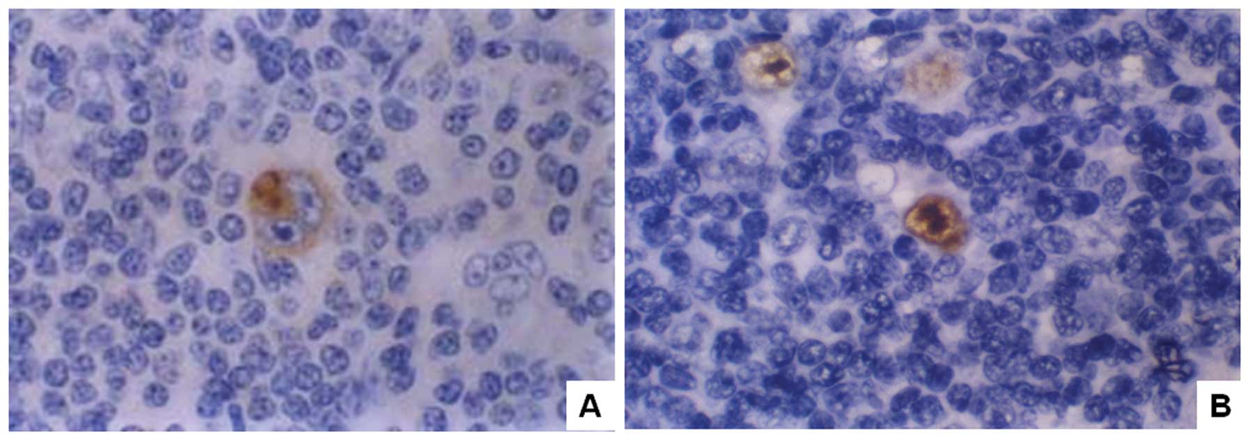
LMP1 and EBER1 expression
The LMP1-positive cases demonstrated staining of the
membrane and plasma of H/RS cells (Fig. 1A). Of the 59 cases, 39 (66.1%) were
shown to be LMP1 positive with the proportions of LR, MC, NS and LD
subtypes revealed as 70.0 (21/30), 66.7 (12/18), 50.0 (4/8) and
66.7% (2/3), respectively. By contrast, the H/RS nuclei were dyed
using EBER1 ISH as described previously (4,5)
(Fig. 1B). Of the 59 cases, 40
(67.8%) were revealed to be EBER1-positive using EBER1 ISH
detection (Table I). Notably,
among the 18 LMP1-positive cases, 3 weakly LMP1-positive cases
(cases 31, 33 and 56) could not be stained in the repeated EBER1
ISH attempts, while another 4 EBER1-positive cases (cases 2, 13, 15
and 43) were LMP1-negative (Table
II). Significantly, we found that 26/27 (96.3%) cases of young
patients (<18 years old) were LMP1- and EBER1-positive, while,
by contrast, only 13/32 (40.6%) adult patients were positive for
LMP1 and EBER1 (Table III).
| Table IISummaries of clinicopathological
findings and EBV results. |
Table II
Summaries of clinicopathological
findings and EBV results.
| Cases | Age/gender | Histological
type | LMP1 | EBER1 | PCR |
|---|
| 2 | 57/F | MC | − | + | + |
| 8 | 36/M | NS | − | − | + |
| 11 | 19/M | LR | − | − | + |
| 13 | 9/M | MC | − | + | + |
| 15 | 49/M | LR | − | + | + |
| 31 | 28/F | NS | + | − | + |
| 33 | 21/M | LR | + | − | + |
| 43 | 34/M | LR | − | + | + |
| 56 | 9/F | LD | + | − | + |
| Table IIIComparison of EBER1 and LMP1 between
the young and adult HL patients. |
Table III
Comparison of EBER1 and LMP1 between
the young and adult HL patients.
| Age (years) | LMP1 (%) | EBER1 (%) | Total |
|---|
| <18 | 26/27 (96.3)a | 26/27 (96.3)a | 27 |
| >18 | 13/32 (40.6)b | 14/32 (43.8)b | 32 |
| Total | 39/59 (66.1) | 40/59 (67.8) | 59 |
EBV gene expression
Using PCR, 44 of 59 (74.6%) cases were identified as
positive for EBV BamHI-W fragment amplification, including
24/30 cases of LR, 13/18 cases of MC, 4/8 cases of NS subtype and
3/3 cases of the LD subtype (Table
I and Fig. 2). Seven cases
(cases 2, 13, 15, 31, 33, 43 and 56) which were identified as
either LMP1- or EBER1-negative, were all recognized as positive
using the PCR detection method (Table
II).
 | Figure 2Results of PCR for 253 bp of the EBV
BamHI-W fragment. M, marker; lane 1, positive control; lane
2, negative control; lanes 3–8, samples; lanes 3, 4, 5 and 8,
positive; lanes 6 and 7, negative. PCR, polymerase chain reaction;
EBV, Epstein-Barr virus. |
Discussion
Previous studies have shown that EBV examination may
aid in making a correct diagnosis, developing treatment and finding
an exact prognosis for EBV-associated diseases (3), thus efficient EBV detection is
extremely important. In order to compare the detection rates of the
EBV identification methods, three techniques, ISH for EBV early
RNA1 (EBER1) sequences, IHC for LMP1 and PCR for EBV BamHI-W
fragments, were used to detect the EBV status of 59 HL cases from
China using paraffin-embedded tissues.
The results revealed that >66% of cases were
identified as EBV-positive, which is a much higher percentage than
that produced by Huang et al from Northern China (39%, EBER
ISH method) (6) and Fatima et
al from Pakistan (60%, EBV-LMP1 method) (4). The reason for this high incidence may
mostly be as our cases include more young patients (27 out of 59
cases). Out of our 27 young cases, 26 (96.3%) exhibited positive
EBV results using either the LMP1 or EBER detection methods. It has
been reported that the frequency of EBV-positive cases in children
is extremely high (between 83 and 100%) in developing countries
(7). For example, there were 96.6%
EBV-positive cases reported in Indian children and 90.3% in
Brazilian children (8,9), which we will examine later in this
discussion.
Out of the three EBV detection methods employed, we
found that the PCR method yields a higher EBV-positive detection
rate (74.6%, 44/59) than that of the LMP1 (66.1%, 39/59) or EBER1
(67.8%, 40/59) methods (Table I).
We also found two cases that were negative for LMP1 and EBER but
that were positive using PCR detection (Table II). The reason for this may be
that the target DNA is able to be amplified by thousands of times
by the PCR procedure, thus the PCR method may have a higher
sensitivity than the other two methods. However, the PCR method was
unable to provide definite information concerning the cellular
localization of the EBV-positive cells.
As for the other two methods, it appeared that there
was no significant difference between the LMP1 (66.1%, 39/59) and
EBER ISH (67.8%, 40/59) techniques. Notably, we found that three
samples that were LMP1-positive proved to be EBER1-negative, while
another four EBER1-positive cases were LMP1-negative (Table II). Repeated experiments revealed
the same results. All these cases appeared EBV-positive when using
the PCR detection method. Although these two methods were less
sensitive than the PCR method, they provided more information about
the localization of EBV-positive cells. We therefore recommend that
at least two methods (PCR and either the LMP1 or EBER methods) be
performed simultaneously to obtain the most accurate results for
EBV infection detection.
In our experiment, we found that the majority of
EBER1 and LMP1 expression occurs in an all-or-nothing manner in
H/RS cells, but that in certain cases only a small section of the
focal H/RS cells were EBV-positive. One interpretation of this may
be that some cells are destroyed during the sample preparation
process (10–13). EBER is RNA that is preserved in
paraffin-embedded tissue, which is easy to destroy during tissue
preparation. To avoid false-negative rates, we recommend that
several factors be considered prior to deciding that LMP1 or EBER
expression is negative. Positive and negative controls should be
performed during the experiments and all slide fields should be
scanned in the diagnosis. For focal H/RS cells to be deemed
EBV-positive, EBER and LMP1 detection methods should be
simultaneously performed if possible.
EBV association in HL also depends on age, subtype
of HL, location and other characteristics of the study population
(3). Notably, almost all of our
young cases (26/27, 96.3%) were LMP1- and EBER1-positive, which is
a much higher percentage than that of African (68%) (14), Brazilian (77%) or Mexican (65%)
(15) cases and it is similar to
results of Honduran (100%) (16)
and Peruvian (100%) (17) cases.
Our results support the view that an association of EBV with
childhood HL may vary as a function of histological subtype and
socio-economic status (18,19).
Concerning the HL subtype, most investigators regard the MC subtype
as most frequently EBV-associated (70%), followed by LR (50%), NS
(20%) and LD (<5%) subtypes (3,19).
Our results are slightly different, LR was observed to be the most
frequent (73.3%), followed by MC (72.2%), LD (66.7%) and NS (37.5%)
according to the results of our EBER detection. This difference may
be attributed to the choice of HL cases.
In conclusion, in the present study, we compared
three EBV detection methods in 59 cases of HL. The results
demonstrated that the PCR method is the most sensitive of the
three, but that it is unable to provide definite information with
regard to cellular localization of the EBV-positive cells, while
the LMP and EBER methods provided such information. We recommend
that at least two methods be performed simultaneously to obtain the
most accurate results for EBV infection.
Acknowledgements
Our thanks to Dr Ben-Chen Zhou. from the General
Hospital of PLA and Dr Wang from Guangzhou Children’s Hospital for
their generosity with the HL samples.
References
|
1
|
Flavell KJ and Murray PG: Hodgkin’s
disease and the Epstein-Barr virus. Mol Pathol. 53:262–269.
2000.
|
|
2
|
Ambinder RF: Epstein-barr virus and
Hodgkin lymphoma. Hematology Am Soc Hematol Educ Program.
2007:204–209. 2007. View Article : Google Scholar : PubMed/NCBI
|
|
3
|
Gulley ML, Glaser SL, Craig FE, et al:
Guidelines for interpreting EBER in situ hybridization and LMP1
immunohistochemical tests for detecting Epstein-Barr virus in
Hodgkin lymphoma. Am J Clin Pathol. 117:259–267. 2002. View Article : Google Scholar : PubMed/NCBI
|
|
4
|
Fatima S, Ahmed R and Ahmed A: Hodgkin
lymphoma in Pakistan: an analysis of subtypes and their correlation
with Epstein Barr virus. Asian Pac J Cancer Prev. 12:1385–1388.
2011.PubMed/NCBI
|
|
5
|
Qi ZL, Zhao T, Zhou XH, Zhang JH, Han XQ
and Zhu MG: Expressions of latent membrane protein 1, p53 and bcl-2
proteins and their significance in Hodgkin lymphoma. Di Yi Jun Yi
Da Xue Xue Bao. 23:225–227. 2003.PubMed/NCBI
|
|
6
|
Huang X, Nolte I, Gao Z, et al:
Epidemiology of classical Hodgkin lymphoma and its association with
Epstein Barr virus in Northern China. PLoS One. 6:e211522011.
View Article : Google Scholar : PubMed/NCBI
|
|
7
|
Audouin J, Diebold J, Nathwani B, et al:
Epstein-Barr virus and Hodgkin’s lymphoma in Cairo, Egypt. J
Hematop. 3:11–18. 2010.
|
|
8
|
Dinand V, Dawar R, Arya LS, et al:
Hodgkin’s lymphoma in Indian children: prevalence and significance
of Epstein-Barr virus detection in Hodgkin’s and Reed-Sternberg
cells. Eur J Cancer. 43:161–168. 2007.
|
|
9
|
Barros MH, Hassan R and Niedobitek G:
Disease patterns in pediatric classical Hodgkin lymphoma: a report
from a developing area in Brazil. Hematol Oncol. 29:190–195. 2011.
View Article : Google Scholar : PubMed/NCBI
|
|
10
|
Harris NL: Hodgkin’s disease:
classification and differential diagnosis. Mod Pathol. 12:159–175.
1999.
|
|
11
|
Murray PG, Billingham LJ, Hassan HT, et
al: Effect of Epstein-Barr virus infection on response to
chemotherapy and survival in Hodgkin’s disease. Blood. 94:442–447.
1999.
|
|
12
|
Gulley ML: Molecular diagnosis of
Epstein-Barr virus-related diseases. J Mol Diagn. 3:1–10. 2001.
View Article : Google Scholar : PubMed/NCBI
|
|
13
|
Valente G, Secchiero P, Lusso P, et al:
Human herpesvirus 6 and Epstein-Barr virus in Hodgkin’s disease: a
controlled study by polymerase chain reaction and in situ
hybridization. Am J Pathol. 149:1501–1510. 1996.
|
|
14
|
Engel M, Essop MF, Close P, et al:
Improved prognosis of Epstein-Barr virus associated childhood
Hodgkin’s lymphoma: study of 47 South African cases. J Clin Pathol.
53:182–186. 2000.
|
|
15
|
Zarate-Osorno A, Roman LN, Kingma DW, et
al: Hodgkin’s disease in Mexico: prevalence of Epstein-Barr
sequences and correlation with histologic subtype. Cancer.
75:1360–1366. 1995.
|
|
16
|
Ambinder RF, Browing PJ, Lorenzana I, et
al: Epstein-Barr virus and childhood Hodgkin’s disease in Honduras
and the United States. Blood. 81:462–467. 1993.
|
|
17
|
Chang KL, Albújar PF, Chen YY, et al: High
prevalence of Epstein-Barr virus in the Reed-Sternberg cells of
Hodgkin’s disease occurring in Peru. Blood. 81:496–501. 1993.
|
|
18
|
Khan G, Norton AJ and Slavin G:
Epstein-Barr virus in Hodgkin disease. Relation to age and subtype.
Cancer. 71:3124–3129. 1993. View Article : Google Scholar : PubMed/NCBI
|
|
19
|
Glaser SL, Lin RJ, Stewart SL, et al:
Epstein-Barr virus-associated Hodgkin’s disease: epidemiologic
characteristics in international data. Int J Cancer. 70:375–382.
1997.
|