Introduction
Diabetic nephropathy is the leading cause of end
stage renal disease (1–3). A number of studies have reported that
hyperglycemia activates multiple downstream signaling pathways in
the diabetic kidney leading to extracellular matrix accumulation,
endothelium dysfunction, glomerular hyperfiltration and eventually
induction of glomerular hypertrophy, increased glomerular basement
membrane thickness and interstitial fibrosis (4–5).
Mesangial cell response to pathological stimuli is associated with
the main events of glomerular injury and is important for
resistance to glycoxidative stress through an antioxidant response
(6). A previous study reported
that alterations in glucose associated with diabetes mellitus
induce mesangial cell apoptosis and contribute to diabetic
nephropathy (7). However, the
mechanism by which mesangial cell apoptosis is regulated in
diabetic individuals remains unclear.
The endoplasmic reticulum (ER) is recognized as an
organelle involved in the folding of secretory and membrane
proteins. An additional emerging function of the ER is the
regulation of apoptosis in the cell (8). A number of stimuli, including
ischemia, hypoxia, hyperglycemia, gene mutation and elevated
protein synthesis, are collectively referred to as ER stress and
all potentially cause ER dysfunction (9,10).
ER stress has been reported to be critical to the pathogenesis of a
number of acute and chronic kidney diseases, including renal
ischemia, acute kidney injury and diabetic kidney disease (11). Stimuli that increase the demand on
the ER to synthesize proteins or degrade improperly folded proteins
cause this stress. Several components of the diabetic milieu, i.e.,
high glucose, free fatty acids, albumin, oxidative activity and
inflammation, induce ER stress in numerous tissues (12–14).
However, prolonged activation of ER stress ultimately initiates the
apoptotic pathway. C/EBP homologous protein (CHOP) is an important
protein in ER stress-mediated apoptosis (15). A number of previous studies have
demonstrated that the ER is the major cell death organelle in
cadmium-induced mesangial cell apoptosis (16), glomerular and tubular damage in
kidney disease and renal injury (17,18).
Therefore, we hypothesized that mesangial cells
suffer from high glucose, induction of CHOP-mediated apoptosis in
the kidney and eventually develop diabetic nephropathy. In the
present study, mesangial cells were cultured in a high glucose
medium to determine the association of CHOP-mediated cell apoptosis
in vitro.
Materials and methods
Cell culture
Mesangial cells of the renal gloraerule (HBZY;
cx0310, Wuhan Boster Bio-Engineering Limited Company, Wuhan, China)
were seeded into 24-well cell culture plates in DMEM/F12 with 10%
FBS and 0.05 mM glutamine to observe survival and neurite
outgrowth. All culture reagents were purchased from Gibco-BRL
(Grand Island, NY, USA). Mesangial cells were cultured to day 6 and
then the medium was replaced with a high glucose DMEM/F12 medium
for 24 h. The high glucose group received 100 mM glucose and the
control group received basal 25 mM glucose. The study was approved
by the ethics committee of School of Medicine, Zhejiang University,
Hangzhou, China.
TUNEL staining analysis
TUNEL staining was performed to assess apoptotic
cells in cultured mesangial cells according to the manufacturer’s
instructions. Cells labeled with brown were counted as positive
cells under a microscope.
Immunohistochemistry and
immunofluorescence of CHOP and caspase-3 protein in cultured
mesangial cells
For immunohistochemical analysis, slides of cultured
mesangial cells of the two groups were washed in 0.01 M phosphate
buffered saline (PBS) containing 0.3% Triton X-100 (pH 7.4, PBS-T),
then immersed in 2% normal goat serum in PBS for 2 h at 37°C.
Slides were incubated overnight at 4°C with polyclonal CHOP and
caspase-3 antibodies (both 1:100; Santa Cruz Biotechnology, Santa
Cruz, CA, USA) in PBS containing 1% bovine serum albumin. Following
this, slides were washed in PBS (3×5 min), incubated with
biotinylated goat-anti-rabbit IgG (1:200; Wuhan Boster
Bio-Engineering Limited Company) in PBS for 2 h at room
temperature, washed in PBS-T (3×5 min) and incubated in
avidin-biotin-peroxidase complex solution (1:100; Wuhan Boster
Bio-Engineering Limited Company) for 2 h at room temperature.
Slides were then rinsed again in PBS-T (3×5 min). Immunolabelling
was visualized with 0.05% diaminobenzdine and 0.3%
H2O2 in PBS. Following staining, the sections
were counterstained with hematoxylin and then dehydrated with
ethanol and xylene prior to application of a coverslip with
Permount. Rat immunoglobulin IgG (1:200; Biomeda Corporation,
Foster City, CA, USA) was used instead of primary antibody as a
negative control. Immunofluorescent microscopy was performed using
the Zeiss LSM-510 confocal microscope (63X/1.2 W; Carl Zeiss
Microscopy, LLC, Thornwood, NY, USA).
Western blot analysis of CHOP and
caspase-3 protein in cultured mesangial cells
Total protein samples were obtained from cultured
cells and the concentration of protein was determined by the Lowry
method using a Bio-Rad DC protein assay kit (Hercules, CA, USA).
Cell lysates containing equal amounts of protein (50 μg) were
resolved by 8–10% SDS-polyacrylamide gel electrophoresis and then
transferred onto Millipore nitrocellulose membranes (Billerica, MA,
USA). Next, the blots were blocked with a solution containing 5%
skimmed milk in Tris-buffered saline with 0.05% Tween-20 (TBST) for
1 h at room temperature and treated with primary antibodies
(polyclonal CHOP and caspase-3 antibodies; 1:2,000; Santa Cruz
Biotechnology) in TBST overnight at 4°C, washed for 1 h with TBST
and further probed with secondary HRP-conjugated anti-rabbit,
anti-mouse or anti-goat IgG (1:2,000) in TBST for 1 h at room
temperature. Immune complexes were visualized using an ECL
detection system according to the manufacturer’s instructions.
Statistical analysis
Sections were observed and analyzed at ×400
magnification with UTHSCSA Image Tools 3.0 (University of Texas
Medical School, San Antonio, TX, USA) and the number and optical
density of the CHOP and caspase-3-positive cells were determined. A
probability of 95% was considered to indicate a statistically
significant difference. Data are presented as mean ± SD.
Results
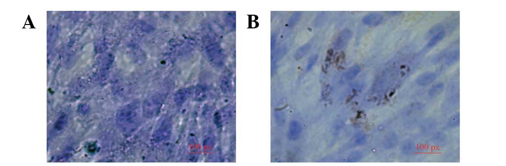
TUNEL staining assay in two groups
TUNEL-positive cells revealed brown staining in the
nucleus. The number of TUNEL-positive cells increased in mesangial
cells in high glucose medium compared with normal (25.2±7.7 vs.
3.3±1.6%; P<0.05; Fig. 1).
Results indicate that mesangial cells are selectively susceptible
to apoptosis under hyperglycemic conditions.
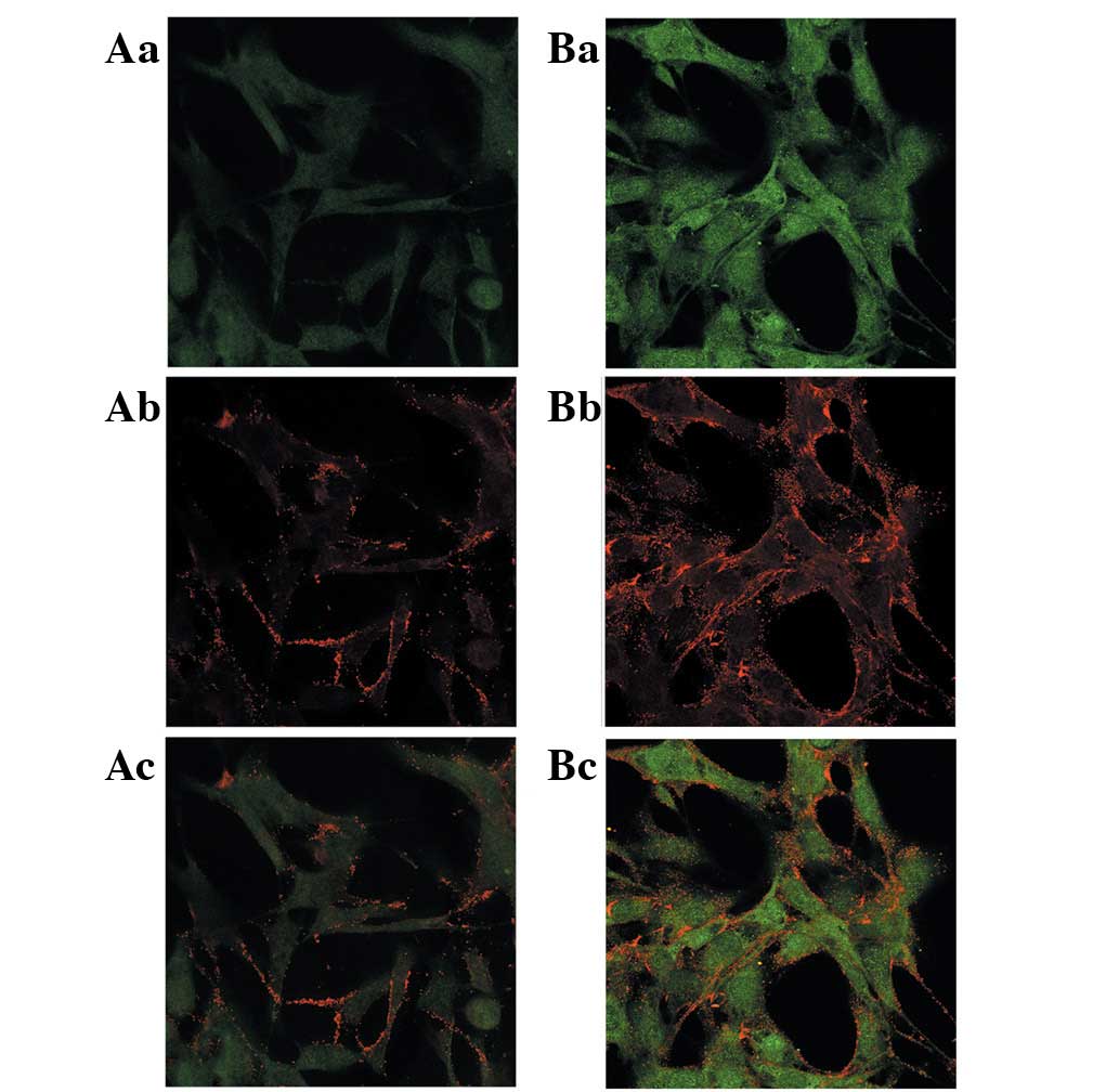
Immunofluorescence
The CHOP positive fluorescence signal was localized
to the nucleus and an upregulation of CHOP staining was observed in
the mesangial cells cultured in high glucose medium while cells
cultured in normal medium were negative for CHOP expression.
Expression of caspase-3 was also increased in high glucose treated
cells (Fig. 2; P<0.05).
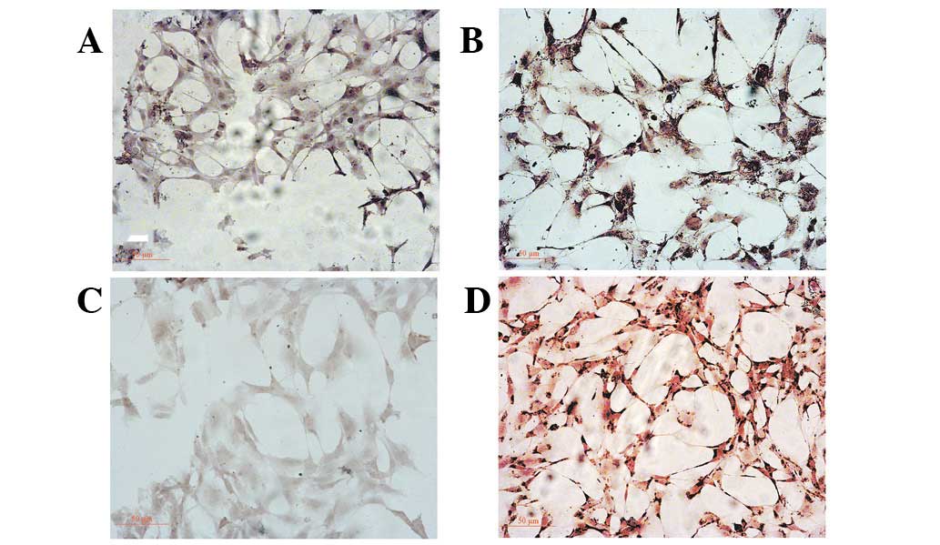
Immunochemistry analysis of CHOP and
caspase-3 expression
Immunochemical analysis revealed that CHOP and
caspase-3 were expressed at high levels in mesangial cells cultured
in high glucose medium while cells in the normal group exhibited
modest or weak immunoreactivity for CHOP and caspase-3 protein
(Fig. 3). The percentage of CHOP-
and caspase-3-positive cells in mesangial cells in high glucose
medium was found to be significantly higher (42.9±14.2 and
58.1±17.6%) than that of the normal group (13.7±3.5 and 6.8±1.1%;
P<0.05)
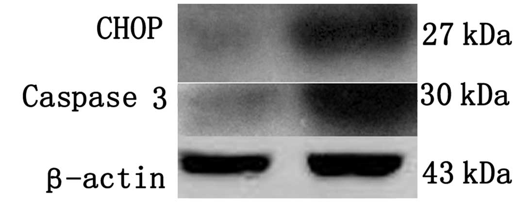
Western blot analysis of CHOP and
caspase-3
Western blot analysis revealed single bands of 27
kDa. Densitometric analysis of bands corresponding to CHOP and
caspase-3 demonstrated a significant increase in relative protein
content in mesangial cells in high glucose medium (375.3±78.4 and
350.2±64.2%) compared with the normal group (Fig. 4; P<0.05).
Discussion
CHOP was previously identified as an ER
stress-induced transcription factor that functions as a significant
mediator of apoptosis in response to ER stress (19). CHOP gene induction is primarily
mediated through the PERK/eIF2α/ATF4 UPR pathway, although
IRE1α/XBP1 and ATF6α pathways also contribute (20,21).
CHOP is a well-known inhibitor of gene transcription and functions
primarily through dimerization with and negative regulation of
other leucine zipper transcription factors of the C/EBP and CREB
families. Overexpression of CHOP leads to growth arrest and
apoptosis (22). Thus, induction
of CHOP indicates that ER-initiated apoptosis is induced (23) and our previous study revealed that
CHOP plays a role in renal injury (18). Caspase-3 is a downstream target of
CHOP and the UPR-independent pathways were demonstrated to
culminate in executioner caspase-3 activation and apoptosis in a
previous study of proteasome inhibitor-mediated apoptosis (24). Caspase-3 is the final caspase of
the apoptotic cascade which activates cytolytic enzymes responsible
for apoptosis (25).
The mesangium occupies a central anatomical position
in the glomerulus, located between the fenestrated endothelial
lining of the capillary lumen and the glomerular basement membrane.
This structure is involved in the pathology of all types of
chronic, progressive kidney diseases (26). The mesangium is composed of
mesangial cells embedded in the mesangial matrix, providing
structural support to capillary loops and modulating glomerular
filtration (27). Mesangial cell
responses to pathological stimuli are associated with the main
events of glomerular injury, leukocyte infiltration, cell
proliferation and NF-κB and MCP-1 expression (26,28).
The present study demonstrated that the number of TUNEL-positive
mesangial cells was significantly increased under high glucose
conditions and CHOP was simultaneously induced. The induction of
CHOP in mesangial cells promoted ER-initiated apoptosis under these
conditions, induced interstitial fibrosis and enhanced progression
of diabetic nephropathy.
In conclusion, the current study demonstrates that
ER stress mediates apoptosis in mesangial cells under
hyperglycemia. The results indicate that ER-initiated apoptosis may
contribute to mesangial cell apoptosis and play a role in the
development of diabetic nephropathy. However, further studies
should be performed to determine the intracellular signaling
pathways involved in ER stress-induced fibrosis in diabetic
nephropathy.
Acknowledgements
The present study was supported by the National
Natural Science Foundation of China (no.30971124).
References
|
1
|
Tokodai K, Amada N, Kikuchi H, Haga I,
Takayama T and Nakamura A: Outcomes of renal transplantation after
end-stage renal disease due to diabetic nephropathy: a
single-center experience. Transplant Proc. 44:77–79. 2012.
View Article : Google Scholar : PubMed/NCBI
|
|
2
|
van den Berg E, Hospers FA, Navis G, et
al: Dietary acid load and rapid progression to end-stage renal
disease of diabetic nephropathy in Westernized South Asian people.
J Nephrol. 24:11–17. 2011.PubMed/NCBI
|
|
3
|
Martínez-Castelao A, De Alvaro F and
Górriz JL: Epidemiology of diabetic nephropathy in Spain. Kidney
Int Suppl. S20–S24. 2005.
|
|
4
|
Samnegard B, Jacobson SH, Jaremko G, et
al: C-peptide prevents glomerular hypertrophy and mesangial matrix
expansion in diabetic rats. Nephrol Dial Transplant. 20:532–538.
2005. View Article : Google Scholar : PubMed/NCBI
|
|
5
|
Brezniceanu ML, Liu F, Wei CC, et al:
Attenuation of interstitial fibrosis and tubular apoptosis in db/db
transgenic mice overexpressing catalase in renal proximal tubular
cells. Diabetes. 57:451–459. 2008. View Article : Google Scholar : PubMed/NCBI
|
|
6
|
Nitti M, Furfaro AL, Patriarca S, et al:
Human mesangial cells resist glycoxidative stress through an
antioxidant response. Int J Mol Med. 27:213–219. 2011.PubMed/NCBI
|
|
7
|
Khera T, Martin J, Riley S, Steadman R and
Phillips AO: Glucose enhances mesangial cell apoptosis. Lab Invest.
86:566–577. 2006.PubMed/NCBI
|
|
8
|
Ferri KF and Kroemer G: Organelle-specific
initiation of cell death pathways. Nat Cell Biol. 3:E255–E263.
2001. View Article : Google Scholar : PubMed/NCBI
|
|
9
|
Ron D: Translational control in the
endoplasmic reticulum stress response. J Clin Invest.
110:1383–1388. 2002. View Article : Google Scholar : PubMed/NCBI
|
|
10
|
Sheikh-Ali M, Sultan S, Alamir AR, Haas MJ
and Mooradian AD: Hyperglycemia-induced endoplasmic reticulum
stress in endothelial cells. Nutrition. 26:1146–1150. 2010.
View Article : Google Scholar : PubMed/NCBI
|
|
11
|
Liu G, Sun Y, Li Z, et al: Apoptosis
induced by endoplasmic reticulum stress involved in diabetic kidney
disease. Biochem Biophys Res Commun. 370:651–656. 2008. View Article : Google Scholar : PubMed/NCBI
|
|
12
|
Kaneto H, Matsuoka TA, Nakatani Y, et al:
Oxidative stress, ER stress and the JNK pathway in type 2 diabetes.
J Mol Med (Berl). 83:429–439. 2005. View Article : Google Scholar : PubMed/NCBI
|
|
13
|
Zhong Y, Wang JJ and Zhang SX:
Intermittent but not constant high glucose induces ER stress and
inflammation in human retinal pericytes. Adv Exp Med Biol.
723:285–292. 2012. View Article : Google Scholar : PubMed/NCBI
|
|
14
|
Lai E, Bikopoulos G, Wheeler MB,
Rozakis-Adcock M and Volchuk A: Differential activation of ER
stress and apoptosis in response to chronically elevated free fatty
acids in pancreatic beta-cells. Am J Physiol Endocrinol Metab.
294:E540–E550. 2008. View Article : Google Scholar : PubMed/NCBI
|
|
15
|
Pino SC, O’Sullivan-Murphy B, Lidstone EA,
et al: CHOP mediates endoplasmic reticulum stress-induced apoptosis
in Gimap5-deficient T cells. PLoS One. 4:e54682009. View Article : Google Scholar : PubMed/NCBI
|
|
16
|
Wang SH, Shih YL, Lee CC, et al: The role
of endoplasmic reticulum in cadmium-induced mesangial cell
apoptosis. Chem Biol Interact. 181:45–51. 2009. View Article : Google Scholar : PubMed/NCBI
|
|
17
|
Inagi R: Endoplasmic reticulum stress in
the kidney as a novel mediator of kidney injury. Nephron Exp
Nephrol. 112:e1–e9. 2009. View Article : Google Scholar : PubMed/NCBI
|
|
18
|
Xu L, Han F, Mandal A, Rao GN and Zhang X:
Diazoxide attenuates hypothermic preservation-induced renal injury
via down-regulation of CHOP and caspase-12. Nephrol Dial
Transplant. 25:3859–3867. 2010. View Article : Google Scholar : PubMed/NCBI
|
|
19
|
Marciniak SJ, Yun CY, Oyadomari S, et al:
CHOP induces death by promoting protein synthesis and oxidation in
the stressed endoplasmic reticulum. Genes Dev. 18:3066–3077. 2004.
View Article : Google Scholar : PubMed/NCBI
|
|
20
|
Suzuki H and Matsuoka M: TDP-43 toxicity
is mediated by the unfolded protein response-unrelated induction of
C/EBP homologous protein expression. J Neurosci Res. 90:641–647.
2012. View Article : Google Scholar : PubMed/NCBI
|
|
21
|
Cox DJ, Strudwick N, Ali AA, Paton AW,
Paton JC and Schroder M: Measuring signaling by the unfolded
protein response. Methods Enzymol. 491:261–292. 2011. View Article : Google Scholar : PubMed/NCBI
|
|
22
|
Jauhiainen A, Thomsen C, Strömbom L, et
al: Distinct cytoplasmic and nuclear functions of the stress
induced protein DDIT3/CHOP/GADD153. PLoS One. 7:e332082012.
View Article : Google Scholar : PubMed/NCBI
|
|
23
|
Myoishi M, Hao H, Minamino T, et al:
Increased endoplasmic reticulum stress in atherosclerotic plaques
associated with acute coronary syndrome. Circulation.
116:1226–1233. 2007. View Article : Google Scholar : PubMed/NCBI
|
|
24
|
Loughlin DT and Artlett CM: Precursor of
advanced glycation end products mediates ER-stress-induced
caspase-3 activation of human dermal fibroblasts through NAD(P)H
oxidase 4. PLoS One. 5:e110932010. View Article : Google Scholar : PubMed/NCBI
|
|
25
|
Porter AG and Janicke RU: Emerging roles
of caspase-3 in apoptosis. Cell Death Differ. 6:99–104. 1999.
View Article : Google Scholar : PubMed/NCBI
|
|
26
|
Gómez-Guerrero C, Hernández-Vargas P,
López-Franco O, Ortiz-Muñoz G and Egido J: Mesangial cells and
glomerular inflammation: from the pathogenesis to novel therapeutic
approaches. Curr Drug Targets Inflamm Allergy. 4:341–351.
2005.PubMed/NCBI
|
|
27
|
Rodriguez-Barbero A, L’Azou B, Cambar J
and López-Novoa JM: Potential use of isolated glomeruli and
cultured mesangial cells as in vitro models to assess
nephrotoxicity. Cell Biol Toxicol. 16:145–153. 2000. View Article : Google Scholar
|
|
28
|
Chen P, Shi Q, Xu X, Wang Y, Chen W and
Wang H: Quercetin suppresses NF-κB and MCP-1 expression in a high
glucose-induced human mesangial cell proliferation model. Int J Mol
Med. 30:119–125. 2012.
|