|
1
|
Rogers HW, Weinstock MA, Harris AR, et al:
Incidence estimate of nonmelanoma skin cancer in the United States,
2006. Arch Dermatol. 146:283–287. 2010. View Article : Google Scholar : PubMed/NCBI
|
|
2
|
Ullrich A, Coussens L, Hayflick JS, et al:
Human epidermal growth factor receptor cDNA sequence and aberrant
expression of the amplified gene in A431 epidermoid carcinoma
cells. Nature. 309:418–425. 1984. View
Article : Google Scholar : PubMed/NCBI
|
|
3
|
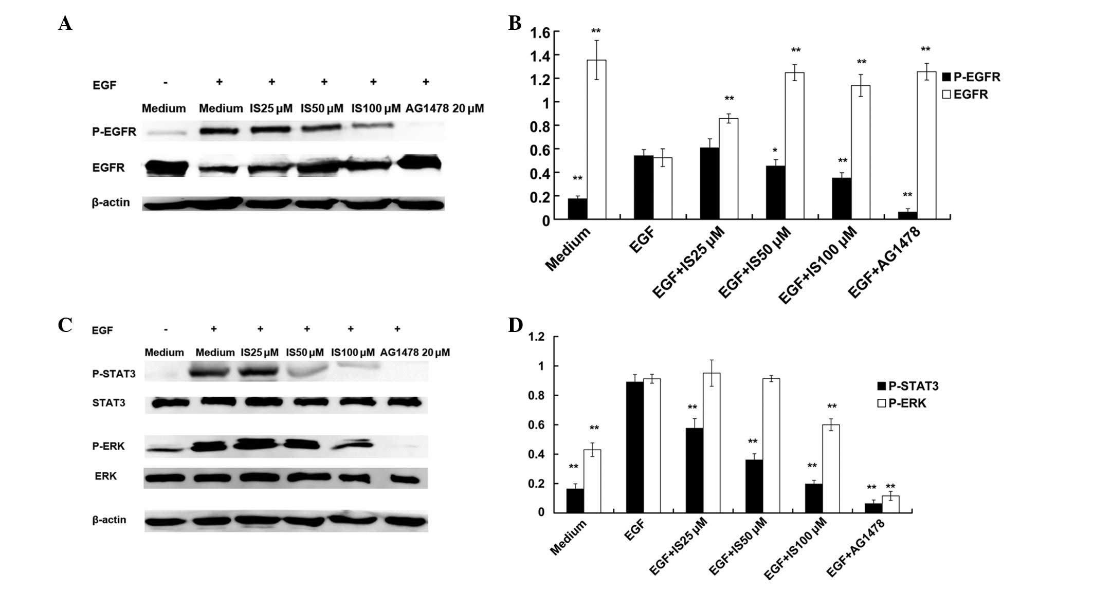
Uribe P and Gonzalez S: Epidermal growth
factor receptor (EGFR) and squamous cell carcinoma of the skin:
molecular bases for EGFR-targeted therapy. Pathol Res Pract.
207:337–342. 2011. View Article : Google Scholar : PubMed/NCBI
|
|
4
|
Lurje G and Lenz HJ: EGFR signaling and
drug discovery. Oncology. 77:400–410. 2009. View Article : Google Scholar : PubMed/NCBI
|
|
5
|
Zhang DW, Cheng Y, Wang NL, et al: Effects
of total flavonoids and flavonol glycosides from Epimedium koreanum
Nakai on the proliferation and differentiation of primary
osteoblasts. Phytomedicine. 15:55–61. 2008. View Article : Google Scholar : PubMed/NCBI
|
|
6
|
Xia Q, Xu D, Huang Z, et al: Preparation
of icariside II from icariin by enzymatic hydrolysis method.
Fitoterapia. 81:437–442. 2010. View Article : Google Scholar : PubMed/NCBI
|
|
7
|
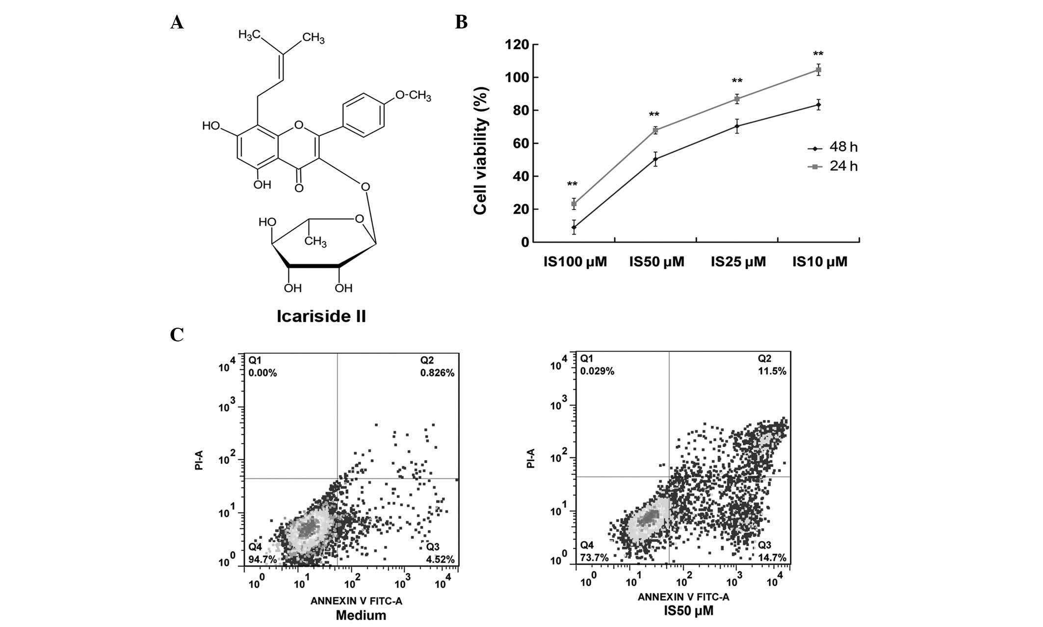
Lee KS, Lee HJ, Ahn KS, et al:
Cyclooxygenase-2/prostaglandin E2 pathway mediates icariside II
induced apoptosis in human PC-3 prostate cancer cells. Cancer Lett.
280:93–100. 2009. View Article : Google Scholar : PubMed/NCBI
|
|
8
|
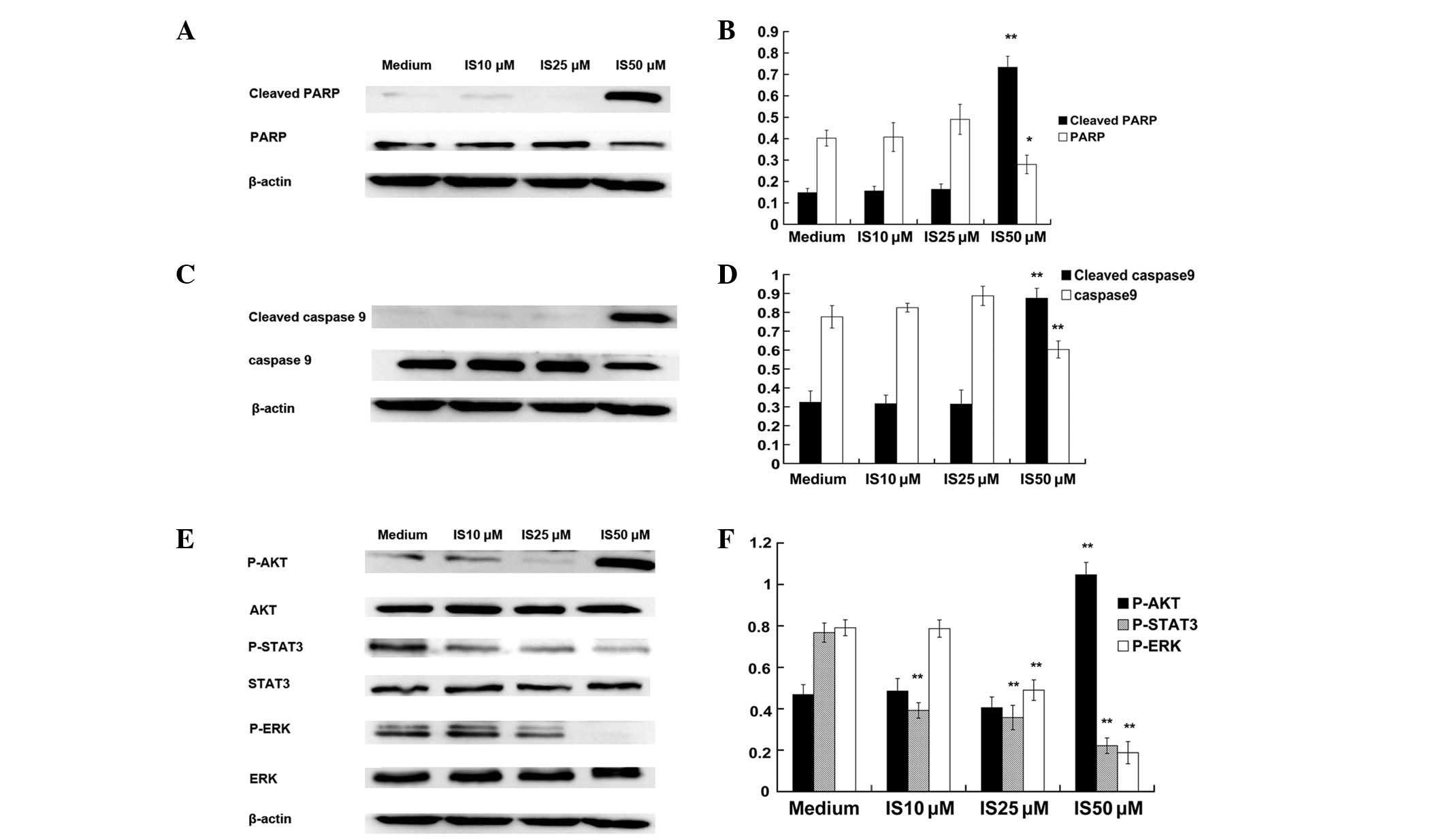
Kim SH, Ahn KS, Jeong SJ, et al: Janus
activated kinase 2/signal transducer and activator of transcription
3 pathway mediates icariside II-induced apoptosis in U266 multiple
myeloma cells. Eur J Pharmacol. 654:10–16. 2011. View Article : Google Scholar : PubMed/NCBI
|
|
9
|
Kang SH, Jeong SJ, Kim SH, et al:
Icariside II induces apoptosis in U937 acute myeloid leukemia
cells: role of inactivation of STAT3-related signaling. PloS One.
7:e287062012. View Article : Google Scholar : PubMed/NCBI
|
|
10
|
Song J, Shu L, Zhang Z, et al: Reactive
oxygen species-mediated mitochondrial pathway is involved in
Baohuoside I-induced apoptosis in human non-small cell lung cancer.
Chem Biol Interact. 199:9–17. 2012. View Article : Google Scholar : PubMed/NCBI
|
|
11
|
Fan TJ, Han LH, Cong RS and Liang J:
Caspase family proteases and apoptosis. Acta Biochim Biophys Sin
(Shanghai). 37:719–727. 2005. View Article : Google Scholar : PubMed/NCBI
|
|
12
|
Ravandi F, Talpaz M and Estrov Z:
Modulation of cellular signaling pathways: prospects for targeted
therapy in hematological malignancies. Clin Cancer Res. 9:535–550.
2003.PubMed/NCBI
|
|
13
|
Chalandon Y and Schwaller J: Targeting
mutated protein tyrosine kinases and their signaling pathways in
hematologic malignancies. Haematologica. 90:949–968.
2005.PubMed/NCBI
|
|
14
|
Nickischer D, Laethem C, Trask OJ Jr, et
al: Development and implementation of three mitogen-activated
protein kinase (MAPK) signaling pathway imaging assays to provide
MAPK module selectivity profiling for kinase inhibitors: MK2-EGFP
translocation, c-Jun, and ERK activation. Methods Enzymol.
414:389–418. 2006. View Article : Google Scholar
|
|
15
|
Davies MA, Stemke-Hale K, Lin E, et al:
Integrated molecular and clinical analysis of AKT activation in
metastatic melanoma. Clin Cancer Res. 15:7538–7546. 2009.
View Article : Google Scholar : PubMed/NCBI
|
|
16
|
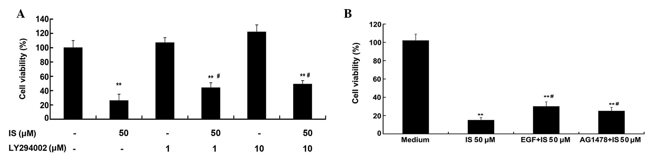
Krasilnikov M, Ivanov VN, Dong J and Ronai
Z: ERK and PI3K negatively regulate STAT-transcriptional activities
in human melanoma cells: implications towards sensitization to
apoptosis. Oncogene. 22:4092–4101. 2003. View Article : Google Scholar
|
|
17
|
Normanno N, Campiglio M, Maiello MR, et
al: Breast cancer cells with acquired resistance to the EGFR
tyrosine kinase inhibitor gefitinib show persistent activation of
MAPK signaling. Breast Cancer Res Treat. 112:25–33. 2008.
View Article : Google Scholar : PubMed/NCBI
|
|
18
|
Shimizu M, Deguchi A, Lim JT, et al:
(−)-Epigallocatechin gallate and polyphenon E inhibit growth and
activation of the epidermal growth factor receptor and human
epidermal growth factor receptor-2 signaling pathways in human
colon cancer cells. Clin Cancer Res. 11:2735–2746. 2005.
|
|
19
|
Zhang X, Ling MT, Feng H, et al: Id-I
stimulates cell proliferation through activation of EGFR in ovarian
cancer cells. Br J Cancer. 91:2042–2047. 2004.PubMed/NCBI
|
|
20
|
Gadgeel SM, Ali S, Philip PA, et al:
Response to dual blockade of epidermal growth factor receptor
(EGFR) and cycloxygenase-2 in nonsmall cell lung cancer may be
dependent on the EGFR mutational status of the tumor. Cancer.
110:2775–2784. 2007. View Article : Google Scholar : PubMed/NCBI
|
|
21
|
Lin YJ and Zhen YS: Rhein lysinate
suppresses the growth of breast cancer cells and potentiates the
inhibitory effect of Taxol in athymic mice. Anticancer Drugs.
20:65–72. 2009. View Article : Google Scholar : PubMed/NCBI
|
|
22
|
Lee DH, Szczepanski MJ and Lee YJ:
Magnolol induces apoptosis via inhibiting the EGFR/PI3K/Akt
signaling pathway in human prostate cancer cells. J Cell Biochem.
106:1113–1122. 2009. View Article : Google Scholar : PubMed/NCBI
|
|
23
|
Nagaprashantha LD, Vatsyayan R, Singhal J,
et al: Anti-cancer effects of novel flavonoid vicenin-2 as a single
agent and in synergistic combination with docetaxel in prostate
cancer. Biochem Pharmacol. 82:1100–1109. 2011. View Article : Google Scholar : PubMed/NCBI
|
|
24
|
Singh F, Gao D, Lebwohl MG and Wei H:
Shikonin modulates cell proliferation by inhibiting epidermal
growth factor receptor signaling in human epidermoid carcinoma
cells. Cancer Lett. 200:115–121. 2003. View Article : Google Scholar : PubMed/NCBI
|