Introduction
Chemotherapy today remains the central therapeutic
approach for the majority of cancer patients, as a treatment that
places emphasis on the quality of life and the preservation of
organ function (1,2). However, the application of
chemotherapy is limited due to numerous obstacles, including
adverse effects and multidrug resistance (MDR), which ultimately
reduces treatment efficacy and are associated with disease
progression (3,4).
MDR is one of the main causes of treatment failure
and high mortality rates in disease, for patients with inherent
resistance to drugs and for those who acquire resistance during
treatment (5–7). In cancer, MDR is a multifactorial
process, defined as a simultaneous resistance to several different
types of commonly used antineoplastic agents, which has a severe
impact on the therapeutic effect of these treatments (8). Therefore, clarifying the mechanisms
and investigating the key elements regulating MDR, is critical for
improving chemotherapy efficacy in the treatment of cancer.
Extensive studies investigating drug resistance and resistance
chemosensitivities have identified numerous genes and proteins
associated with MDR. For example, certain membrane proteins are
important in the development of MDR, notably MDR1 (MDR1/P-gp), MDR
protein (MRP) and breast cancer resistance protein (BCRP), which is
a member of the ATP binding cassette (ABC) transporter family that
encode efflux pumps (9–12). Overexpression of these transporters
has been reported to be correlated with the chemosensitivities of
numerous chemotherapeutic agents (13). Nevertheless, MDR has not been
prevented or effectively controlled in clinical practice. Further
exploration for new regulators of MDR is urgently required, to
improve the efficacy and application of chemotherapy as a cancer
treatment.
Twist1 is a highly conserved transcription
factor which belongs to a basic helix-loop-helix family. Previous
studies confirmed that overexpression of Twist1 was identified in
multiple types of cancer in humans, with numerous damaging
consequences, including promoting the immigration and invasion of
cancer cells, and decreasing sensitivity to chemotherapy (14–16).
Overexpression of Twist1 may be key to tumor drug resistance, but
the precise mechanisms underlying this effect remain elusive. At
present, no studies have investigated the role of Twist1 in
taxol-exerted MDR on FaDu cells, or reported the possible role of
Twist1 on FaDu cell apoptosis sensitivity. In an attempt to
ascertain the role of Twist1 during MDR and clarify its mechanism
of apoptosis sensitivity, a MDR cell line of FaDu cells was
established and the stable transfections targeted to Twist1
overexpression and Twist1 silenced expression in FaDu cells were
conducted. Chemosensitivity was studied in MDR cells and cells with
variable expression levels of Twist1.
Materials and methods
Cells and reagents
The human hypopharyngeal carcinoma cell line FaDu
was obtained from the American Type Culture Collection (ATCC;
Manassas, VA, USA). Media and serum were purchased from Gibco
(Invitrogen Life Technologies, Carlsbad, CA, USA). Anti-Twist1,
activated caspase-3, activated caspase-9, Bcl-2, Bax and β-actin
antibodies were purchased from Santa Cruz Biotechnology (Santa
Cruz, CA, USA). The BCA protein assay kit was a product of Shenergy
Biocolor Bioscience & Technology Company (Shanghai, China). The
RevertAid First Strand cDNA Synthesis kit was obtained from
Fermentas (Burlington, Ontario, Canada). All reagents were
purchased from Sigma (St. Louis, MO, USA).
Cell culture
FaDu/T was developed and determined as previously
described (17). FaDu and FaDu/T
cells were cultured as a monolayer on Dulbecco’s Modified Eagle’s
Medium (DMEM; Gibco) containing 10% fetal calf serum, 100 U/ml
penicillin and 100 mg streptomycin at 37°C in a humidified
atmosphere composed of 95% air and 5% CO2.
Assessment of cell viability and
IC50 determination
Cells (5×10⁴/ml) sub-cultured in a
96-well cell culture cluster (Corning, Tewksbury, MA, USA) were
treated with different concentrations of taxol. MTT (5 mg/ml, 20
μl) was added to each well 4 h prior to the indicated time points.
Following 4 h of incubation at 37°C, the medium was removed and the
precipitate was dissolved in dimethylsulfoxide. Then, the optical
density (OD) values were measured at 570 nm using an ELISA reader
(Multiskan MK3, Shanghai Bio-excellent, Shanghai, China). Relative
cell viability was calculated according to the following formula:
Cell relative viability (%) =
ODexperiment/ODcontrol × 100% (OD blank was
used to zero). The IC50 was defined as the drug
concentration required to decrease the cell viability to 50% of the
control (no drug) value.
Morphological observation for the
apoptosis of cells
FaDu, FaDu/T and FaDu cells treated with taxol (200
nM) for 24 h were seeded (15×104/well) in 24-well dishes
containing 1 ml culture medium to observe the morphological
changes. Acridine orange (AO) staining and Hoechst/PI double
staining were conducted as previously described (17).
Plasmid constructions of pcDNA3.1-Twist1
and generation of microRNA-Twist1
Entire coding cDNA fragments of Twist1 were
amplified by RT-PCR and sub-cloned into the multi-cloning site of
pcDNA3.1 vector (pcDNA3.1-Twist1). The primers for human full
length Twist1 amplification were as follows: Forward (F),
5′-CGAAGCTTGAGAGATGATGCAGGACGTGTC-3′; rev erse (R),
5′-GGAATTCCTAGTGGGACGCGGACATG-3′.
Confirmation of final constructs by DNA
sequencing
MicroRNA-Twist1 was generated using the Block-iT™
PolII miR RNAi expression vector kit with EmGFP (Invitrogen Life
Technologies) according to the manufacturer’s instructions. Four
pairs of oligo sequences targeted to Twist1 silencing were tested
and a scrambled microRNA was used as a control. The sequences
targeting the Twist1 gene-coding region were annealed and
inserted into the pcDNA6.2-GW/EmGFPmiR vector to generate the
microRNA interfering expression vector. We selected one pair of the
sequence in which Twist1 was silenced most effectively and
constructed the stable transfectant with this sequence. Briefly,
the most effective sequence of microRNA-Twist1 and scrambled
control was as follows: Twist1-oligo-F,
5′-TGCTGCTGCCGGTCTGGCTCTTCCTCGTTTTGGCCACTGACTGACGAGGAAGACAGACCGG
CAG-3′, Twist1-oligo-R, 5′-CCTGCTGCCGTCTGTCTTCCTCGTCAGTC
AGTGGCCAAAACGAGGAAGAGCCAGACCGGCAGC-3′; control-F:
5′-tgctgAAATGTACTGCGCGTGGAGACGTTTTGGCCACTGACTGACGTCTCCACGCAGTACATTT-3′,
control-R:
5′-cctgAAATGTACTGCGTGGAGACGTCAGTCAGTGGCCAAAACGTCTCCACGCGCAGTACATTTc-3′.
Generation of stable transfectants
Cell transfection was conducted using Lipofectamine
2000 (Invitrogen Life Technologies) according to the manufacturer’s
instructions. Briefly, cells were grown to 80–90% confluence
without antibiotics. Vectors containing the different constructs
(10 μg) were diluted in opti-MEM (250 μl) and then mixed with the
transfection solution for 20 min at room temperature. Following
washing, cells were incubated with the transfection mixture at 37°C
for 6–8 h and then were allowed to grow in fresh media.
Stable transfectant with pcDNA3.1/Twist1, named as
‘FaDu/Twist1+’, was isolated by selection with 500 mg/ml of G418
(Amresco, Solon, OH, USA) for 2~4 weeks. Cells with pcDNA3.1 vector
were established as a negative control at the same time.
Stable transfectant with microRNA-Twist1, named as
‘FaDu/Twist1-miRNA’, was isolated by selection with blasticidin
(2.5 μg/ml) and GFP under blue excitation. At the same time, cells
with the above scrambled microRNA were built as a control as
described above.
RNA extraction and RT-PCR
Total RNA was extracted using TRIzol (Invitrogen
Life Technologies). Expression of Twist1 mRNA was determined by
RT-PCR with M-MuL V reverse transcription (Takara Bio, Inc., Shiga,
Japan). All operations were conducted under the guidance of the
manufacturer’s instructions. The primers were as follows: Twist1-F,
5′-GGAGTCCGCAGTCTTACGAG-3′; Twist1-R, 5′-TCTGGAGGACCTGGTACAGG-3′;
β-actin-F, 5′-CTCCTTAATGTCACGCACGATTT-3′; β-actin-R,
5′-GTGGGGCGCCCCAGGCACCA-3′.
Protein extraction and western blot
analysis
Protein extraction and western blotting were
conducted as previously described (17). Bands for β-actin, Twist1, cleaved
caspase-3, cleaved caspase-9, Bcl-2 and Bax were visualized at
apparent molecular weights of 43, 170, 28, 17, 37, 26 and 23 kDa,
respectively. The relative OD ratio was calculated with Image J
software (National Institutes of Health, Bethesda, MD, USA) by
comparison to β-actin from three independent experiments.
Intracellular calcium measurements
Cells with variable expression of Twist1 (FaDu,
FaDu/T-200 nM, FaDu/Twist1+ and FaDu/Twist1-miRNA) were
sub-cultured (5×10⁴/ml) in 6-well cell culture clusters
(Corning). Following overnight growth, cells were treated with
taxol (200 nM) for 24 h. Ca2+ concentrations were
examined using Flou-3/AM (5 μM, 37°C for 30 min). Equal PBS was
used as a control. At the end of the incubation, cells were
examined under flow cytometry. The experiment was repeated three
times.
Statistical analysis
Data are presented as the mean ± standard error of
the mean (SEM). Statistical calculations were performed using SPSS
16.0 software package (SPSS, Inc., Chicago, IL, USA). One-way
analysis of variance (ANOVA) was applied to analyze the comparison
of the means greater than or equal to three groups. P<0.05 was
considered to indicate a statistically significant difference.
Results
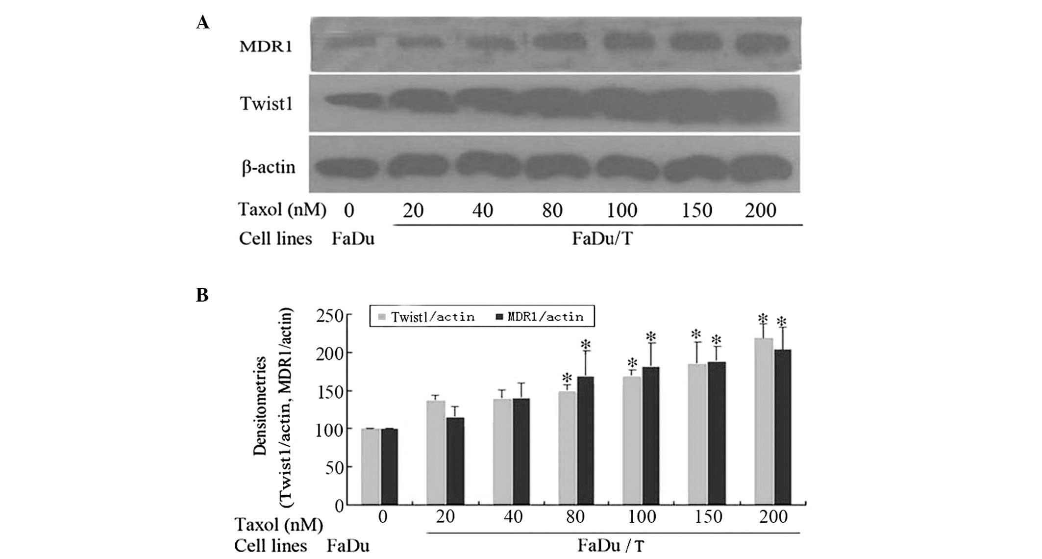
Twist1 and MDR1/P-gp levels increase in a
MDR-dependent manner in FaDu/T cells
Compared with FaDu cells, FaDu/T cells expressed
higher levels of Twist1 and MDR1/P-gp in a MDR-dependent manner. A
significant difference was identified from the FaDu/T cells, whose
endurance to taxol was 80 nM (Fig.
1).
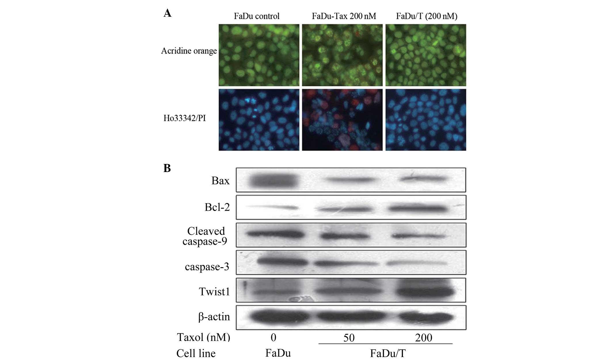
Apoptosis sensitivity in FaDu/T
cells
As illustrated in Fig.
2A, by AO staining, FaDu cells had a polygonal shape, but cells
treated with taxol (200 nM) for 24 h became rounded and exhibited
cytoplasmic contraction and chromatin condensation. Apoptotic
bodies, the main morphological characteristic of apoptosis, were
also present. However, FaDu/T cells had a similar morphology to
FaDu cells, with intact polygonal nuclei.
For Ho.33342/PI double staining, blue intact nuclei
can be observed, as in FaDu cells, red staining was interpreted as
necrosis, while blue nuclear fragmentation was an indication of
apoptosis. Compared with FaDu cells, red nuclei and blue nuclear
fragmentation could be detected in cells treated with taxol (200
nM) for 24 h. However, FaDu/T cells (the endurability to taxol was
200 nM) demonstrated a similar morphology to FaDu cells, with blue
intact nuclei. Our data indicated that FaDu/T cells exhibited
anti-apoptosis activity when stimulated by taxol (Fig. 2A).
Changes in apoptosis-related proteins in
FaDu/T cells
Compared with FaDu cells, FaDu/T cells demonstrated
apoptotic resistance, with cleaved caspase-3, cleaved caspase-9,
Bcl-2 and Bax, all altered to resist apoptosis (Fig. 2B).
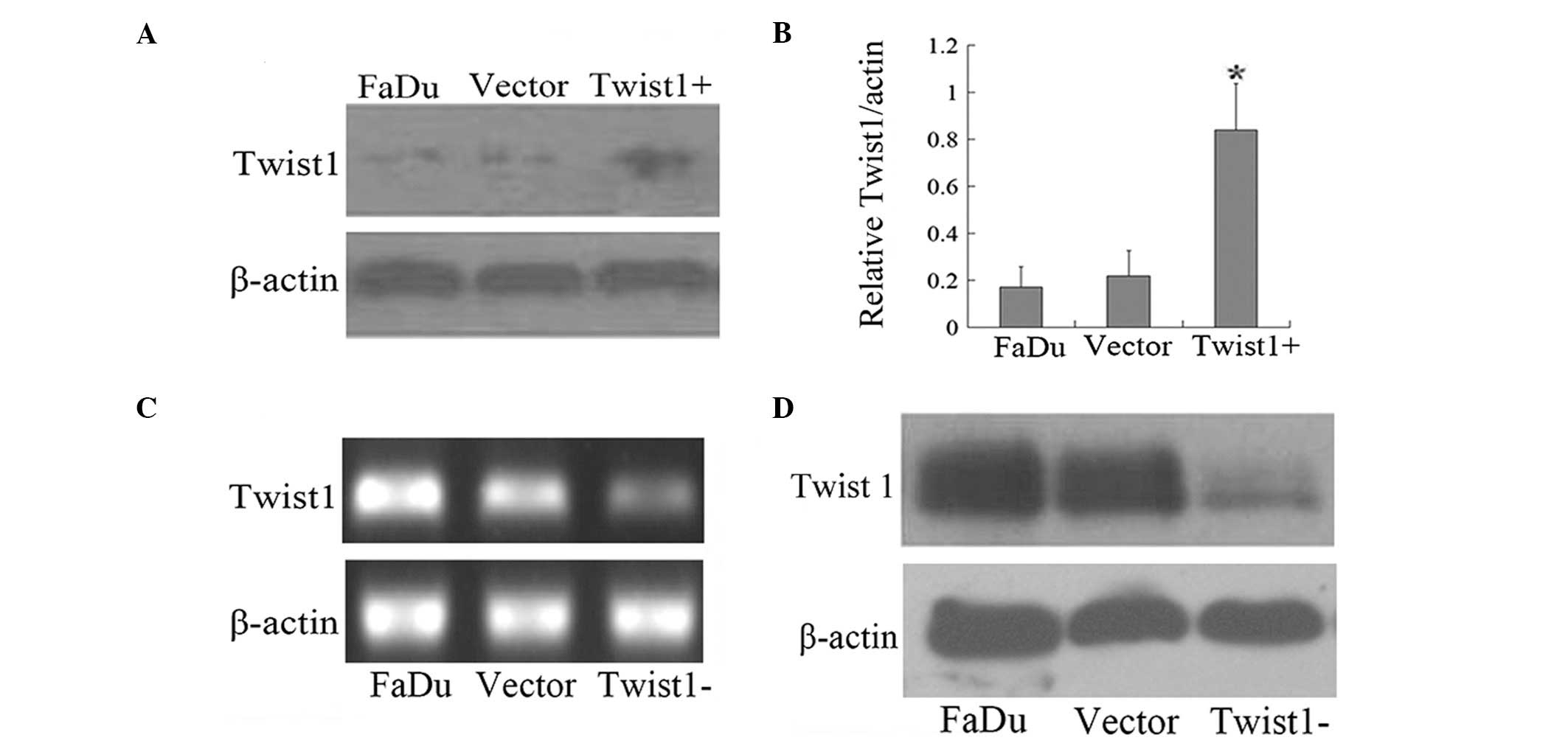
Generation and determination of stable
transfectants of Twist1 in FaDu Cells
As summarized in Fig.
3, FaDu/Twist1+ cells exhibited a higher Twist1 expression
level compared with FaDu cells and negative control cells (Fig. 3A and B; P<0.05). mRNA and
protein levels of Twist1 in FaDu/Twist1-miRNA cells were detected,
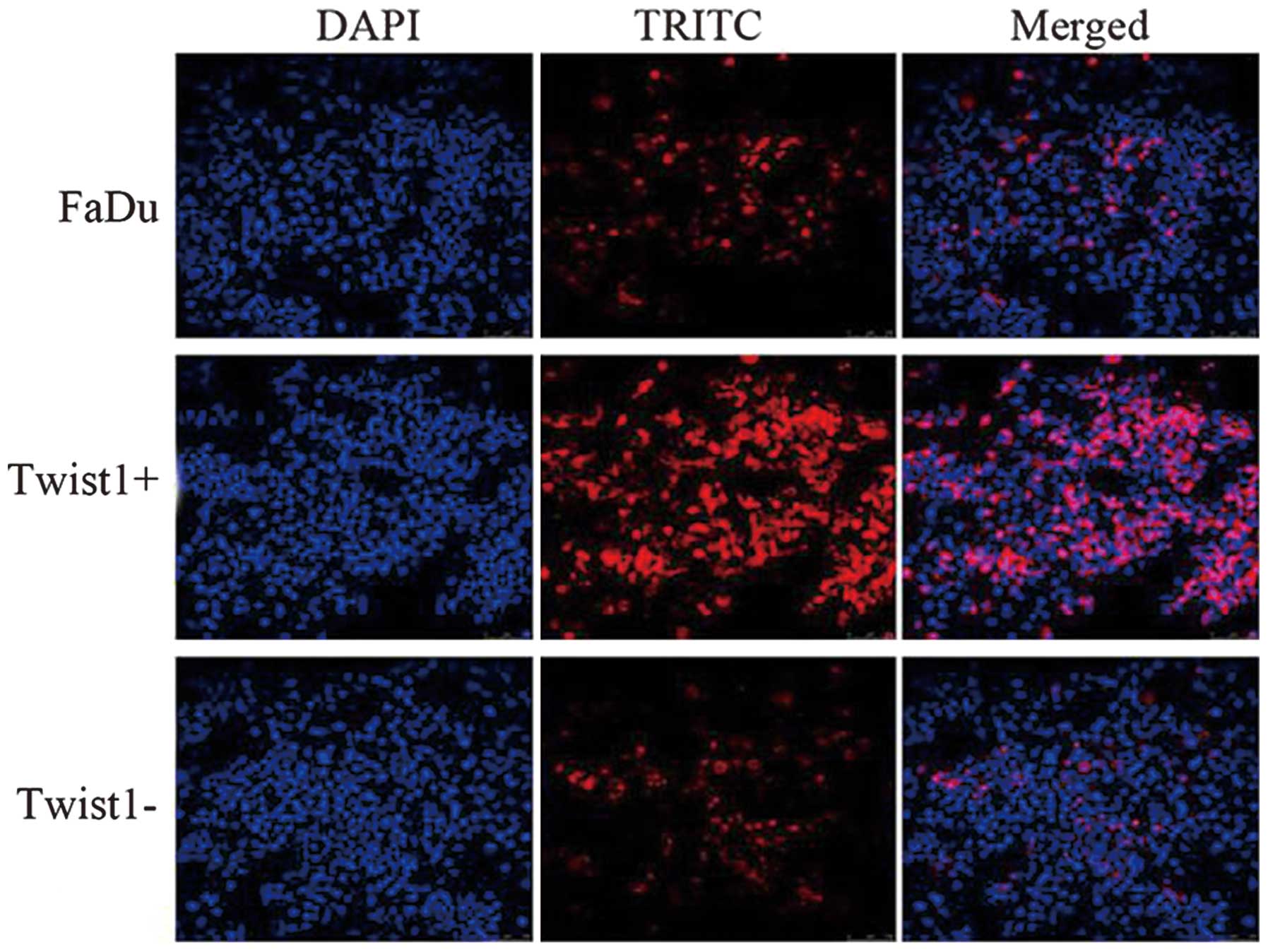
compared with the negative control cells and FaDu cells (Fig. 3C and D). Laser scanning confocal
microscopy detected a consistent expression of Twist1 with above in
FaDu cells, FaDu/Twist1+ cells and FaDu/Twist1-miRNA cells,
respectively (Fig. 4).
Twist1 regulates the chemosensitivity of
FaDu cells to taxol
MDR1/P-gp was elevated or downregulated accompanied
by corresponding changes in Twist1, which demonstrated that Twist1
may positively regulate MDR1/P-gp expression levels (Fig. 5A and B). The IC50 was
further analyzed in the transfectants. As illustrated in Fig. 5C, IC50 of FaDu/Twist1+
cells was 0.208±0.042 μM, and for FaDu-Twist1-miRNA cells was
0.085±0.012 μM, which were significantly different compared with
FaDu cells (IC50=0.134±0.022 μM; P<0.05). The data
suggests that overexpression of Twist1 protected the cell from
taxol damage and decreased the chemosensitivity of FaDu.
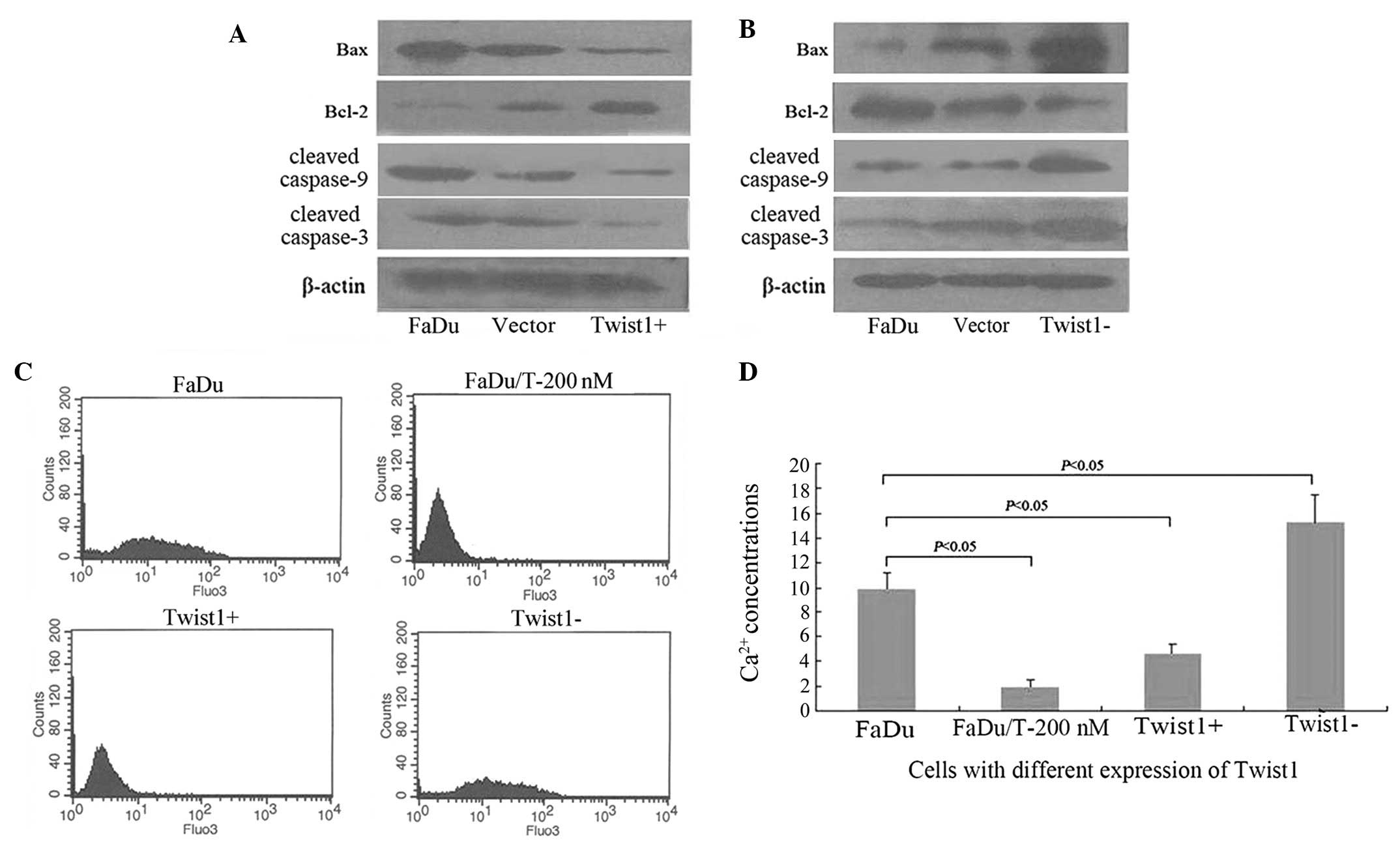
Changes in apoptosis-related proteins and
Ca2+ release
In FaDu/Twist1+ cells, cleaved caspase-3, cleaved
caspase-9, Bcl-2 and Bax, all demonstrated alterations with the
purpose of resisting apoptosis (Fig.
6A). By contrast, in FaDu/Twist1-miRNA cells, cleaved
caspase-3, cleaved caspase-9, Bcl-2 and Bax all changed
accordingly, with the purpose of promoting apoptosis (Fig. 6B). Results proved that Twist1
regulated the apoptosis sensitivity of FaDu cells. Overexpression
of Twist1 decreased the sensitivity of apoptosis in FaDu cells.
Twist1 overexpression inhibits
Ca2+ release induced by taxol
Mean counts of Ca2+ released in FaDu,
FaDu/T-200 nM, FaDu/Twist1+ and FaDu/Twist1-miRNA cells were
9.89±1.35, 1.96±0.57, 4.7±0.66 and 15.22±2.24, respectively
(Fig. 6C). As summarized in
Fig. 6D, statistical analysis
revealed that the Ca2+ concentration was significantly
elevated under taxol for 24 h in FaDu cells, which may be inhibited
in FaDu/T-200 nM. By contrast, Ca2+ elevation induced by
taxol could be buffered by Twist1 overexpression and significantly
aggravated by Twist1 silencing (P<0.05). These results suggested
that Ca2+ was reduced during MDR, and that Twist1 may
regulate the sensitivity of cells to factors inducing
Ca2+ release.
Discussion
MDR is a multi-factorial process defined as the
simultaneous resistance to several different types of commonly used
antineoplastic agents. The main mechanism of MDR involves the
exclusion of drugs from the cell by overexpression of either
MDR1/P-glycoprotein (MDR1/P-gp; a type of glycoprotein responsible
for drug exclusion) or various members of the MRP family (8). A number of other factors are
considered to be involved, including the alteration of levels or
properties of drug targets, increasing detoxification due to
enhanced activity of glutathione S-transferase, preventing
activation of drug to its active form and enhancing repair
capability of the cell following injury (18,19).
Although extensive studies have investigated MDR, the effect
observed in clinical practice is of little significance. MDR
remains as the central reason for chemotherapy failure in cancer
therapeutics. Therefore, further exploration for the development of
a new regulator of MDR is urgently required.
Twist1 is a highly conserved transcription
factor and is a member of a basic helix-loop-helix family. In our
previous study, we reported that Twist1 may be critically involved
in taxol-induced apoptosis of Hep-2 cells (20). Several other studies have also been
exploring the associations of chemosensitivity and Twist1, in an
attempt to further our understanding of the mechanisms of MDR
(21,22). Zhuo et al raised the
possibility of Twist1 depletion as a promising approach to lung
cancer therapy, in a short interfering RNA study directed against
Twist1 on A549 (23). However,
studies identifying the function of Twist1 in the chemosensitivity
of hypopharyngeal carcinomas are lacking. Our recent findings
demonstrated that the MDR cell line of Hep-2, induced by taxol, was
more invasive than its parent cell line, which was to a certain
extent, mediated through the overexpression of MDR1/P-gp/P-pg
(24). In the present study, the
upregulation of MDR1/P-gp and Twist1 in MDR FaDu/T cells was
detected, which suggest the expression levels of MDR1/P-gp and
Twist1 were positively correlated with MDR progression in
hypopharyngeal carcinomas. On the assumption that certain
correlations may exist between MDR1/P-gp and Twist1, DNA
recombinant and cell transfection experiments were conducted.
Stable transfections characterized by overexpression of Twist1 and
Twist1 silencing were established. It was identified that Twist1
overexpression led to the upregulation of MDR1/P-gp, and reversely,
Twist1 silencing led to the decrease of MDR1/P-gp. The results
reliably proved that Twist1 modulated the MDR1/P-gp expression
level. These findings are consistent with several other studies
identifying similar effects in other cell types. Overexpression of
Twist1, Snail and FOXC2 are considered to increase the promoter
activity of ABC transporters (25).
As the main regulator of MDR, manipulation of
MDR1/P-gp expression may effect chemosensitivity. Specific
functions must be ultimately implemented. Therefore, the
chemosensitivities of stable transfectants with variable levels of
Twist1, were detected by MTT. IC50 values were used to
assess chemosensitivities and the results proved our hypothesis was
appropriate. The IC50 values in cells with
overexpression of Twist1 was markedly increased, meaning that
Twist1 upregulation decreased the chemosensitivity of cells and
Twist1 silencing reversely increased it. Similar findings in
advanced and/or metastatic bladder (BCa) and prostate (PCa) cancer
types support these findings (26). They identified that Twist1 may be
utilized as a molecular target to restore chemosensitivity in BCa
and prostate PCa cancer types.
In hypothesizing how Twist1 modulates
chemosensitivities, it may be possible that numerous changes occur
with Twist1 expression, such as alteration of drug metabolism,
derangement of intracellular pathway signaling, cross-talk between
different membrane receptors and modification of apoptotic
signaling. In the present study, the focus was on the modification
of apoptosis (20). It has been
confirmed that proteins of the Bcl-2 family, which generally
repress apoptosis (Bcl-2 and Bcl-xL) or promote apoptosis (Bax, Bak
and Bad), are important in regulating the activation of caspases
(27–29). These proteins, to a certain degree,
affect caspase activation by controlling the release of cytochrome
C from the mitochondria, which in turn interacts with the adapter
protein Apaf-1, resulting in the activation of pro-Caspase-9
(30). The disturbance of
intracellular free Ca2+ ions is another key event when
cells are exposed to damaging stress (31–33).
In the present study, apoptosis was inhibited in MDR cells. Bcl-2
expression was upregulated, while expression of Bax, cleaved
caspase-3 and caspase-9 were decreased. Detection of stable
transfectants demonstrated that apoptosis sensitivity decreased in
cells which overexpressed Twist1, with Bcl-2 increased and Bax
cleaved caspase-3 and caspase-9 all downregulated. Furthermore,
after Twist1 was silenced by targeted micro-RNA, the apoptosis
sensitivity of these cells increased, with all the above
apoptotic-related proteins altered to sensitize apoptosis. Also, it
was identified that taxol led to an increase in intracellular free
cytosolic Ca2+, which may be partially attenuated by
Twist1 overexpression and aggravated by Twist1 silencing. These
data strongly indicated that Twist1 expression may regulate
apoptosis sensitivity, Bcl-2 and caspase family proteins (which
were involved in Twist1-mediated processes) and taxol-triggered
apoptosis in FaDu cells, at least in part, in the participation of
intracellular Ca2+. Studies on pancreatic cancer also
concluded a similar phenomenon (34). Evidence in the nasopharyngeal
carcinoma cell line HNE1 revealed a downregulation of Twist1 may
increase drug sensitivity of HNE1 to taxol by inducing apoptosis
(35). To the best of our
knowledge, this is the first study to report the function of Twist1
in apoptosis sensitivity of hypopharyngeal cancer, yet the report
about the regulation of Twist1 to Ca2+ influx has not
appeared. It was identified that the Ca2+ influx caused
by taxol may be attenuated by Twist1 overexpression. This may
provide novel molecular mechanisms of targeted gene therapy of head
and neck squamous cell cancer to Twist 1, and combining the
sensitizer of Ca2+ infux may enhance the
chemosensitivity of Twist1-targeted chemotherapy.
Acknowledgements
The author would like to thank Dr. Edward C. Mignot,
formerly of Shandong University, for linguistic advice. This study
was supported by Shandong Provincial International Science and
Technology Cooperation Project of China (no. 2010GHZ20202).
References
|
1
|
Mehta PS and Harrison LB: Function and
organ preservation in adult cancers of the head and neck. Expert
Rev Anticancer Ther. 7:361–371. 2007. View Article : Google Scholar : PubMed/NCBI
|
|
2
|
Vokes EE: Induction chemotherapy for head
and neck cancer: recent data. Oncologist. 15(Suppl 3): 3–7. 2010.
View Article : Google Scholar : PubMed/NCBI
|
|
3
|
Shukla S, Wu CP and Ambudkar SV:
Development of inhibitors of ATP-binding cassette drug
transporters: present status and challenges. Expert Opin Drug Metab
Toxicol. 4:205–223. 2008. View Article : Google Scholar : PubMed/NCBI
|
|
4
|
Neyns B, Tosoni A, Hwu WJ and Reardon DA:
Dose-dense temozolomide regimens: antitumor activity, toxicity, and
immunomodulatory effects. Cancer. 116:2868–2877. 2010. View Article : Google Scholar : PubMed/NCBI
|
|
5
|
Baguley BC: Multiple drug resistance
mechanisms in cancer. Mol Biotechnol. 46:308–316. 2010. View Article : Google Scholar : PubMed/NCBI
|
|
6
|
O’Connor R: The pharmacology of cancer
resistance. Anticancer Res. 27:1267–1272. 2007.
|
|
7
|
Kruse AL and Grätz KW: Oral carcinoma
after hematopoietic stem cell transplantation - a new
classification based on a literature review over 30 years. Head
Neck Oncol. 1:292009.PubMed/NCBI
|
|
8
|
Longley DB and Johnston PG: Molecular
mechanisms of drug resistance. J Pathol. 205:275–292. 2005.
View Article : Google Scholar : PubMed/NCBI
|
|
9
|
Dizdarevic S and Peters AM: Imaging of
multidrug resistance in cancer. Cancer Imaging. 11:1–8. 2011.
View Article : Google Scholar
|
|
10
|
Vasiliou V, Vasiliou K and Nebert DW:
Human ATP-binding cassette (ABC) transporter family. Hum Genomics.
3:281–290. 2009. View Article : Google Scholar : PubMed/NCBI
|
|
11
|
Cianfriglia M, Cenciarelli C, Barca S,
Tombesi M, Flego M and Dupuis ML: Monoclonal antibodies as a tool
for structure-function studies of the MDR1-P-glycoprotein. Curr
Protein Pept Sci. 3:513–530. 2002. View Article : Google Scholar : PubMed/NCBI
|
|
12
|
van den Heuvel-Eibrink MM, van der Holt B,
Burnett AK, et al: CD34-related coexpression of MDR1 and BCRP
indicates a clinically resistant phenotype in patients with acute
myeloid leukemia (AML) of older age. Ann Hematol. 86:329–337.
2007.PubMed/NCBI
|
|
13
|
Kuo MT: Roles of multidrug resistance
genes in breast cancer chemoresistance. Adv Exp Med Biol.
608:23–30. 2007. View Article : Google Scholar : PubMed/NCBI
|
|
14
|
Zhu K, Chen L, Han X and Wang J and Wang
J: Short hairpin RNA targeting Twist1 suppresses cell proliferation
and improves chemosensitivity to cisplatin in HeLa human cervical
cancer cells. Oncol Rep. 27:1027–1034. 2012.PubMed/NCBI
|
|
15
|
Qin Q, Xu Y, He T, Qin C and Xu J: Normal
and disease-related biological functions of Twist1 and underlying
molecular mechanisms. Cell Res. 22:90–106. 2012. View Article : Google Scholar : PubMed/NCBI
|
|
16
|
Zhan X, Feng X, Kong Y, Chen Y and Tan W:
JNK signaling maintains the mesenchymal properties of multi-drug
resistant human epidermoid carcinoma KB cells through snail and
twist1. BMC Cancer. 13:1802013. View Article : Google Scholar
|
|
17
|
Ma J, Lu S, Yu L, et al: FaDu cell
characteristics induced by multidrug resistance. Oncol Rep.
26:1189–1195. 2011.PubMed/NCBI
|
|
18
|
Liang Y, Meleady P, Cleary I, McDonnell S,
Connolly L and Clynes M: Selection with melphalan or paclitaxel
(Taxol) yields variants with different patterns of multidrug
resistance, integrin expression and in vitro invasiveness. Eur J
Cancer. 37:1041–1052. 2001. View Article : Google Scholar
|
|
19
|
Laborde E: Glutathione transferases as
mediators of signaling pathways involved in cell proliferation and
cell death. Cell Death Differ. 17:1373–1380. 2010. View Article : Google Scholar : PubMed/NCBI
|
|
20
|
Yu L, Li HZ, Lu SM, et al: Alteration in
TWIST expression: possible role in paclitaxel-induced apoptosis in
human laryngeal carcinoma Hep-2 cell line. Croat Med J. 50:536–542.
2009. View Article : Google Scholar : PubMed/NCBI
|
|
21
|
Lemma S, Karihtala P, Haapasaari KM, et
al: Biological roles and prognostic values of the
epithelial-mesenchymal transition-mediating transcription factors
Twist, ZEB1 and Slug in diffuse large B-cell lymphoma.
Histopathology. 62:326–333. 2013. View Article : Google Scholar
|
|
22
|
Banerjee A, Qian P, Wu ZS, et al: Artemin
stimulates radio- and chemo-resistance by promoting
TWIST1-BCL-2-dependent cancer stem cell-like behavior in mammary
carcinoma cells. J Biol Chem. 287:42502–42515. 2012. View Article : Google Scholar
|
|
23
|
Zhuo WL, Wang Y, Zhuo XL, Zhang YS and
Chen ZT: Short interfering RNA directed against TWIST, a novel zinc
finger transcription factor, increases A549 cell sensitivity to
cisplatin via MAPK/mitochondrial pathway. Biochem Biophys Res
Commun. 369:1098–1102. 2008. View Article : Google Scholar : PubMed/NCBI
|
|
24
|
Li L, Jiang AC, Dong P, Wang H, Xu W and
Xu C: MDR1/P-gp and VEGF synergistically enhance the invasion of
Hep-2 cells with multidrug resistance induced by taxol. Ann Surg
Oncol. 16:1421–1428. 2009. View Article : Google Scholar : PubMed/NCBI
|
|
25
|
Saxena M, Stephens MA, Pathak H and
Rangarajan A: Transcription factors that mediate
epithelial-mesenchymal transition lead to multidrug resistance by
upregulating ABC transporters. Cell Death Dis. 2:e1792011.
View Article : Google Scholar : PubMed/NCBI
|
|
26
|
Wallerand H, Robert G, Pasticier G, et al:
The epithelial-mesenchymal transition-inducing factor TWIST is an
attractive target in advanced and/or metastatic bladder and
prostate cancers. Urol Oncol. 28:473–479. 2010. View Article : Google Scholar : PubMed/NCBI
|
|
27
|
Piotrowska H, Kucinska M and Murias M:
Biological activity of piceatannol: leaving the shadow of
resveratrol. Mutat Res. 750:60–82. 2012. View Article : Google Scholar : PubMed/NCBI
|
|
28
|
Wyllie AH: “Where, O death, is thy sting?”
A brief review of apoptosis biology. Mol Neurobiol. 42:4–9.
2010.
|
|
29
|
Tomiyama A, Tachibana K, Suzuki K, et al:
MEK-ERK-dependent multiple caspase activation by mitochondrial
proapoptotic Bcl-2 family proteins is essential for heavy ion
irradiation-induced glioma cell death. Cell Death Dis. 1:e602010.
View Article : Google Scholar
|
|
30
|
Qin H, Srinivasula SM, Wu G,
Fernandes-Alnemri T, Alnemri ES and Shi Y: Structural basis of
procaspase-9 recruitment by the apoptotic protease-activating
factor 1. Nature. 399:549–557. 1999. View
Article : Google Scholar : PubMed/NCBI
|
|
31
|
Shang L, Liu J, Zhu Q, et al: Gypenosides
protect primary cultures of rat cortical cells against oxidative
neurotoxicity. Brain Res. 1102:163–174. 2006. View Article : Google Scholar : PubMed/NCBI
|
|
32
|
Wang JQ, Chen Q, Wang X, et al:
Dysregulation of mitochondrial calcium signaling and superoxide
flashes cause mitochondrial genomic DNA damage in Huntington
disease. J Biol Chem. 288:3070–3084. 2013. View Article : Google Scholar : PubMed/NCBI
|
|
33
|
Demirci S, Kutluhan S, Naziroğlu M, Uğuz
AC, Yürekli VA and Demirci K: Effects of selenium and topiramate on
cytosolic Ca(2+) influx and oxidative stress in neuronal PC12
cells. Neurochem Res. 38:90–97. 2013.PubMed/NCBI
|
|
34
|
Naziroglu M: Molecular role of catalase on
oxidative stress-induced Ca(2+) signaling and TRP cation channel
activation in nervous system. J Recept Signal Transduct Res.
32:134–141. 2012.
|
|
35
|
Zhang X, Wang Q, Ling MT, Wong YC, Leung
SC and Wang X: Anti-apoptotic role of TWIST and its association
with Akt pathway in mediating taxol resistance in nasopharyngeal
carcinoma cells. Int J Cancer. 120:1891–1898. 2007. View Article : Google Scholar : PubMed/NCBI
|