Introduction
Colorectal cancer (CRC) is the third most common
type of cancer worldwide, with ~1.4 million novel cases diagnosed
in 2012 (1). At present, the
treatment of CRC involves surgical resection, chemotherapy,
radiotherapy and immunotherapy (2). However, the therapeutic efficacy,
particularly for advanced CRC, is limited (3).
Caudal-related homeobox protein 2 (CDX2), an
intestinal transcription factor, is critically involved in the
development, proliferation and differentiation of intestinal
epithelial cells (4–7). In addition, CDX2 is a
tumor-suppressor gene in colorectal cancer (8–10),
and reduces mobility and antagonizes dissemination of colon cancer
cells in vitro and in vivo (11). By contrast, the presence of reduced
CDX2 expression levels is a predictor for poor overall survival
amongst patients with colorectal cancer (12). Therefore, forced overexpression of
CDX2 under a cytomegalovirus (CMV) promoter in colon cancer cells
is used to inhibit LoVo colon cancer cell invasion (13) and gastric cancer progression
(14).
However, due to the fact that non-specific
expression of CDX2 may lead to the generation of side effects,
regulated colorectal cancer cell-specific expression of CDX2 is
necessary. The human telomerase reverse transcriptase (hTERT)
promoter is active in the majority of cancer cells but not normal
cells (15,16). Therefore, this promoter has
previously been used to target A549 human lung adenocarcinoma cells
(17) and human gastric cancer
MKN45 cells (18).
Hypoxia is a major feature of solid tumors (19,20)
and induces hypoxia-inducible factor-1α (HIF-1α) expression, which
binds to the hypoxia-response elements (HREs) of various target
genes (21) and activates their
transcription in order to regulate glucose transport and
angiogenesis, and potentially to enhance the survival of tumor
cells (22,23). Previous studies have reported that
targeted genes may be significantly upregulated by five copies of
HREs under hypoxic conditions (24,25).
At present, the effects of CDX2 overexpression,
under the control of five copies of HREs and the hTERT promoter, on
human colorectal cancer cell proliferation in vitro remain
unclear. In the current study it was hypothesized that CDX2
overexpression specifically inhibits human colorectal cancer cell
proliferation under hypoxic conditions.
Materials and methods
Polymerase chain reaction (PCR)
amplification of target DNA
The hTERT gene promoter and CDX2 gene were amplified
from a DNA library of hTERT(+) CRC cells and pEGFP-C1-CDX2
(26), respectively, by PCR using
specific primers (Table I). hTERT
was obtained using the hTERT forward and reverse1 primer, whereas
5HRE + hTERT used the forward 5HRE primer and the hTERT reverse2
primer. For the hTERT promoter, the PCR cycling conditions were as
follows: Amplification at 98°C for 2 min, 30 cycles of 98°C for 10
sec, 55°C for 15 sec and 72°C for 30 sec, followed by an extension
step at 72°C for 10 min. For the CDX2 promoter, the conditions were
as follows: Amplification at 98°C for 2 min, 35 cycles of 98°C for
20 sec, 59°C for 30 sec and 72°C for 1 min, followed by an
extension step at 72°C for 10 min. This was performed using the
PTC-100 (Bio-Rad Laboratories, Inc., Hercules, CA, USA).
 | Table IPrimer sequences. |
Table I
Primer sequences.
| Gene | Primer | Base sequence
5′-3′ | PCR product (base
pairs) |
|---|
| 5HRE | Forward | CGACGCGTATTATGCTAGTCCAC | 221 |
| hTERT | Forward | CCCAAGCTTCACAGACGCCCAGGACCGCGCTTC | 327 |
| Reverse1 | AACTGCAGCCACGTGCGCCCACGTGCGCCCAC | |
| Reverse2 | CCGCTCGAGCCACGTGCGCCCACGTGCGCCCAC | |
| CDX2 | Forward | CGGAATTCATGTACGTGAGCTACCTCCTGGACAAGGAC | 943 |
| Reverse | CGGGATCCGTCTGGGTGACGGTGGGGTTTAGCACCCCCCCAGTTG | |
The PCR products were resolved on a 1% agarose gel
electrophoresis (Shaanxi Pioneer Biotech Co., Ltd., Xi’an, China).
The hTERT promoter (p) and CDX2 products were digested with
HindIII/PstI and EcoRI/BamHI,
respectively, and confirmed by DNA sequence analysis at Sangon
Biotech Co. Ltd. (Shanghai, China).
Construction of lentiviral vectors
The hTERT promoter was first cloned into the
pLenhanced green fluorescent protein (EGFP)-N1-5HRE-CEAp (27) plasmid (Translational Medical
Center, First Affiliated Hospital of Xi’an Jiaotong University,
Xi’an, China) at the HindIII and PstI sites by
replacing CEAp with the restriction endoenzymes for 16 h at 37°C,
in order to derive a recombinant plasmid named
pLEGFP-N1-5HRE-hTERTp. An incision enzyme and Taq DNA polymerase
from Takara Bio, Inc. (Otsu, Japan) were used. Subsequently, the
5HRE-hTERTp fragment, which was digested with the restriction
endoenzymes BgIII and PstI in buffer at 37°C for 16
h, was cloned into the lentiviral vector pLVX-EGFP-3FLAG
(Translational Medical Center, First Affiliated Hospital of Xi’an
Jiaotong University) by replacing the CMV promoter in the plasmid
to generate the recombinant plasmid pLVX-5HRE-hTERTp-EGFP-3FLAG
(designated as 5Hh), into which the amplified CDX2 fragment was
cloned by replacing EGFP to produce the recombinant plasmid
pLVX-5HRE-hTERTp-CDX2-3FLAG (designated as 5HhC). The identity of
the final recombinant lentiviral vector construct was confirmed by
restriction endonuclease digestion and DNA sequence analysis at
Sangon Biotech Co., Ltd.
Cell lines and cell cultures
Human epithelial kidney HEK 293T, human proximal
tubular HK-2 and human CRC LoVo cells (Translational Medical
center, First Affiliated Hospital of Xi’an Jiaotong University)
were cultured in Dulbecco’s modified Eagle’s medium (DMEM) from
Gibco Life Technologies (Carlsbad, CA, USA), supplemented with 10%
fetal bovine serum obtained from GE Healthcare Life Sciences
(Logan, UT, USA) in a humidified atmosphere with 5% CO2
at 37°C.
Generation and titration of
lentiviruses
Recombinant lentiviruses were produced by
co-transfecting 293T cells with the lentiviral expression plasmid
5HhC or the control plasmid 5Hh, in addition to the pCD/NL-BH*DDD
and pLTR-G plasmids (Shanghai Sunbio Technology Co., Ltd.,
Shanghai, China) using Trans-EZ reagent (Shanghai Sunbio Technology
Co., Ltd.). The 293T cells (6×10⁵) were cultured in a
10-cm tissue culture plate with opti-MEM (Gibco Life Technologies).
Transfection was performed when the cell density reached 30–40%
confluence. Solution A was prepared by adding 0.5 ml (0.5 mg/ml)
5HhC or 5Hh plasmid and 1 ml (0.2 mg/ml) pCD/NL-BH*DDD or 0.5 ml
(0.2 mg/ml) pLTR-G plasmids (diluted with opti-MEM media) to 18 ml
opti-MEM media. Solution B was prepared by adding 0.5 ml Trans-EZ
to 18 ml opti-MEM media. The transfection solution was prepared by
adding solution B slowly to solution A. The mixture was agitated
and then incubated at room temperature for 20 min. The 293T cells
were plated in six-well plates prior to incubation with 3 ml
prepared transfection mixture in a humidified incubator with 5%
CO2 at 37°C for 30 min. The culture media was then
replaced with fresh DMEM. Infectious lentiviruses were harvested at
48 h post-transfection by collecting the medium and centrifuging at
500 × g and 4°C for 10 min, then filtering the supernatant
containing the lentiviruses through a 0.45 µm polyvinylidene
fluoride filter unit (EMD Millipore, Billerica, MA, USA) to
concentrate it. The infectious titer was determined by quantitative
PCR (qPCR). The total RNA of the transfected cells was extracted
using TRIzol Reagent (Invitrogen Life Technologies,Carlsbad, CA,
USA). Reverse transcription and PCR were performed using a Takara
Bio, Inc. RNA PCR (AMV) kit with the CDX2 primers presented in
Table I. The results were
normalized against the level of the internal control. β-actin PCR
amplifications were performed at 98°C for 2 min, 40 cycles of 50°C
for 2 min, 95°C for 10 min and 95°C for 15 sec, followed by an
extension step at 60°C for 1 min (ABI PRISM 7000 Sequence Detection
System). qPCR was performed in triplicate for each experiment,
including for the non-template controls.
Infection of LoVo cells with
lentiviruses
LoVo cells were cultured in six-well plates
(5×10⁵cells/well) overnight, infected with 5HhC or
control 5Hh lentiviruses and exposed to puromycin (800
µg/ml; Gibco Life Technologies) for two weeks. The 5HhC/LoVo
or 5Hh/LoVo cells were then routinely cloned (28).
Immunofluorescence assay
To detect CDX2 expression by the 5HhC lentivirus,
the hTERT(+) LoVo and hTERT(-) HK-2 cells were infected with 5HhC
lentiviruses in a humidified incubator with 5% CO2 at
37°C for 48 h and the CDX2 expression was examined using an
immunofluorescence assay. The 5HhC-infected LoVo and HK-2 cells
were cultured and fixed with 4% paraformaldehyde (Shaanxi Pioneer
Biotech Co., Ltd.) for 30 min. The cells were stained with a
primary monoclonal rabbit anti-FLAG antibody (1:200; F2555;
Sigma-Aldrich, St. Louis, MO, USA) at 4°C overnight prior to
incubation at 37°C for 30 min with a fluorescein
isothiocyanate-conjugated goat anti-rabbit immunoglobulin (Ig) G
secondary antibody (1:5,000; Wuhan Sanying Biotechnology, Wuhan,
China). The cells were then mounted with DAPI solution (Santa Cruz
Biotechnology, Inc., Dallas, TX, USA) and observed under a confocal
fluorescence microscope (Leica DMi8; Leica Microsystems GmbH,
Wetzlar, Germany) to evaluate FLAG expression.
MTT assay
The effects of CoCl2 on the viability of
LoVo cells were detected using an MTT assay. The LoVo cells, at a
density of 5×10³cells/well, were cultured for 24 h and
then treated with 100, 200, 300, 400 or 500 µmol/l
CoCl2 (Sigma-Aldrich) for 1, 3, 5 or 7 days. The
experiment was performed in triplicate. Subsequently, the plates
were washed extensively with serum-free DMEM to remove
CoCl2 and dead cells, and were exposed to 20 µl
(5 g/l) MTT (Sigma-Aldrich) for 4 h. The resulting formazan
crystals were dissolved in 200 µl dimethyl sulfoxide
(Sigma-Aldrich) and the absorbance was measured at 490 nm with a
microplate reader (Victor3; PerkinElmer, Inc., Waltham, MA, USA).
The cytotoxicity of CoCl2 to LoVo cells was evaluated by
determination of the survival rate of LoVo cells (calculated as
A490 of treatment group/A490 of untreated group).
Western blot analysis
The 5HhC/LoVo cells were cultured under a normoxic
(absence of CoCl2) or hypoxic conditions (100, 200, 300,
400 or 500 µmol/l CoCl2) for 24 h and then
cultured for 12, 24 or 36 h with the optimal concentration (300
µmol/l) of CoCl2. The relative ratios of
recombinant CDX2 protein to control β-actin were determined by
western blot analysis. Briefly, the 5HhC lentivirus-infected LoVo
cells (1×10⁶ cells) were lysed with 150 µl lysis
buffer (50 mM Tris, 150 mM NaCl, 5 mM EDTA, 5 mM EGTA and 1% SDS;
pH 7.5; (Shaanxi Pioneer Biotech Co., Ltd.), followed by gentle
sonication (Bio-Rad Laboratories, Inc.). Following quantification
with Bradford reagent (Thermo Fisher Scientific, Waltham, MA, USA),
the protein lysates (80 µg/lane) of each sample were
subjected to SDS-PAGE (Shaanxi Pioneer Biotech Co., Ltd.) with a
machine from EMD Millipore on 10% gels and transferred to
polyvinylidene fluoride membranes (EMD Millipore). Subsequent to
being blocked with 5% fat-free dry milk, the membranes were
incubated with 1:1,000 diluted monoclonal rabbit anti-CDX2 (2475-1;
Epitomics, Burlingame, CA, USA) and 1:3,000 diluted polyclonal
rabbit anti-β-actin (BS10005; Bioworld Technology, Inc., St. Louis
Park, MN, USA) at 4°C, overnight prior to incubation with
horseradish peroxidase-conjugated goat anti-rabbit IgG secondary
antibody (OriGene Technologies, Inc., Beijing, China) for 30 min at
37°C, followed by visualization with enhanced chemiluminescence
(Amresco LLC, Solon, OH, USA).
Reverse transcription (RT)-PCR
analysis
The 5HhC/LoVo cells were cultured under normoxic or
hypoxic conditions and the transcription of recombinant CDX2 mRNA
was determined by RT-PCR using the primers listed in Table I. In brief, total RNA was extracted
from the infected cells cultured under normoxia or hypoxia using
TRIzol (Gibco Life Technologies, Grand Island, NY, USA), according
to the manufacturer’s instructions. The resulting RNAs were treated
with RNase-free DNase (Promega Corp., Madison, WI, USA) and reverse
transcribed into cDNA using an RT-PCR kit (Invitrogen Life
Technologies) according to the manufacturer’s instructions. PCR
amplifications were performed in duplicate at 98°C for 2 min and
subjected to 35 cycles of 98°C for 20 sec, 59°C for 30 sec and 72°C
for 1 min, followed by an extension at 72°C for 10 min. The PCR
products were resolved by agarose gel electrophoresis using a
ChemiDoc System (ChemiDoc™ MP System 170-8280; Bio-Rad
Laboratories, Inc.).
Cloning assay
A total of 200 5HhC/LoVo and 200 5Hh cells plated on
60-mm cell culture dishes were cultured in a humidified incubator
with 5% CO2 at 37°C for three weeks. The cloning cells
were fixed with methanol (Shaanxi Pioneer Biotech Co., Ltd.) at
room temperature for 15 min and then stained by Giemsa solution.
The clones containing ≥50 cells [counted using a Leica M125
microscope (Leica Microsystems GmbH, Heidelberg, Germany)] were
regarded as true clones (29).
Statistical analysis
All experiments were repeated three times. The data
from all experiments was pooled, and the results were expressed as
the mean ± standard deviation. Differences in mean values were
analyzed by one-way analysis of variance followed by Student’s
t-test with SPSS software version 13.0 (SPSS, Inc., Chicago, IL,
USA). P<0.05 was considered to indicate a statistically
significant difference.
Results
Recombinant plasmids were successfully
generated
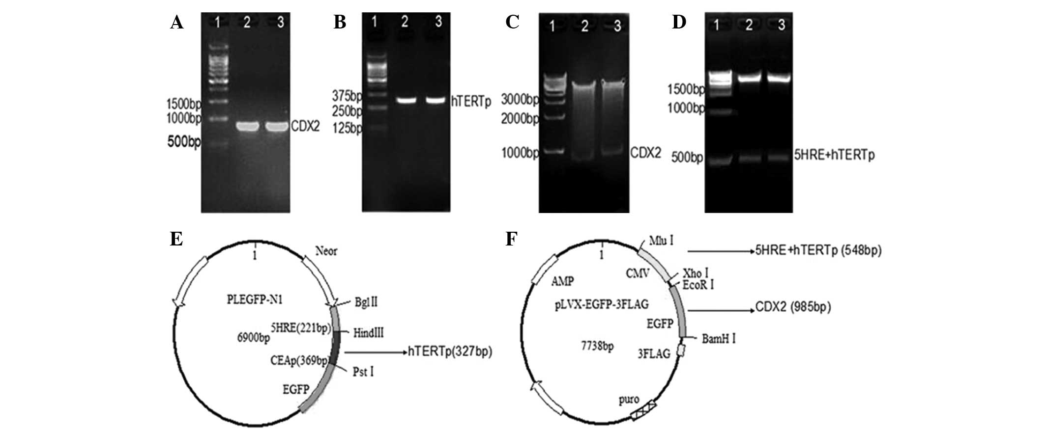
The 327 base pairs (bp) hTERTp and 985 bp CDX2 DNA
fragments were successfully amplified by PCR (Fig. 1A and B). The restricted enzyme
digestions (Fig. 1C and D) and
sequencing demonstrated that the recombinant plasmids
pLEGFP-N1-5HRE-hTERTp and pLVX-5HRE-hTERTp-CDX2-3FLAG (Fig. 1E and F) were also successfully
constructed.
5HhC-infected LoVo cells, but not HK-2
cells, express FLAG in vitro
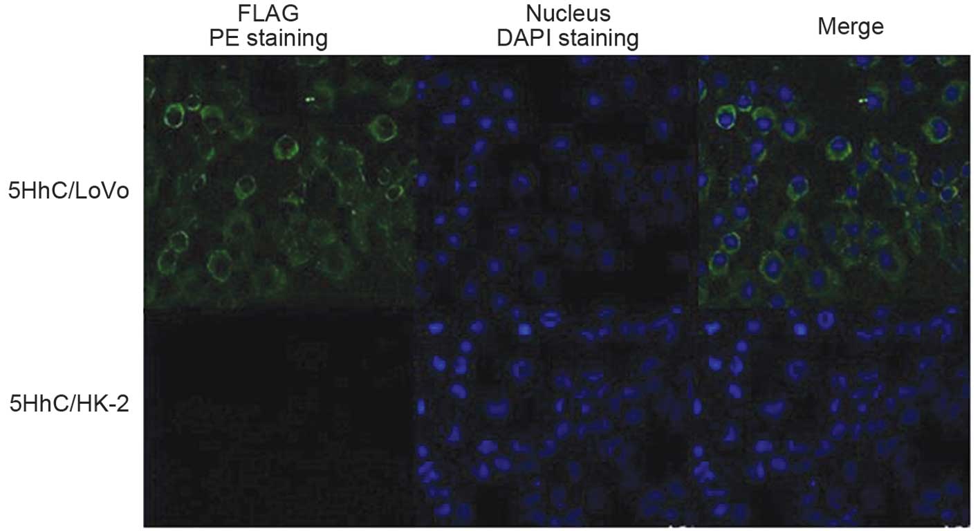
An immunofluorescence assay was conducted to examine
FLAG expression of 5HhC in the hTERT(+) LoVo and hTERT(−) HK-2 cell
lines. It was observed that FLAG was expressed on the membrane and
cytoplasm of 5HhC-infected LoVo cells, but not 5HhC-infected HK-2
cells (Fig. 2). This indicated
that hTERT specifically directed exogenous gene expression in
hTERT(+), but not hTERT(−) cell lines.
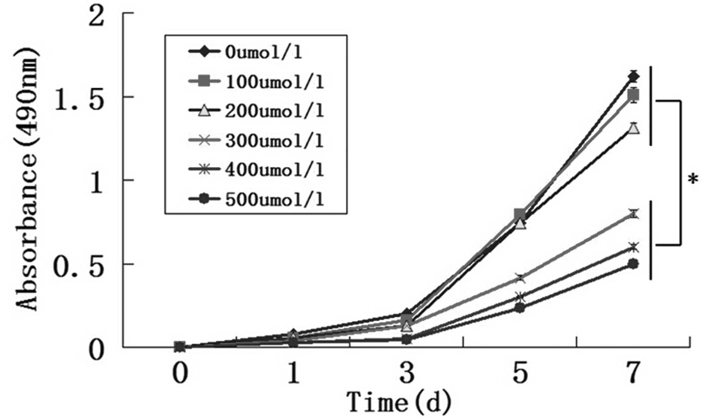
High concentrations of CoCl2
attenuate LoVo cell proliferation in vitro
The cytotoxic effect of CoCl2 on LoVo
cells was investigated using an MTT assay. The results demonstrated
that cell viability was not influenced by CoCl2 at 100
or 200 µmol/l (P>0.05), but the cell viability was
suppressed with 300–500 µmol/l CoCl2 (P<0.05),
particularly at day 7 (Fig.
3).
High CDX2 expression levels with 5HhC are
induced in the hTERT(+) cell line under hypoxic conditions
The levels of recombinant CDX2 protein relative to
control β-actin expression following CoCl2 hypoxic
treatment over time was compared between 5HhC/LoVo and LoVo cells.
Following 24 h of hypoxic treatment, the relative expression levels
of CDX2 were upregulated at CoCl2 concentrations from
100–500 µmol/l (P<0.05), with the greatest effect on CDX2
expression observed following treatment with 300 µmol/l
CoCl2, compared with that of 5HhC/LoVo cells without
CoCl2 treatment (P<0.05; Fig. 4A). In 5HhC/LoVo cells treated with
300 µmol/l CoCl2, the highest CDX2 expression
levels were observed following 24 h of treatment when compared with
those at 0, 12 or 36 h (P<0.05; Fig. 4B). Subsequently, 5HhC/LoVo cells
were treated for 24 h under normoxic or hypoxic conditions (with
300 µmol/l CoCl2). The relative expression levels
of CDX2 mRNA in 5HhC/LoVo cells under hypoxic conditions were
observed to be significantly higher when compared with those under
normoxic conditions (P<0.05; Fig.
4C). The data presented indicated that the CDX2 mRNA and
protein expression levels were upregulated in 5HhC/LoVo cells,
particularly under hypoxic conditions.
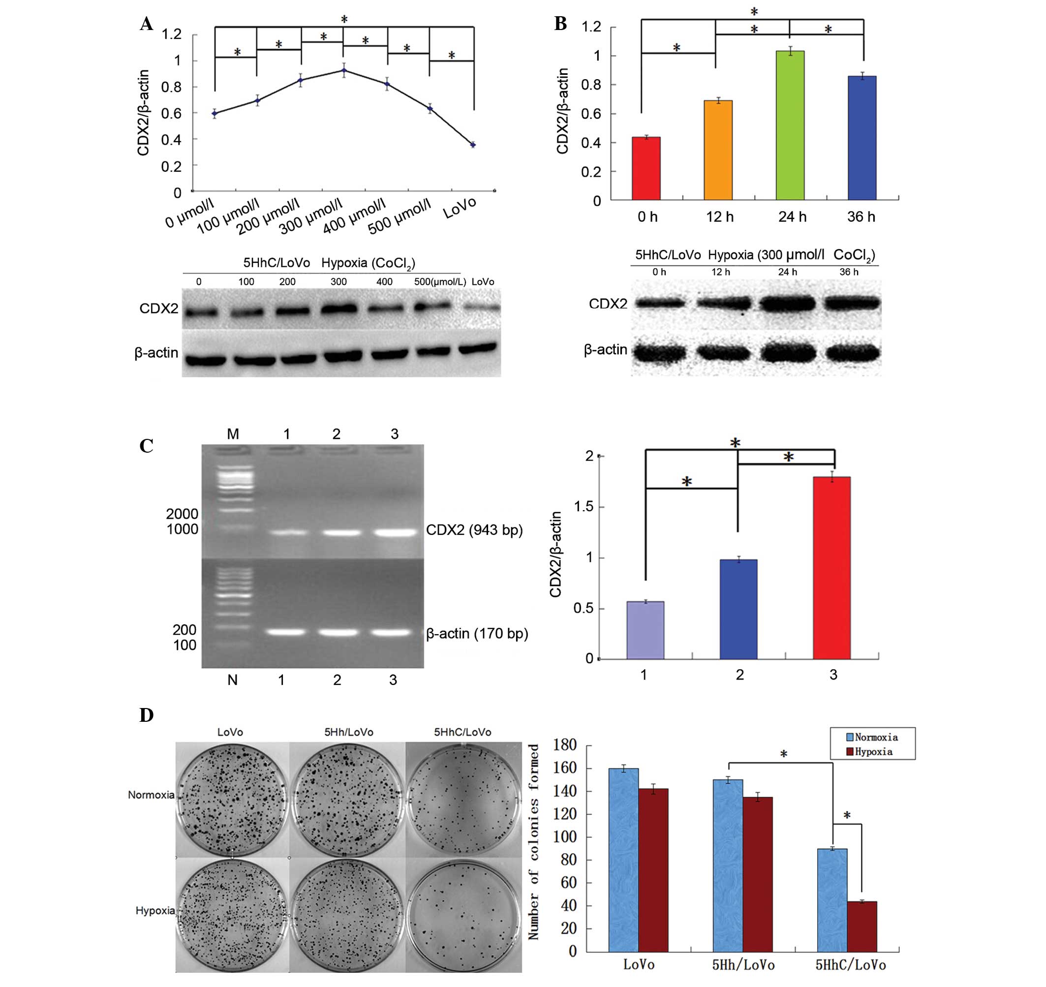 | Figure 4Expression levels of CDX2 in 5HhC
LoVo cells and the effect of CDX2 overexpression on LoVo cell
proliferation. (A) Western blot analysis of CDX2 expression. The
5HhC/LoVo cells and LoVo cells were cultured under normoxic or
hypoxic conditions (100–500 µmol/l CoCl2) for 24
h. (B) Western blot analysis of CDX2 expression. The 5HhC/LoVo
cells were cultured under hypoxic conditions (300 µmol/l
CoCl2) for 0, 12, 24 or 36 h. (C) Reverse
transcription-polymerase chain reaction analysis of CDX2 mRNA
expression. The 5HhC/LoVo cells were cultured under a normoxic or
hypoxic conditions (300 µmol/l CoCl2) for 24 h.
M, DNA marker; 1, LoVo cells; 2, 5HhC/LoVo cells under normoxic
conditions; 3, 5HhC/LoVo cells under hypoxic conditions. (D) Clone
formation of LoVo, 5Hh/LoVo or 5HhC/LoVo cells. Each group had a
hypoxic control (200 µmol/l CoCl2; red bars). All
data presented are representative images of each group of cells
from three separate experiments. The results are presented as the
mean ± standard deviation (*P<0.05). CDX2,
caudal-related homeobox; 5HhC, pLVX-5HRE-hTERTp-CDX2-3FLAG; 5Hh,
pLVX-5HRE-hTERTp-EGFP-3FLAG; bp, base pairs. |
To further assess the potential effects of CDX2
expression with 5HhC on the proliferation of LoVo cells, a cloning
assay was conducted with 5HhC/LoVo and LoVo cells. The
proliferation results demonstrated that the clone numbers and size
in 5HhC/LoVo cells were significantly reduced compared with those
of LoVo cells (P<0.05). Notably, the proliferation levels of
5HhC/LoVo cells under hypoxic conditions were significantly lower
compared with those under normoxic conditions (P<0.05; Fig. 4D). These results indicated that a
hypoxic environment resulted in upregulation of CDX2 expression
with 5HhC to inhibit LoVo cell proliferation via the 5 HRE
enhancers.
Discussion
In the present study, a recombinant lentivirus with
enhanced CDX2 expression driven by hTERTp and 5 HRE enhancers was
generated, and it was observed that CDX2 was highly expressed in
hTERT positive cells under hypoxic conditions, which attenuated CRC
cell proliferation in vitro.
It was identified that CDX2 expression was detected
in hTERT(+) LoVo cells, but not in hTERT(−) HK-2 cells, suggesting
that the 5HhC vector was hTERT-specific in vitro. The hTERT
promoter, which is active in the majority of cancer cell lines but
not in normal cells, has been previously reported as a useful tool
for tumor transcriptional targeting (30–32).
In addition, it has been demonstrated to effectively target A549
human lung adenocarcinoma cells and MKN45 human gastric cancer
cells (16).
In order to enhance CDX2 expression under the hTERT
promoter, five copies of HRE were positioned upstream of the hTERT
promoter. It was observed that 5HhC/LoVo cells under hypoxic
conditions, produced by treatment with 300 µmol/l
CoCl2 for 24 h, exhibited the greatest expression levels
of CDX2 compared with those under normoxic conditions without
CoCl2. A previous study reported that the development of
CRC was dependent on the tumor microenvironment (33). The observations of the current
study were in agreement with a previous study, in which five copies
of HRE under the control of a CMV promoter induced bacterial
cytosine deaminase expression in the bacterial cytosine
deaminase/5-fluorocytosine gene therapy system under hypoxic
conditions, enhancing the efficacy of radiotherapy in a tumor
xenograft (34). In the present
study, it was observed that 5HhC/LoVo cells under hypoxic
conditions exhibited significantly inhibited LoVo cell
proliferation compared with those under normoxic conditions,
suggesting that CDX2 expression was induced under hypoxic
conditions to suppress CRC cell proliferation. This is consistent
with the role of CDX2 as a tumor-suppressor gene in colorectal
cancer (35). A
non-transcriptional function of CDX2 is that it suppresses
tumorigenesis via p27Kip1 stabilization, and low levels of CDX2
have been reported to accelerate colon tumorigenesis by reducing
p27Kip1 levels (36). Notably,
mutation of a subdomain in the N-terminus of CDX2 has been reported
to abrogate the anti-proliferative effects of CDX2, which may be
via inhibition of β-catenin/T-cell factor (TCF) transcriptional
activity by disrupting the β-catenin-TCF protein complex in colon
cancer cells (37). CDX2 tumor
suppression may also function via the extracellular
signal-regulated kinase 1/2 pathway in CRC (38). Clinically, a reduction in CDX2
expression is correlated with poor overall survival amongst
patients with colorectal cancer (39).
In conclusion, a recombinant lentivirus with the
hTERT promoter and five HREs promoted expression of the tumor
suppressor CDX2 in CRC cells under hypoxic conditions, and was
effective at targeting CRC cells in vitro. CDX2 expression
under the hTERT promoter and five HREs may provide a potential tool
for the treatment and gene therapy of CRC.
Acknowledgments
The current study was supported by grants from the
National Natural Science Foundation of China (grant nos. 81101874,
81172362 and 81172359), the Science and Technology Project of
Shaanxi Province (grant no. 2011-K12-19), the Science and
Technology Plan and Integrated Innovation Engineering Project of
Shaanxi Province (grant no. 2013KTCQ03-08) and the Clinical
Innovation Fund of the First Affiliated Hospital of Xi’an Jiaotong
University (grant nos. 12ZD12 and 12ZD21).
Abbreviations:
|
CDX2
|
caudal-related homeobox protein 2
|
|
hTERT
|
human telomerase reverse
transcriptase
|
|
HREs
|
hypoxia-response elements
|
|
CMV
|
cytomegalovirus
|
|
CRC
|
colorectal cancer
|
|
MTT
|
3-(4,5-dimethylthiazol-2yl)-2,5-diphenyltetrazolium bromide
|
References
|
1
|
László L: Predictive and prognostic
factors in the complex treatment of patients with colorectal
cancer. Magy Onkol. 54:383–394. 2010. View Article : Google Scholar : PubMed/NCBI
|
|
2
|
Akkad J, Bochum S and Martens UM:
Personalized treatment for colorectal cancer: novel developments
and putative therapeutic strategies. Langenbecks Arch Surg.
400:129–143. 2015. View Article : Google Scholar : PubMed/NCBI
|
|
3
|
O’Connor JP, Rose CJ, Jackson A, et al:
DCE-MRI biomarkers of tumour heterogeneity predict CRC liver
metastasis shrinkage following bevacizumab and FOLFOX-6. Br J
Cancer. 105:139–145. 2011. View Article : Google Scholar
|
|
4
|
Akhavan-Niaki H and Samadani AA: Molecular
insight in gastric cancer induction: an overview of cancer stemness
genes. Cell Biochem Biophys. 68:463–473. 2014. View Article : Google Scholar
|
|
5
|
Natoli M, Christensen J, El-Gebali S,
Felsani A and Anderle P: The role of CDX2 in Caco-2 cell
differentiation. Eur J Pharm Biopharm. 85:20–25. 2013. View Article : Google Scholar : PubMed/NCBI
|
|
6
|
Lin ME, Huang D, Deng BH, Lv YS, Rong L
and Yao YS: Expression and functional role of Cdx2 in intestinal
metaplasia of cystitis glandularis. J Urol. 190:1083–1089. 2013.
View Article : Google Scholar : PubMed/NCBI
|
|
7
|
Zheng J, Sun X, Wang W and Lu S:
Hypoxia-inducible factor-1alpha modulates the down-regulation of
the homeodomain protein CDX2 in colorectal cancer. Oncol Rep.
24:97–104. 2010.PubMed/NCBI
|
|
8
|
Aoki K, Tamai Y, Horiike S, Oshima M and
Taketo MM: Colonic polyposis caused by mTOR-mediated chromosomal
instability in Apc+/Delta716 Cdx2+/− compound mutant mice. Nat
Genet. 35:323–330. 2003. View
Article : Google Scholar : PubMed/NCBI
|
|
9
|
Chawengsaksophak K, James R, Hammond VE,
Köntgen F and Beck F: Homeosis and intestinal tumours in Cdx2
mutant mice. Nature. 386:84–87. 1997. View
Article : Google Scholar : PubMed/NCBI
|
|
10
|
Olsen AK, Coskun M, Bzorek M, Kristensen
MH, Danielsen ET, Jørgensen S, Olsen J, Engel U, Holck S and
Troelsen JT: Regulation of APC and AXIN2 expression by intestinal
tumor suppressor CDX2 in colon cancer cells. Carcinogenesis.
34:1361–1369. 2013. View Article : Google Scholar : PubMed/NCBI
|
|
11
|
Gross I, Duluc I, Benameur T, Calon A,
Martin E, Brabletz T, Kedinger M, Domon-Dell C and Freund JN: The
intestine-specific homeobox gene Cdx2 decreases mobility and
antagonizes dissemination of colon cancer cells. Oncogene.
27:107–115. 2008. View Article : Google Scholar
|
|
12
|
Hong KD, Lee D, Lee Y, Lee SI and Moon HY:
Reduced CDX2 expression predicts poor overall survival in patients
with colorectal cancer. Am Surg. 79:353–360. 2013.PubMed/NCBI
|
|
13
|
Zheng JB, Sun XJ, Qi J, Li SS, Wang W, Ren
HL, Tian Y, Lu SY and Du JK: Effects of homeodomain protein CDX2
expression on the proliferation and migration of lovo colon cancer
cells. Pathol Oncol Res. 17:743–751. 2011. View Article : Google Scholar : PubMed/NCBI
|
|
14
|
Xie Y, Li L, Wang X, Qin Y, Qian Q, Yuan X
and Xiao Q: Overexpression of Cdx2 inhibits progression of gastric
cancer in vitro. Int J Oncol. 36:509–516. 2010.PubMed/NCBI
|
|
15
|
Wang W, Jin B, Li W, Xu CX, Cui FA, Liu B,
Yan YF, Liu XX and Wang XL: Targeted antitumor effect induced by
hTERT promoter mediated ODC antisense adenovirus. Mol Biol Rep.
37:3239–3247. 2010. View Article : Google Scholar
|
|
16
|
Bougel S, Renaud S, Braunschweig R,
Loukinov D, Morse HC III, Bosman FT, Lobanenkov V and Benhattar J:
PAX5 activates the transcription of the human telomerase reverse
transcriptase gene in B cells. J Pathol. 220:87–96. 2010.
View Article : Google Scholar
|
|
17
|
Zhang P, Tan J, Yang DB, et al: Gene
therapy using the human telomerase catalytic subunit gene promoter
enables targeting of the therapeutic effects of vesicular
stomatitis virus matrix protein against human lung adenocarcinoma.
Exp Ther Med. 4:859–864. 2012.PubMed/NCBI
|
|
18
|
Hioki M, Kagawa S and Fujiwara T, Sakai R,
Kojima T, Watanabe Y, Hashimoto Y, Uno F, Tanaka N and Fujiwara T:
Combination of oncolytic adenovirotherapy and Bax gene therapy in
human cancer xenografted models. Potential merits and hurdles for
combination therapy. Int J Cancer. 122:2628–2633. 2008. View Article : Google Scholar : PubMed/NCBI
|
|
19
|
Gout S and Huot J: Role of cancer
microenvironment in metastasis: focus on colon cancer. Cancer
Microenviron. 1:69–83. 2008. View Article : Google Scholar
|
|
20
|
Jubb AM, Buffa FM and Harris AL:
Assessment of tumour hypoxia for prediction of response to therapy
and cancer prognosis. J Cell Mol Med. 14:18–29. 2010. View Article : Google Scholar
|
|
21
|
Law AY, Ching LY, Lai KP and Wong CK:
Identification and characterization of the hypoxia-responsive
element in human stanniocalcin-1 gene. Mol Cell Endocrinol.
314:118–127. 2010. View Article : Google Scholar
|
|
22
|
Zhang J, Shi Q, Chen X, et al:
Hypoxia-regulated neurotrophin-3 expression by multicopy hypoxia
response elements reduces apoptosis in PC12 cells. Int J Mol Med.
30:1173–1179. 2012.PubMed/NCBI
|
|
23
|
Hu J, Stiehl DP, Setzer C, Wichmann D,
Shinde DA, Rehrauer H, Hradecky P, Gassmann M and Gorr TA:
Interaction of HIF and USF signaling pathways in human genes
flanked by hypoxia-response elements and E-box palindromes. Mol
Cancer Res. 9:1520–1536. 2011. View Article : Google Scholar : PubMed/NCBI
|
|
24
|
Tian Y, Sun XJ and Wang W and Wang W:
TSST-1 regulated synergistically by 5HRE and CEAp activates
lymphocytes to kill CEA-positive tumor cells specifically. Xi Bao
Yu Fen Zi Mian Yi Xue Za Zhi. 26:525–529. 2010.In Chinese.
PubMed/NCBI
|
|
25
|
Sun XJ, Lu L, Lu SY, Zhou PH and Wang W:
The construction of 5HRE inducible TSST-1 gene therapy vector
targeting for CEA-positive tumors. Chin J Cell Mol Immunol.
25:863–865. 2009.
|
|
26
|
Ren HL, Sun XJ, Zheng JB, Wang Z, Wang W
and Cheng L: Over-expression CDX2 inhibits growth of colorectal
transplanted tumors in mude mice. Chin J Cancer Biotherapy.
18:434–436. 2011.
|
|
27
|
Wang W, Sun X, Lu L, Zheng JB, Tian Y and
Wang W: Cytotoxicity of lymphocytes activated by superantigen
toxic-shock-syndrome toxin-1 against colorectal cancer LoVo cells.
Mol Cell Biochem. 376:1–9. 2013. View Article : Google Scholar : PubMed/NCBI
|
|
28
|
Li X, Wang K, Ren Y, et al: MAPK signaling
mediates sinomenine hydrochloride-induced human breast cancer cell
death via both reactive oxygen species-dependent and -independent
pathways: an in vitro and in vivo study. Cell Death Dis.
5:e13562014. View Article : Google Scholar : PubMed/NCBI
|
|
29
|
Ji J and Zheng PS: Expression of Sox2 in
human cervical carcinogenesis. Hum Pathol. 41:1438–1447. 2010.
View Article : Google Scholar : PubMed/NCBI
|
|
30
|
Xu Y, Hou J, Liu Z, Yu H, Sun W, Xiong J,
Liao Z, Zhou F, Xie C and Zhou Y: Gene therapy with tumor-specific
promoter mediated suicide gene plus IL-12 gene enhanced tumor
inhibition and prolonged host survival in a murine model of Lewis
lung carcinoma. J Transl Med. 9:392011. View Article : Google Scholar : PubMed/NCBI
|
|
31
|
Zhang H, Liao ZK, Sun WJ, Huang C, Xiong
J, Zhou FX, Xie CH and Zhou YF: Enhanced suicide gene therapy using
a tumor-specific promoter in combination with cisplatin. Mol Med
Rep. 2:1017–1022. 2009.PubMed/NCBI
|
|
32
|
Nemunaitis J, Tong AW, Nemunaitis M, et
al: A phase I study of telomerase-specific replication competent
oncolytic adenovirus (telomelysin) for various solid tumors. Mol
Ther. 18:429–434. 2010. View Article : Google Scholar :
|
|
33
|
Taketo MM: Roles of stromal
microenvironment in colon cancer progression. J Biochem.
151:477–481. 2012. View Article : Google Scholar : PubMed/NCBI
|
|
34
|
Liu J, Harada H, Ogura M, Shibata T and
Hiraoka M: Adenovirus-mediated hypoxia-targeting cytosine deaminase
gene therapy enhances radiotherapy in tumour xenografts. Br J
Cancer. 96:1871–1878. 2007. View Article : Google Scholar : PubMed/NCBI
|
|
35
|
Hryniuk A, Grainger S, Savory JG and
Lohnes D: Cdx1 and Cdx2 function as tumor suppressors. J Biol Chem.
289:33343–33354. 2014. View Article : Google Scholar : PubMed/NCBI
|
|
36
|
Aoki K, Kakizaki F, Sakashita H, Manabe T,
Aoki M and Taketo MM: Suppression of colonic polyposis by
homeoprotein CDX2 through its nontranscriptional function that
stabilizes p27Kip1. Cancer Res. 71:593–602. 2011. View Article : Google Scholar : PubMed/NCBI
|
|
37
|
Guo RJ, Funakoshi S, Lee HH, Kong J and
Lynch JP: The intestine-specific transcription factor Cdx2 inhibits
beta-catenin/TCF transcriptional activity by disrupting the
beta-catenin-TCF protein complex. Carcinogenesis. 31:159–166. 2010.
View Article : Google Scholar :
|
|
38
|
Krueger F, Madeja Z, Hemberger M, McMahon
M, Cook SJ and Gaunt SJ: Down-regulation of Cdx2 in colorectal
carcinoma cells by the Raf-MEK-ERK 1/2 pathway. Cell Signal.
21:1846–1856. 2009. View Article : Google Scholar : PubMed/NCBI
|
|
39
|
Olsen J, Espersen ML, Jess P, Kirkeby LT
and Troelsen JT: The clinical perspectives of CDX2 expression in
colorectal cancer: a qualitative systematic review. Surg Oncol.
23:167–176. 2014. View Article : Google Scholar : PubMed/NCBI
|