Introduction
Malignant cancers are the second leading cause of
mortality, and within this group, hepatocellular carcinoma (HCC) is
the most highly malignant tumor, associated with poor patient
prognoses and high rates of morbidity and mortality (1). There is an increasing incidence of
HCC in China, where it is responsible for 90% of primary liver
cancer cases, and represents the second most common cause of
mortality. However, the therapeutic strategies available for the
treatment of HCC have remained limited (2–4).
Therefore the development of novel and improved anticancer agents,
particularly those of natural product origin, is urgently required.
Plants represent a source of numerous phytochemical compounds and
secondary metabolites that have a major role in conferring their
clinical properties. Almost 60% of the drugs used at present are
derived from naturally occurring compounds, plants therefore
represent a significant source of potential anticancer agents
(5). To date, the use of medicinal
plants in the treatment of cancer has been steadily increasing, due
to their relative availability, affordability and reduced side
effects, in comparison to those of commercially available
chemotherapeutic agents (6,7). In
light of the continued requirement for effective anticancer drugs,
medicinal and aromatic plants may present a source of novel agents,
rich in terms of variety and mechanism of action (6,7).
The development of HCC is preceded by the occurrence
of hepatocellular damage caused by reactive oxygen species (ROS),
and the induction of chronic inflammation associated with
hepatocarcinogenesis. Various adjunctive therapies, including tumor
necrosis factor with melphalan; cisplatin, epirubicin and
5-fluorouracil (5-FU); or doxorubicin, interferon-α and 5-FU have
been used in the treatment of HCC. However, the major limitation to
the use of chemotherapy to treat HCC is the cancer resistance
mechanism, which occurs as a result of upregulation of multi-drug
resistance protein (MDR) and a decrease in the expression levels of
apoptotic proteins. For these reasons, more effective methods of
chemotherapy are required in order to control cancer and apoptosis
induction and facilitate successful cancer treatment (8–11).
The induction of apoptosis in cancer cells is considered an
effective strategy for the elimination of targeted cancerous cells.
There are two major pathways that trigger apoptotic signaling, the
mitochondria-dependent pathway and death receptor-dependent
pathway.
In light of the limited therapeutic approaches
currently available for the treatment of HCC, the present study
aimed to evaluate the anticancer activity of the coumarin
derivative, umbelliferone. The effects of umbelliferone on the cell
cycle, apoptosis and DNA fragmentation were also evaluated in order
to elucidate its mechanism of action. To the best of our knowledge,
the present study constitutes the first such investigation
regarding umbelliferone.
Materials and methods
Experimental procedure
Umbelliferone (2)
was isolated from the dried shoot of Ferula communis
(collected between July and August 2014; Jiuzhaigou, Chengdu,
China). Briefly, the dried, well-chopped Ferula communis
shoot material (5 kg) was extracted using the 95% method at room
temperature three times, and concentrated in vacuo using a
rotary evaporator (2L R206B; Senco Technology Co., Ltd., Shanghai,
China) at 45°C to produce the crude extract (500 g). The extract
was then sequentially partitioned with dichloro-methane and ethyl
acetate. The ethyl acetate soluble fraction (200 g) was subjected
to silica gel column chromatography (Thomson Instruments, Clear
Brook, VA, USA), eluted with a solvent mixture of n-hexane and
ethyl acetate from 9:10 to 50:50 to produce 20 fractions. Fractions
10–20 were further subjected to silica gel column chromatography
eluted with n-hexane and ethyl acetate (1:4) to produce compound 1
(45 mg). The spectroscopic data (1H-NMR,
13C-NMR and EI-MS) (8453 UV-Vis; Agilent Technologies,
Santa Clara, CA, USA) were comparable to the values presented in
the literature (12).
Cell lines
The HepG2 human HCC cell line was obtained from the
Shanghai Cell Biology Institute of the Chinese Academy of Sciences
(Shanghai, China). The cells were maintained in RPMI-1640
(Sigma-Aldrich, St. Louis, MO, USA), supplemented with 10% FBS
(Invitrogen Life Technologies, Carlsbad, CA, USA), 100 U/ml
penicillin and 100 µg/ml streptomycin (Sigma-Aldrich) in a
humidified atmosphere containing 50 µg/ml CO2 at
37°C.
Cell viability assay
The viability of cells was evaluated by a
3-(4,5-dimthylthaizol-2-yl)-2,5, diphenyltetrazolium bromide (MTT;
Aladdin Chemical Co., Shanghai, China) assay. HepG2 cells were
subjected to treatment with umbelliferone at various concentrations
(1, 2, 5, 25 or 50 µM) for 12, 24 or 48 h. Following drug
treatment, 20 µl of 5 mg/ml MTT (pH 4.6) was added to each
well and incubated for a further 3 h. Subsequently, the supernatant
was removed and 100 µl/well dimethyl sulfoxide
(Sigma-Aldrich) was added and agitated for 10 min. The absorbance
at 570 nm was measured with a microplate reader (Bio-Rad
Laboratories, Inc., Hercules, CA, USA), using wells containing no
cells as blank controls. Three independent experiments were
performed. The half-maximal inhibitory concentration values
(IC50) were obtained from the MTT viability curves using
GraphPad Prism 4.0 (GraphPad, La Jolla, CA, USA).
Evaluation of cell morphology following
drug treatment
HepG2 cells were plated in six-well plates
(Guangzhou Jet Biofil, Guangzhou, China) at a density of
1×10⁶ cells/ml and then cultured for 24 h to facilitate
total attachment to the surface of the plates. Subsequently, the
cells were subjected to treatment with various concentrations of
umbelliferone (0, 5, 25 or 50 µM) for 24 h. Following drug
treatment, culture plates were examined with an inverted light
microscope (Eclipse Ti;Nikon Corp., Tokyo, Japan) and images were
captured.
Another group of cells were analyzed with a staining
method using acridine orange (AO) and ethidium bromide (EB)
(Sigma-Aldrich), following incubation. HepG2 cells were treated
with various concentrations of umbelliferone (0, 5, 25 or 50
µM) for 24 h. Subsequently, cells on coverslips were
collected, washed twice with phosphate-buffered saline (PBS),
stained with AO/EB solution (each 50 µg/ml), and examined
and photographed using a fluorescence microscope (Eclipse Ti; Nikon
Corp.).
Cell cycle analysis
The cell cycle was analyzed by FACScan flow
cytometry (FACSCalibur; BD Biosciences, San Jose, CA, USA) at a
wavelength of 488 nm. Briefly, HepG2 cells (1×10⁶ cells)
were treated with various concentrations of umbelliferone (0, 5, 25
or 50 µM) for 48 h. Subsequently, cells were collected,
washed with ice-cold PBS, fixed with 70% alcohol at 4°C for 12 h
and stained with propidium iodide (PI) in the presence of 1% RNase
A (Invitrogen Life Technologies) at 37°C for 20 min prior to flow
cytometric analysis.
Cell apoptosis detection by flow
cytometry
An Apoptosis Detection kit (Multi Sciences,
Shanghai, China) was used to detect cell apoptosis, according to
the manufacturer's instructions. Briefly, HepG2 cells were treated
with various concentrations of umbelliferone at (0, 5, 25 or 50
µM) for 48 h. Cells were subsequently collected, washed with
Annexin-binding buffer and stained with Annexin V-fluorescein
isothiocyanate (FITC; Invitrogen Life Technologies) and PI for 15
min, prior to flow cytometric analysis using a FACScan Flow
Cytometer (BD Biosciences).
DNA fragmentation assay
HepG2 cells were seeded in a 100-mm cell culture
dish (2×10⁶) for 24 h, and treated with 5, 25 or 25
µm umbelliferone for 72 h. The untreated control and treated
cells were harvested and washed with PBS, and the pellets were
lysed with a 200 µl DNA lysis buffer (1% NP-40, 10 mM EDTA,
50 mM Tris-HCl) for 20 min. Following centrifugation at 336 × g for
10 min, the supernatants were diluted into an equal volume of 1.5%
SDS, incubated with 10 mg/ml RNase A at 50°C for 2 h and digested
with 1.5 mg/ml proteinase K (Sigma-Aldrich) for 2 h at 30°C.
Following the addition of 0.5 volumes of 10 M ammonium acetate, the
DNA was precipitated with 2.5 volumes of cold ethanol and collected
by centrifugation at 1,680 × g for 30 min. Samples were
subsequently dissolved in gel loading buffer, separated by
electrophoresis in 1% agarose gel and visualized under UV light
following ethidium bromide staining.
Statistical analysis
All the data were analyzed using analysis of
variance, followed by Dunnett's test for pair-wise comparison. The
analysis of variance was performed using Microsoft Excel (Microsoft
Corporation, Redmond, WA, USA) and Dunnett's test was performed
using GraphPad Prism 4.0. Values are presented as the mean ±
standard deviation. P<0.05 was considered to indicate a
statistically significant difference.
Results
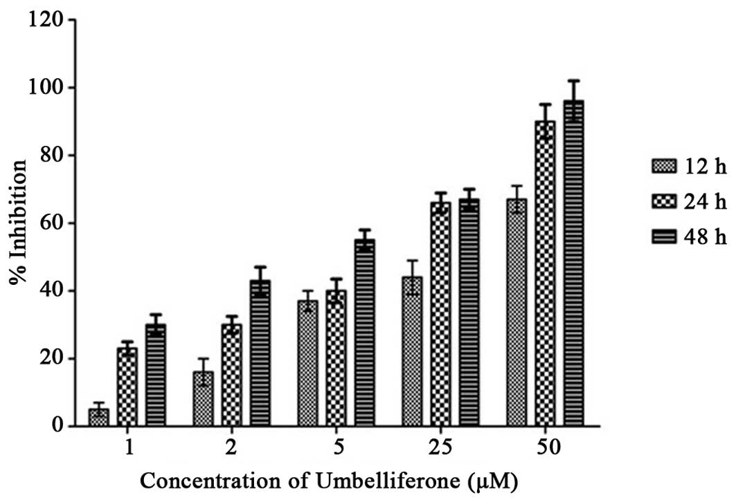
Cytotoxic effect of umbelliferone against
HepG2 cells
HepG2 cells were treated with 1, 2, 5, 25 or 50
µM umbelliferone for 12, 24 and 48 h, prior to the
evaluation of cell viability using an MTT assay. As indicated in
Fig. 1, umbelliferone induced a
dose- and time-dependent reduction in cell viability. The extent of
growth inhibition of various concentrations of umbelliferone on HCC
cells was determined as the percentage of viable treated cells
compared with the percentage of viable cells of untreated
controls.
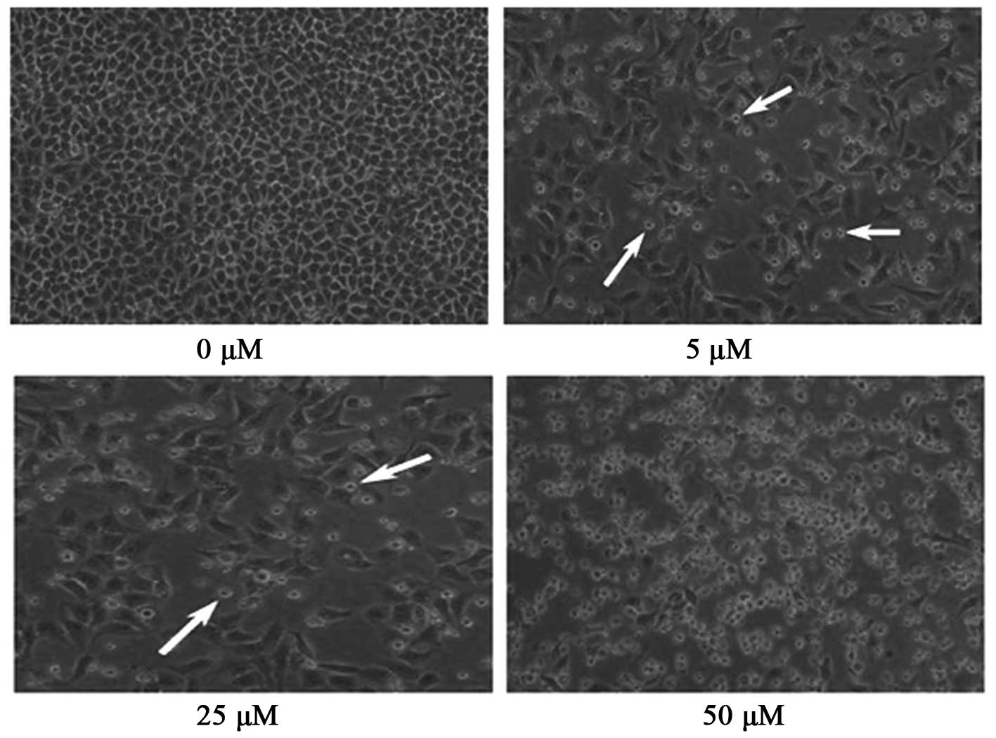
Umbelliferone induces alterations in cell
morphology of HepG2 cancer cells
Malignant cells have developed multiple mechanisms
to facilitate their evasion of apoptosis, the induction of
apoptosis in cancer cells has been suggested to be a potentially
significant strategy useful in cancer therapy and prevention
(13). Apoptosis is a tightly
regulated biochemical process, which is activated in order to
eliminate injured or abnormal cells in multicellular organisms
(14). In order to determine
whether cell death induced by umbelliferone is mediated via
apoptosis, HepG2 cells were treated with various concentrations of
umbelliferone (0, 5, 25 or 50 µM) for 24 h. The cells were
subsequently examined under an inverted light fluorescence
microscope in order to detect the presence of the characteristic
morphological features of apoptosis. As exhibited in Fig. 2, 5
and 25 µM umbelliferone treatment resulted in the appearance
of cell shrinkage and membrane blebbing, when compared with the
morphology of untreated cells (0 µM). Following treatment
with 50 µM umbelliferone, almost all the HepG2 cancer cells
shrank significantly and no cells with normal morphological
features were detected.
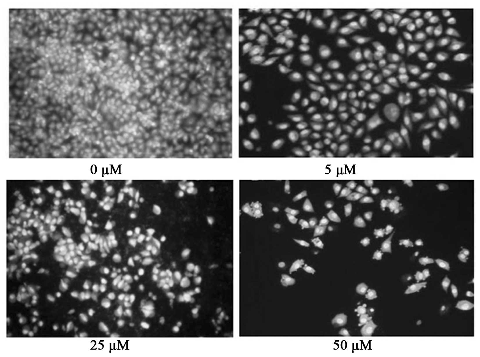
Furthermore, AO and EB double-staining of the HepG2
cells was performed in order to detect cell apoptosis using a
fluorescence microscope. Following AO/EB staining, viable cells (0
µM umbelliferone; Fig. 3)
exhibited large nuclei and intact membranes, which indicated that
no ethidium bromide entered into the cell. However, following
treatment with 5 and 25 µM umbelliferone, the number of
cells exhibiting large nuclei was significantly reduced (Fig. 3). In addition, following treatment
with umbelliferone at a concentration of 50 µM, almost all
cells showed signs of nuclear condensation and apoptotic body
formation.
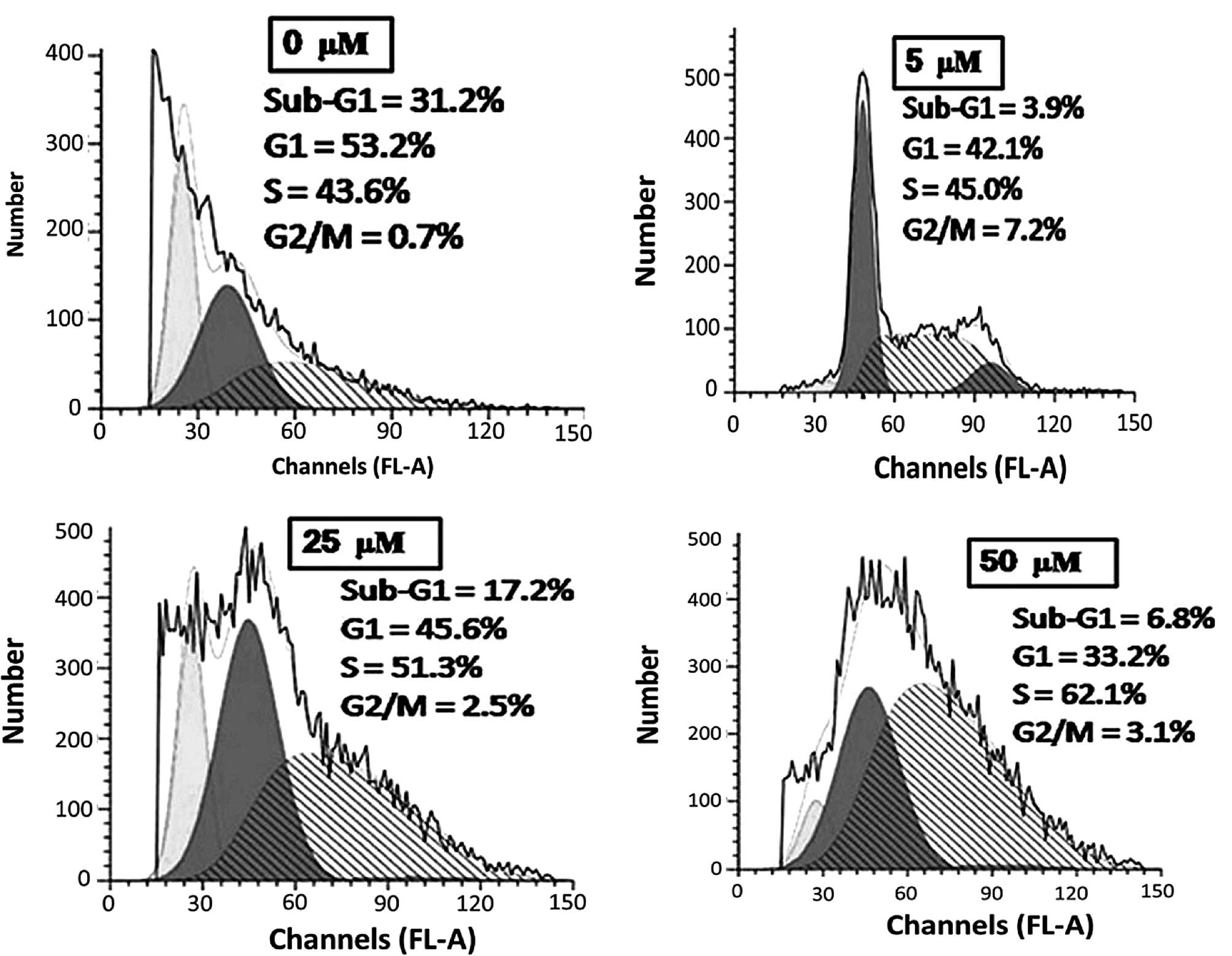
Umbelliferone induces cell cycle arrest
in HepG2 cells
Flow cytometric analysis using PI as a staining
agent was performed following treatment with umbelliferone at
various concentrations (0, 5, 25, and 50 µM) for 24 h, in
order to determine whether umbelliferone induced cell cycle
disturbances in HepG2 cells. As displayed in Fig. 4, following treatment with
umbelliferone at 5, 25 and 50 µM, an increase in the
proportion of cells in S phase (45.0, 51.3 and 62.1%, compared with
43.6% in untreated cells) and a reduction in the fraction of cells
in G1 phase (42.1, 45.6 and 32.2%, compared with 53.2% in the
untreated cells) was observed.
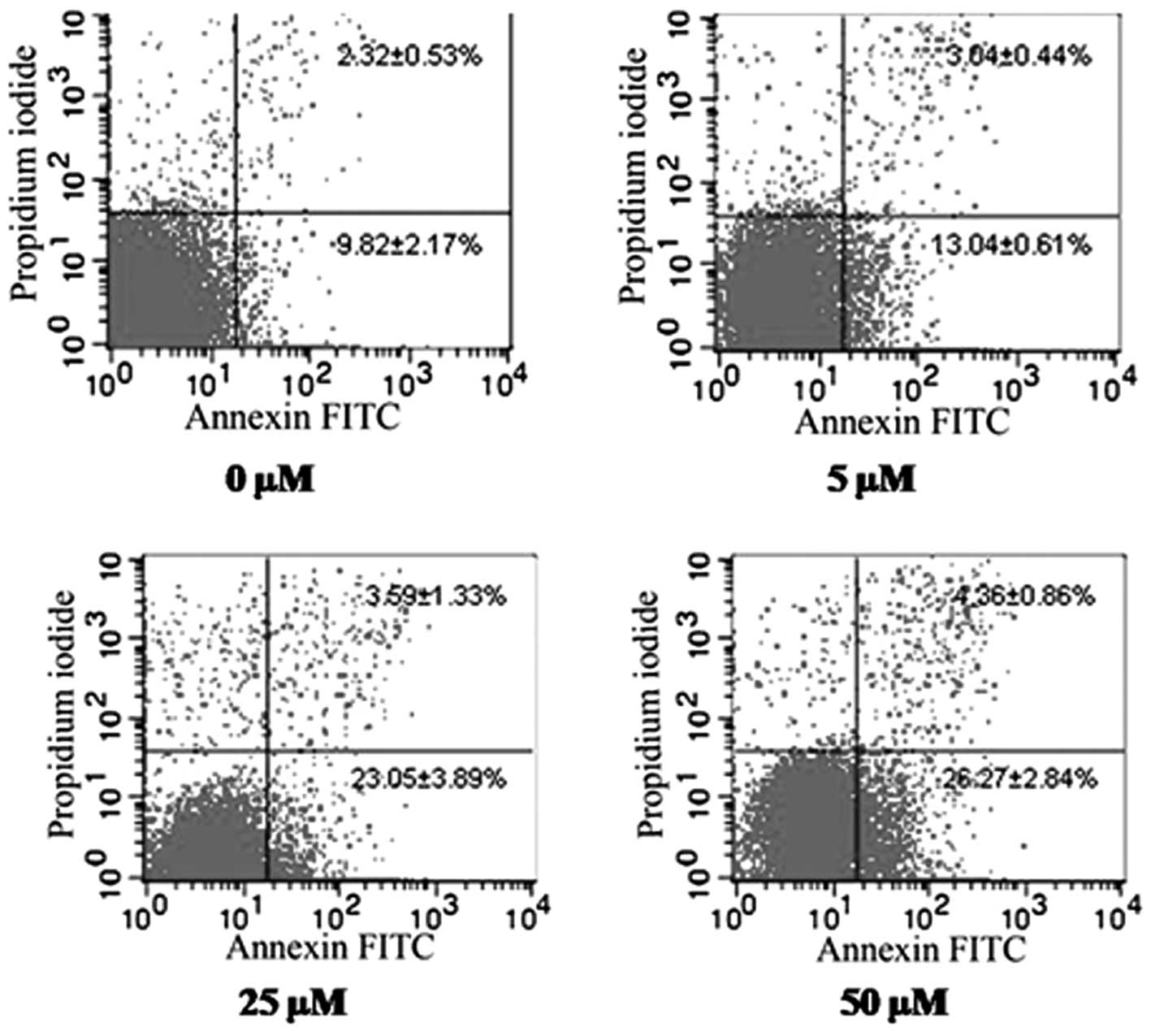
Umbelliferone induces death in HepG2
cells via apoptosis
In the current study, staining was performed using
Annexin V/FITC, a protein that has a high affinity for phosphatidyl
serine (PS) and exhibits fluorescence. PS is a phospholipid
component localized on the cytoplasmic surface of the cell membrane
of normal, viable cells. Following the induction of apoptosis in a
cell, PS is no longer restricted to the cytosolic section of the
membrane and is exposed on the cellular surface. PS translocation
is therefore considered to be a biochemical marker of apoptosis.
Annexin V staining detects the presence of PS and is therefore able
to be used for PS expression analysis (14). Once cells are stained with Annexin
V and PI, the Annexin V/PI stain is only able to enter the cell
when the plasma membrane is sufficiently compromised. This
facilitates distinguishing cells in the early apoptotic phase
(positive for PS, but negative for PI) from the late apoptotic and
necrotic cells (positive for PS and PI) (15,16).
As shown in Fig. 5, increasing
doses of umbelliferone (0–50 µM) resulted in the induction
of apoptotic events, particularly early apoptosis, in a
dose-dependent manner.
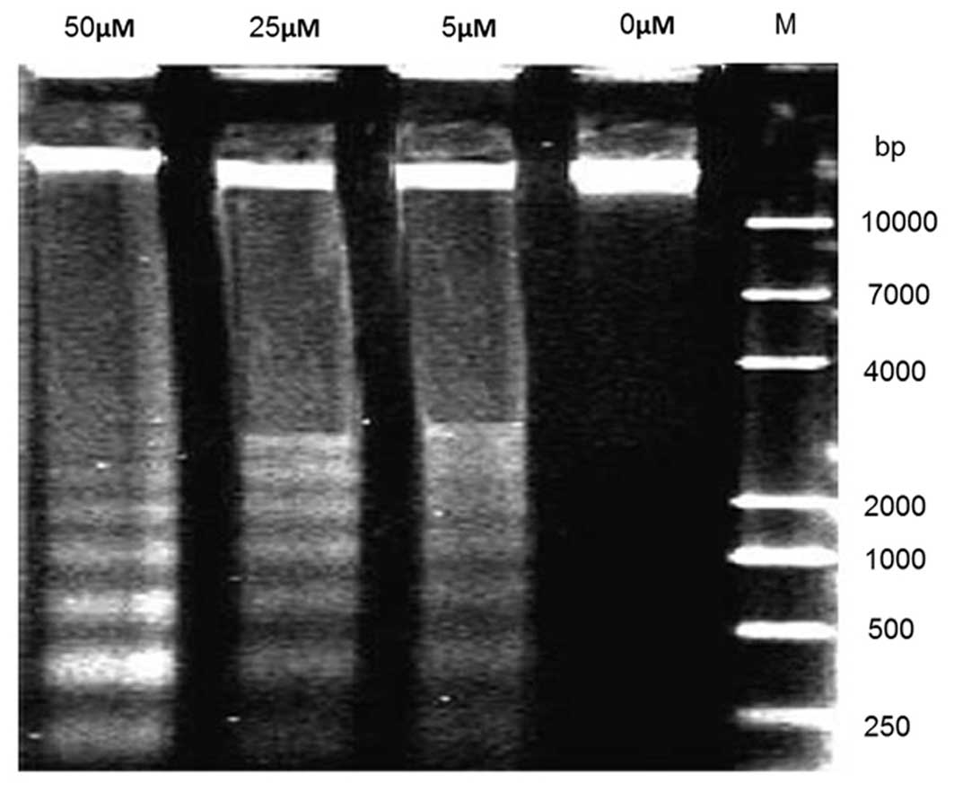
Umbelliferone induces DNA
fragmentation
Pronounced DNA fragmentation was observed in HepG2
cancer cells following treatment with 5, 25 and 50 µM
umbelliferone, for 72 h. However, the control cells (0 µM)
did not produce a clear DNA ladder (Fig. 6).
Discussion
HCC is a type of malignant cancer, associated with a
high incidence and rate of mortality. The therapeutic approaches
currently available for the treatment of HCC remain limited.
Despite of the fact that chemotherapy is one of the most effective
therapeutic approaches for HCC, the associated toxic side effects
mean that it is difficult to tolerate (1). For these reasons, there is an urgent
requirement for the design and development of novel therapeutic
agents, which exhibit higher efficacy and induce fewer serious side
effects (17). Natural
product-based therapies, including the use of natural plant-derived
products and traditional Chinese medicine in cancer therapies, may
aid minimization of the induction of adverse side effects (18). Medicinal and aromatic plants
represent a rich source of anticancer chemotherapeutic drugs, which
exhibit low or no toxicity to normal cells. Increasing attention is
therefore being paid to the search for novel anticancer drugs
derived from natural sources (19).
Umbelliferone (2),
also termed 7-hydroxycoumarin, is a wide-spread natural product of
the coumarin family. Umbelliferone is a yellow-white crystalline
solid, which occurs in numerous well-known plants, including
carrots, coriander, angelica, Hydrangea macrophylla and
Ferula communis. Umbelliferone is currently used in
sunscreens as it is able to absorb ultraviolet light effectively at
multiple wavelengths (20). It has
also been reported to exhibit antinociceptive, anti-inflammatory
and bronchodilator activities, and certain studies have
additionally reported its analgesic activities (21,22).
Umbelliferone has been reported to exhibit antitumor and
immunomodulatory effects against sarcoma 180 in mice, inhibiting
tumor growth and increasing survival time of tumor-bearing animals
(23). Kielbus et al
(24) reported that umbelliferone
inhibited proliferation and migration of laryngeal cancer cells
in vitro. The authors reported that umbelliferone reduced
viability and migration of RK33 laryngeal cancer cells in a
dose-dependent manner (24).
Antitumor activities of umbelliferone have also been reported
against 7,12-dimethylbenz(a)anthracene-induced rat mammary
carcinomas (25).
In conclusion, to the best of our knowledge, the
anticancer activity of umbelliferone against HepG2 cancer cells has
not previously been reported. The current study is therefore the
first to investigate the cytotoxic mechanism of umbelliferone
action. The results revealed that umbelliferone induced apoptosis
in HepG2 cells. Therefore, umbelliferone may have the potential to
be used in the management and treatment of liver cancer, provided
further studies are performed to verify this role.
References
|
1
|
Filipova A, Seifrtova M, Mokry J, Dvorak
J, Rezacova M, Filip S and Diaz-Garcia D: Breast cancer and cancer
stem cells: A mini-review. Tumori. 100:363–369. 2014.PubMed/NCBI
|
|
2
|
Li ZF, Wang ZD, Ji YY, Zhang S, et al:
Induction of apoptosis and cell cycle arrest in human HCC MHCC97H
cells with Chrysanthemum indicum extract. World J Gastroenterol.
15:4538–4546. 2009. View Article : Google Scholar : PubMed/NCBI
|
|
3
|
Lu CX, Nan KJ, Nie YL, Hai YN and Jiao M:
Delisheng, a Chinese medicinal compound, exerts anti-proliferative
and pro-apoptotic effects on HepG2 cells through extrinsic and
intrinsic pathways. Mol Biol Rep. 37:3407–3412. 2010. View Article : Google Scholar :
|
|
4
|
Roomi MW, Roomi NW, Kalinovsky T,
Niedzwiecki A and Rath M: In vivo and in vitro effect of a nutrient
mixture on human hepatocarcinoma cell line SK-HEP-1. Exp Oncol.
32:84–91. 2010.PubMed/NCBI
|
|
5
|
Newman DJ: Natural products as leads to
potential drugs: An old process or the new hope for drug discovery?
J Med Chem. 51:2589–2599. 2008. View Article : Google Scholar : PubMed/NCBI
|
|
6
|
Manosroi J, Sainakham M, Manosroi W and
Manosroi A: Anti-proliferative and apoptosis induction activities
of extract from Thai medicinal plant recipes selected from MANOSROI
II database. J Ethnopharmacol. 141:451–459. 2012. View Article : Google Scholar : PubMed/NCBI
|
|
7
|
Engel N, Oppermanne C, Falodunb A and
Kragle U: Proliferative effects of five traditional Nigerian
medicinal plant extracts on human breast and bone cancer cell
lines. J Ethnopharmacol. 137:1003–1010. 2011. View Article : Google Scholar : PubMed/NCBI
|
|
8
|
Tanaka H, Fujita N, Sugimoto R, Urawa N,
et al: Hepatic oxidative DNA damage is associated with increased
risk for hepatocellular carcinoma in chronic hepatitis C. Br J
Cancer. 98:580–586. 2008. View Article : Google Scholar : PubMed/NCBI
|
|
9
|
Park SH, Lee Y, Han SH, Kwon SY, et al:
Systemic chemotherapy with doxorubicin, cisplatin and capecitabine
for metastatic hepatocellular carcinoma. BMC Cancer. 6:32006.
View Article : Google Scholar : PubMed/NCBI
|
|
10
|
Chen YB, Yan ML, Gong JP, Xia RP, et al:
Establishment of hepatocellular carcinoma multidrug resistant
monoclone cell line HepG2/mdr1. Chinese Med J (Engl). 120:703–707.
2007.
|
|
11
|
Locher C, Conforti R, Aymeric L, Ma Y, et
al: Desirable cell death during anticancer chemotherapy. Ann NY
Acad Sci. 1209:99–108. 2010. View Article : Google Scholar : PubMed/NCBI
|
|
12
|
Jizhong Y and Shengqiang T: Preparative
isolation and purification of two coumarins from Edgeworthia
chrysantha lindl by high speed countercurrent chromatography. J Liq
Chromatogr Relat Technol. 29:1307–1315. 2006. View Article : Google Scholar
|
|
13
|
Taraphdar AK, Roy M and Bhattacharya RK:
Natural products as inducers of apoptosis: Implication for cancer
therapy and prevention. Current Sci. 80:1387–1396. 2001.
|
|
14
|
Yu HY, Zhang XQ, Li X, Zeng FB and Ruan
HL: 2-methoxyjuglone induces apoptosis in HepG2 human
hepatocellular carcinoma cells and exhibits in vivo antitumor
activity in a H22 mouse hepatocellular carcinoma model. J Nat Prod.
76:889–895. 2013. View Article : Google Scholar : PubMed/NCBI
|
|
15
|
Darzynkiewicz Z, Bruno S, Del Bino G,
Gorczyca W, et al: Features of apoptotic cells measured by flow
cytometry. Cytometry. 13:795–808. 1992. View Article : Google Scholar : PubMed/NCBI
|
|
16
|
Nicoletti I, Migliorati G, Pagliacci MC,
Grignani F and Riccardi C: A rapid and simple method for measuring
thymocyte apoptosis by propidium iodide staining and flow
cytometry. J Immunol Meth. 139:271–279. 1991. View Article : Google Scholar
|
|
17
|
Mukherjee AK, Basu S, Sarkar N and Ghosh
AC: Advances in cancer therapy with plant based natural products.
Curr Med Chem. 8:1467–1486. 2001. View Article : Google Scholar : PubMed/NCBI
|
|
18
|
Wang S, Penchala S, Prabhu S, Wang J and
Huang Y: Molecular basis of traditional Chinese medicine in cancer
chemoprevention. Curr Drug Discov Technol. 7:67–75. 2010.
View Article : Google Scholar : PubMed/NCBI
|
|
19
|
Desai AG, Qazi GN, Ganju RK, El-Tame M, et
al: Medicinal plants and cancer chemoprevention. Curr Drug Metab.
9:581–591. 2008. View Article : Google Scholar : PubMed/NCBI
|
|
20
|
Lorenzetti OJ, Boltralik J, Bushy E and
Fortenberr B: The influence of protein vehicles on the
penetrability of sunscreens. J Soc Cosmet Chem. 26:5931975.
|
|
21
|
Leal LK, Ferreira AA, Bezerra GA, Matos FJ
and Viana GS: Antinociceptive, anti-inflammatory and bronchodilator
activities of Brazilian medicinal plants containing coumarin: a
comparative study. J Ethnopharmacol. 70:151–159. 2000. View Article : Google Scholar : PubMed/NCBI
|
|
22
|
Lino CS, Taveira ML, Viana GSB and Matos
FJA: Analgesic and antiinflammatory activities of Justicia
pectoralis Jacq and its main constituents: coumarin and
umbelliferone. Phytother Res. 11:211–215. 1997. View Article : Google Scholar
|
|
23
|
Stefanova TH, Nikolova NJ, Toshkova RA and
Neychev HO: Antitumor and immunomodulatory effect of coumarin and
7-hydroxycoumarin against Sarcoma 180 in mice. J Exp Ther Oncol.
6:107–115. 2007.PubMed/NCBI
|
|
24
|
Kielbus M, Skalicka-Wozniak K, Grabarska
A, Jeleniewicz W, et al: 7-substituted coumarins inhibit
proliferation and migration of laryngeal cancer cells in vitro.
Anticancer Res. 33:4347–4356. 2013.PubMed/NCBI
|
|
25
|
Maucher A and von Angerer E: Antitumour
activity of coumarin and 7-hydroxycoumarin against
7,12-dimethylbenz[a] anthracene-induced rat mammary carcinomas. J
Cancer Res Clin Oncol. 120:502–504. 1994. View Article : Google Scholar
|