Introduction
Apoptosis is a common cause of various diseases. It
is one of the factors that is able to reduce cardiac contractility
and can also lead to heart failure through multiple pathways.
Certain studies have indicated that cardiomyocyte apoptosis
promotes the transition from compensatory cardiac hypertrophy to
heart failure in response to pressure overload. (1–3).
It is well established that the
renin-angiotensin-aldosterone system (RAAS) is activated in
cardiovascular diseases, including hypertension (4), atherosclerosis (5), left ventricular hypertrophy (LVH)
(6) and heart failure (6). Angiotensin II (AngII) is the primary
active peptide hormone of the RAAS, which impairs the homeostasis
between the production and elimination of reactive oxygen species
(ROS) through NADPH oxidase, cause a series of cascade reactions,
and finally leads to cardiomyocyte hypertrophy and apoptosis
(7,8). Ang II also induces H9c2 cardiac cell
hypertrophy, oxidative stress, mitochondrial dysfunction and cell
apoptosis through Ang II type 1 receptor activation (9,10).
Since the mitochondrial membrane potential (MMP) has
central roles in cardiomyocyte apoptosis, Ang II also induces
apoptosis via the mitochondrial-dependent apoptotic pathway
(11); therefore, anti-oxidant
supplementation may ameliorate apoptosis induced by Ang II, which
may be an effective therapeutic method for cardiovascular
disease.
Sanguinarine (SAN), derived from the root of
Sanguinaria canadendid is a benzophenanthridin alkaloid
(12). As a Traditional Chinese
Medicine, SAN has been proved to have significant anti-bacterial,
anti-oxidant, anti-proliferative, anti-tumor and immune enhancing
effects (13). Previous studies
indicated that SAN accelerated cell apoptosis and inhibited cell
proliferation in cancer cells (14,15).
A recent study by our group demonstrated that SAN exerted a
significant protective effect against pressure overload-induced
cardiac remodeling via inhibition of nuclear factor-κB activation
(16). However, whether SAN has
protective effects against Ang II-induced H9c2 cardiac cell
apoptosis has remained elusive. Therefore, the present study
investigated the effect of SAN on the apoptosis, ROS generation and
mitochondrial dysfunction of H9c2 cardiac cells induced by Ang
II.
Materials and methods
Cell culture
The rat cardiomyocyte-derived cell line H9c2 was
obtained from the Cell Bank of the Chinese Academy of Sciences
(GNR5; Shanghai, China). Cells were cultured in 1X Dulbecco's
modified Eagle's medium (DMEM) basic (C11995; Gibco-BRL, Invitrogen
Life Technologies, Carlsbad, CA, USA) supplemented with 10% fetal
bovine serum (FBS; 10099; Gibco-BRL) and 1% penicillin -
streptomycin (PS; 1308300; Gibco-BRL) at 37°C in a humidified
atmosphere containing 5% CO2 (18 M; Sanyo, Osaka,
Japan). Upon reaching 80% confluency, cells were detached with 1 ml
0.25% trypsin-EDTA (1316929; Gibco-BRL) and passaged at a
1:2-ratio. Prior to stimulation, cells were cultured with
serum-free DMEM basic (1X; supplemented with 0.05% PS) for 24 h in
order to eliminate the influence of FBS and synchronize the
cells.
Cell viability assay
Cell viability was measured using a Cell Counting
kit-8 (CCK-8) assay (ER612; Dojindo, Kumamoto, Japan). SAN
(>98%; C2OH14NO4) was purchased
from Shanghai Winherb Medical S&T Development Co., Ltd.
(Shanghai, China). The cells were seeded into 96-well plates at a
density of 1×10⁵ cells/ml and starved for 24 h prior to
being exposed to different concentrations of SAN,
N-acetyl-L-cysteine (NAC, 1 mmol/l; 1009005; Sigma-Aldrich, St
Louis, MO, USA) and co-treatment with Ang II (A9525; Sigma-Aldrich)
for 12 h. After that, 10 µl CCK-8 solution was added to each
well followed by incubation at 37°C for 2.5 h. The samples were
read at 450 nm on a Synergy HT plate reader (Bio-Tek, Winoosky, VT,
USA). The means of the optical density (OD) of the five wells were
used to determine the percentage of viable cells according to the
following formula: Cell viability (%) = OD (treatment group)/OD
(control group) ×100%.
ROS measurement
The level of intracellular ROS generation was
assessed using the fluorescent dye dichlorodihydrofluorescein
diacetate (DCFH-DA; D6883; Sigma-Aldrich). After the indicated
treatments, cells were washed twice with phosphate-buffered saline
(PBS; Beyotime Institute of Biotechnology, Jiangsu, China) and then
incubated with serum-free DMEM basic (1X) containing 10
µmol/l DCFH-DA at 37°C for 30 min. After that, cells were
washed with PBS three times in order to eliminate the residual
DCFH-DA. Cells from each group were analyzed by measuring the
excitation and emission spectrum at 488 and 525 nm, respectively,
using a Synergy HT microplate reader. Data were collected and
analyzed, and the mean fluorescence intensity (FI) of five wells
per group were used to determine the ROS content ratio as a
percentage of the control according to the following formula: ROS
levels (%) = FI (treatment group)/FI (control group) ×100%.
Furthermore, fluorescence microscopy (CX 21FS1C; Olympus, Tokyo,
Japan) was used to confirm the results of the microplate ROS assay.
In brief, after the indicated treatments for 6 h, cells were washed
twice with PBS and then incubated with serum-free DMEM basic (1X)
containing 10 µmol/l DCFH-DA at 37°C for 30 min.
Subsequently, the cells were washed with PBS three times in order
to eliminate the residual DCFH-DA; the climbing glasses of cells
were collected and mounted using SlowFade Gold antifade reagent
with DAPI (Invitrogen Life Technologies). The fluorescence was
visualized using a fluorescence microscope coupled with an image
analysis system (DP2-BSW version 1.3; Olympus).
Measurement of the mitochondrial
transmembrane potential (MMP; ΔΨm)
The change of the mitochondrial membrane potential
(MMP) was assessed using the fluorescent dye
5,5′,6,6′-tetrachloro-1,1′,3,3′-tetraethylimidacarbocyanine iodide
(JC-1; C2005; Beyotime Institute of Biotechnology, Shanghai,
China). After the designated treatment of the cells, 100 µl
serum-free DMEM basic (1X) and 5 µg/ml JC-1 was added to the
cells, followed by incubation at 37°C for 20 min. Following two
washes with PBS, the FI of JC-1 monomer was analyzed by capturing
excitation and emission spectra at 485 and 530 nm, respectively,
with a Synergy HT. JC-1 multimer FI was analyzed via excitation and
emission spectra at 528 and 590 nm, respectively. The mean values
of the five wells were used to determine the percentage of FI
levels according to the following formula: FI levels (%) = FI
(treatment group)/FI (control group) ×100%. In addition, cells
labeled with JC-1 were observed using fluorescence microscopy. JC-1
fluorescence was measured using a single excitation wavelength (485
nm) with dual emission (shift from green at 530 nm to red at 590
nm).
Reverse transcription quantitative
polymerase chain reaction (RT-qPCR)
Total RNA was isolated from H9c2 cardiac cells using
TRIzol reagent (15596-026; Invitrogen Life Technologies). Their
yields and purities were spectrophotometrically estimated using the
absorbance at 260 nm (A260)/A280 and A230/A260 ratios using a
Nanodrop 2000c (Thermo Fisher Scientific, Waltham, MA, USA). The
RNA (2 µg of each sample) was reverse-transcribed into cDNA
using oligo (dT) primers and the Transcriptor First Strand cDNA
Synthesis kit (04896866001; Roche Diagnostics, Basel, Switzerland)
according to the manufacturer's instructions. SYBR Green PCR Master
Mix (04707516001; Roche Diagnostics) was then used to quantify PCR
amplifications using a Light Cycler 480 instrument with designated
software (version 1.5; Roche Diagnostics), the PCR conditions were
as follows: Initial denaturation at 94°C for 2 min, followed by
25–35 amplification cycles consisting of denaturation at 94°C for
40 sec, annealing at 58°C for 45 sec and elongation at 72°C for 1
min. NOX2 mRNA was amplified using the following primers: Forward,
5′-TGA ATC TCA GGC CAA TCA CTTT-3′ and reverse, 5′-AAT GGT CTT GAA
CTC GTT ATCCC-3′. The primers were manufactured by Sangon Biotech
Co., Ltd. (Shanghai, China). The housekeeping gene GAPDH was
employed to normalize gene expression values, using the following
primers: Forward, 5′-GAC ATG CCG CCT GGA GAAAC-3′ and reverse,
5′-AGC CCA GGA TGC CCT TTAGT-3′.
Flow cytometric analysis of
apoptosis
Apoptosis was evaluated using an Annexin
V-fluorescein isothiocyanate (FITC)/propidium iodide (PI) apoptosis
kit (3300222; MultiSciences Biotech, Co., Ltd, Suzhou, China).
After experimental treatment, cells were harvested, washed with
cold PBS and then re-suspended in 500 µl 1X binding buffer
and 5 µl Annexin V-FITC. Following incubation in the dark at
room temperature for 15 min, 10 µl PI was added. Cellular
fluorescence was measured by flow cytometric analysis using a
FACSCalibur flow cytometer (BD Biosciences, Franklin Lakes, NJ,
USA).
Western blot analysis
The cells were lysed in RIPA lysis buffer (Wuhan
Goodbio Technology Co. Ltd., Wuhan, China) containing 50 mM
Tris-Hcl, 150 mM NaCl, 1% Triton X-100, 1% sodium deoxycholate,
0.1% SDS; the cells were then scraped into 1.5-ml centrifuge tubes.
The cell suspension was centrifuged at 3,362 g for 30 min at 4°C,
and the protein concentration was measured using a bicinchoninic
acid protein assay kit (23227; Thermo Fisher Scientific, Cambridge,
MA, USA) using the Synergy HT microplate reader. The cell lysates
(40 µg) were fractionated by 10% SDS-PAGE (12072472;
Invitrogen Life Technologies). After electrophoresis with a Gel
Transfer Device (IB1001; Invitrogen Life Technologies), proteins
were transferred onto a polyvinylidene difluoride membrane
(Millipore, Billerica, MA, USA) and incubated with the appropriate
primary antibodies, including rabbit monoclonal cleaved
(c)-caspase-3 (1:1,000; cat. no. 9664; Cell Signaling Technology,
Danvers, MA, USA), rabbit polyclonal c-caspase-9 (1:1,000; cat. no.
9509P; Cell Signaling Technology), rabbit polyclonal Bcl-2
(1:1,000; cat. no. 2870; Cell Signaling Technology), rabbit
polyclonal Bax (1:1,000; cat. no. 2772; Cell Signaling Technology)
and the membrane was incubated with diluted primary antibody in 5%
w/v nonfat dry milk, 1X Tris-buffered saline, 0.1%
Tween® 20 at 4°C with gentle agitation, overnight.
Thereafter, membranes were incubated with the secondary antibody,
goat anti-rabbit immunoglobulin G (926-32211; LI-COR Biosciences,
Lincoln, NE, USA), for 60 min. The blots were scanned using a
two-color infrared imaging system (Odyssey; LI-COR Biosciences) to
quantify protein expression. Protein expression levels were
normalized to GAPDH (1:1,000; cat. no. 2118, Cell Signaling
Technology).
Caspase-3 and caspase-9 activity
assay
ELISA kits were used to detect the activity of
caspase-3 using the caspase-3 activity kit (C1115; Beyotime
Institute of Biotechnology). Caspase-9 activity was assessed using
the caspase-9 activity kit (H082; Nanjing Jiancheng Bioengineering,
Nanjing, China). The two indicators were measured with the
corresponding detection kit according to the manufacturer's
instructions.
Statistical analysis
Data are expressed as the mean ± standard error of
the mean and analyzed using SPSS 19.0 (SPSS, Inc., Chicago, IL,
USA). Comparisons between two groups were performed using an
unpaired Student's t-test. Differences among groups were de
termined by one-way analysis of variance followed by
Student-Newman-Keuls tests. P<0.05 was considered to indicate a
statistically significant difference between values.
Results
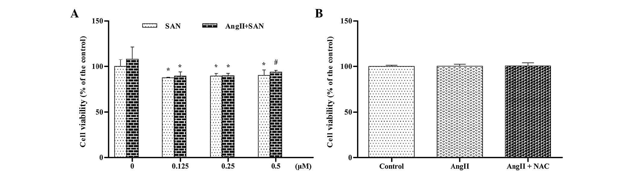
SAN does not affect the viability of H9c2
cardiac cells
The cytotoxicity of SAN and the ROS scavenger NAC
were assessed in the presence or absence of Ang II by CCK-8 assay
(Fig. 1). The viability of H9c2
cardiac cells treated with various concentrations of SAN with or
without Ang II was lower than that of the control group, while cell
viability remained >85% in all groups (Fig. 1A). Furthermore, the viability of
the H9c2 cardiac cells in the Ang II and NAC + Ang II groups was
the same as that in the control group (Fig. 1B). These results indicated that SAN
and NAC exerted no cytotoxic effect on H9c2 cardiac cells.
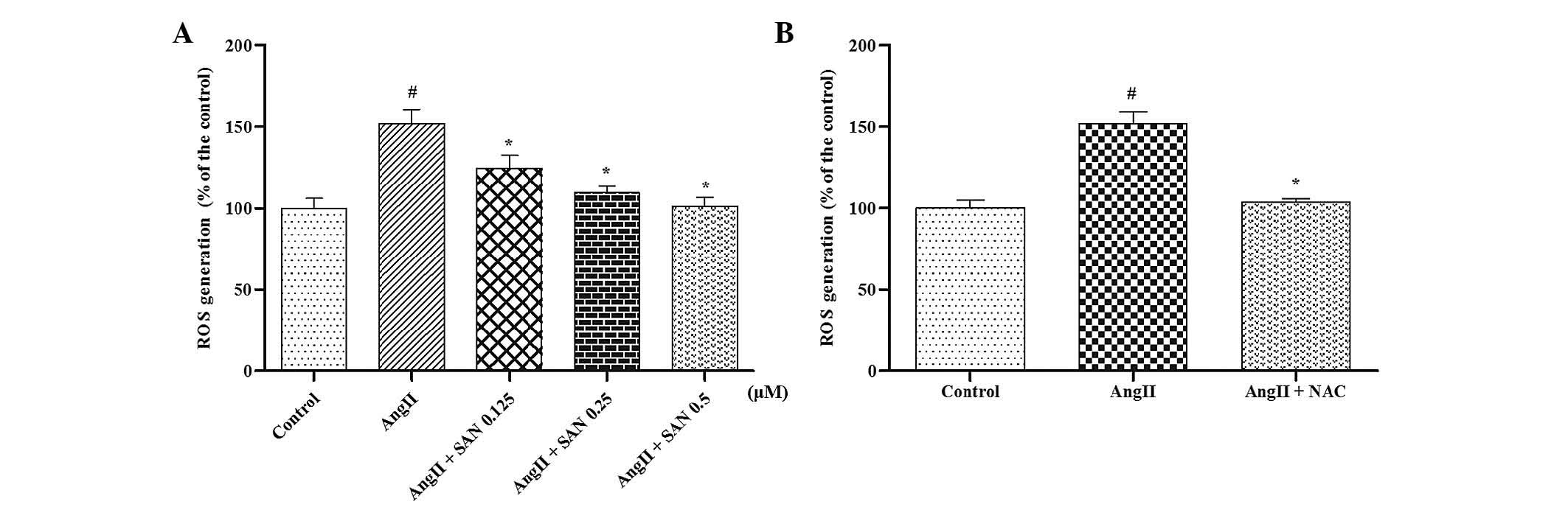
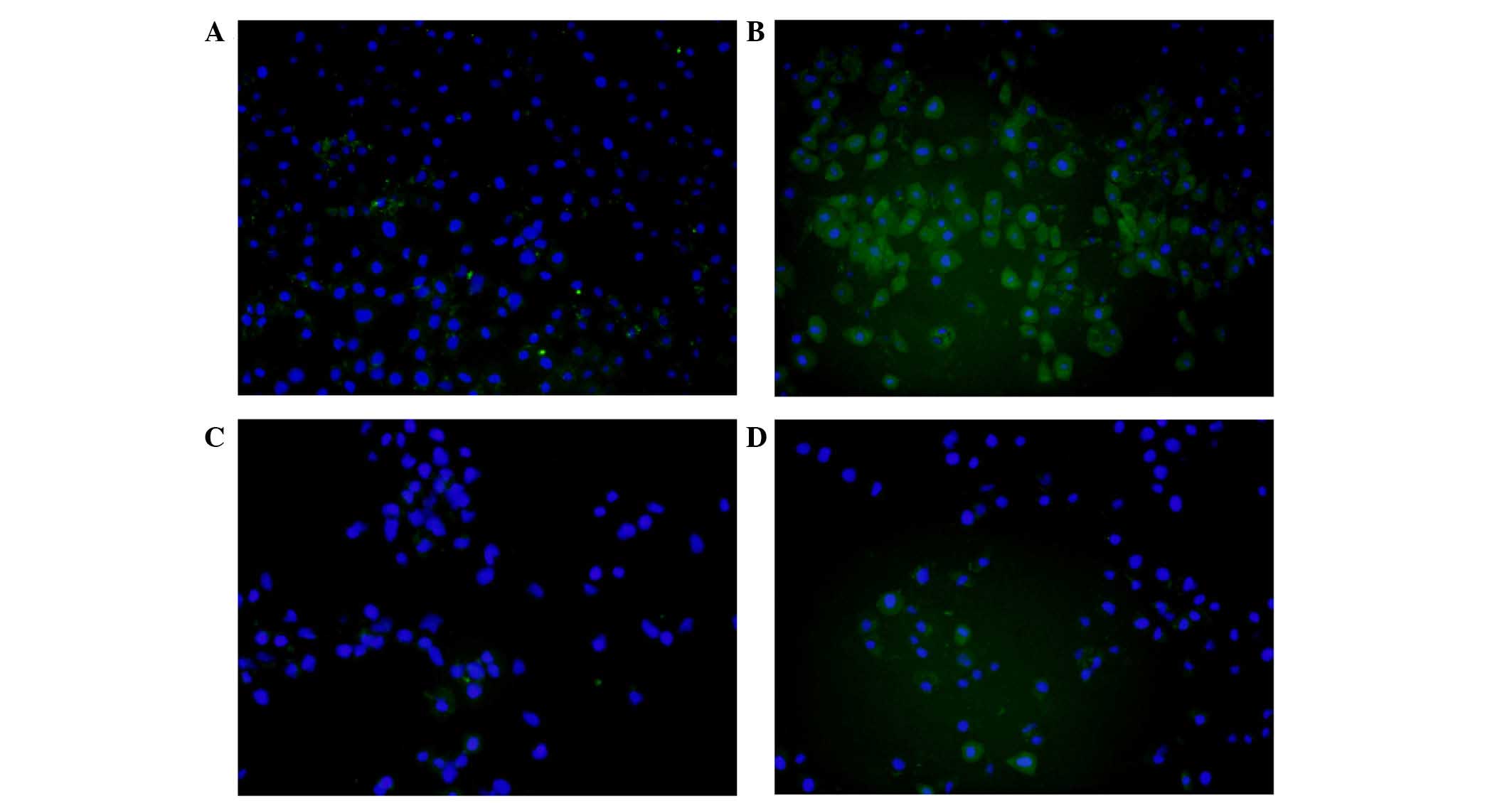
SAN inhibits ROS generation induced by
Ang II
Previous studies showed that Ang II can generate
excess intracellular ROS via stimulation of NADPH oxidase (8). In the present study, a marked
increase in ROS was observed in H9c2 cardiac cells treated with Ang
II, while SAN suppressed ROS generation in a dose-dependent manner
(Fig. 2A). Furthermore, NAC
exerted a similar ROS-decreasing effect following co-treatment with
Ang II (Fig. 2B). These results
were further confirmed by fluorescence microscopy, which showed
that the fluorescence of DCFH-DA observed in the Ang II-treated
group was effectively suppressed following treatment with SAN or
NAC (Fig. 3).
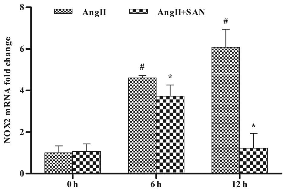
SAN inhibits NOX2 mRNA expression induced
by Ang II
NOX2 is a catalytic subunit of NADPH oxidase and
NOX2 NADPH oxidase has an important role in ROS generation induced
by Ang II. Thus, the present study investigated whether SAN was
able to affect the expression of NOX2 in H9c2 cardiac cells treated
with Ang II. The results showed that Ang II significantly increased
NOX2 mRNA expression in H9c2 cardiac cells in a time-dependent
manner, while the most effective concentration of 0.5 µM SAN
significantly inhibited the elevated expression of NOX2 in a
time-dependent manner (Fig.
4).
SAN ameliorates MMP loss induced by Ang
II
The stability of MMP was measured in H9c2 cardiac
cells after Ang II treatment. The results revealed that the
stability of MMP was significantly impaired by Ang II, as the
amount of JC-1 monomers was increased and that of JC-1 multimers
was decreased in the Ang II-treated group compared with that in the
control group. When H9c2 cardiac cells were co-treated with SAN at
various concentrations, the decreased MMP as well as the amount of
JC-1 monomers and -multimers were restored to normal levels in a
dose-dependent manner (Fig. 5A).
In the positive control group, in analogy to the effect of SAN, NAC
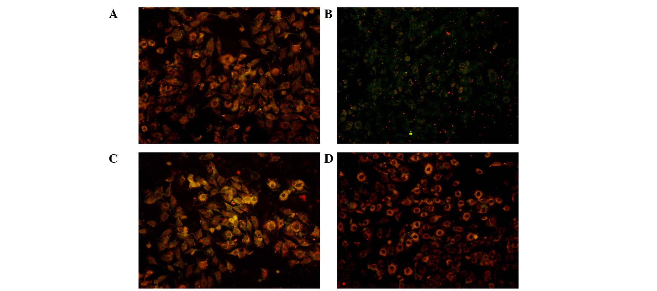
reversed the MMP loss induced by Ang II (Fig. 5B). These results were further
confirmed using fluorescence microscopy. After the indicated
treatments of H9c2 cardiac cells for 24 h, cells were stained with
JC-1 and red/green image densities were measured. The green
fluorescence density, indicating JC-1 monomers the red fluorescence
density indicating multimers. A collapse of the MMP, was markedly
increased in the Ang II group, while treatment with SAN or NAC was
able to shift the green fluorescence to red fluorescence and
therefore a restored MMP (Fig.
6).
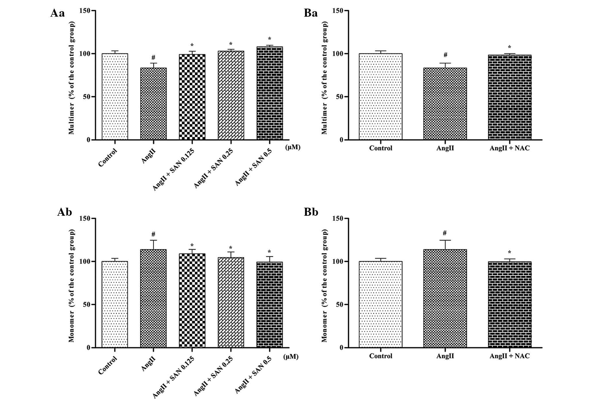 | Figure 5Effects of SAN on MMP loss induced by
Ang II. (A) H9c2 cardiac cells were pre-treated with various
concentrations (0.125, 0.25 and 0.5 µM) of SAN for 40 min
prior to being co-treated with Ang II (10 µM) for 24 h. JC-1
staining indicated that in the Ang II group, the amount of monomers
was increased, while that of multimers was decreased compared with
that in the control group. When H9c2 cardiac cells were co-treated
with the indicated concentrations of SAN, the decreased MMP
returned to normal and (a) the number of JC-1 multimers was
increased, while (b) monomers were decreased. (B) Treatment of the
cells with NAC (1 mM) and Ang II (10 µM) for 24 h caused
similar changes in the MMP changes to that following treatment with
SAN, with (a) increases in JC-1 multimers and (d) decreases in
monomers. Values are expressed as the mean ± standard error of the
mean for three independent experiments. #P<0.01 vs.
control; *P<0.01 vs. Ang II group. SAN, sanguinarine;
Ang, angiotensin; ROS, reactive oxygen species; NAC,
N-acetylcysteine; MMP, mitochondrial membrane potential. |
SAN attenuates Ang II-induced
apoptosis
Previous studies have proved that SAN is able to
regulate cell apoptosis (14).
Thus, the present study investigated the anti-apoptotic effect of
SAN in Ang II-treated H9c2 cardiac cells. Compared with the control
group, Ang II significantly promoted cell apoptosis, as shown by
the Annexin V/PI staining: The early apoptotic rate was
significantly enhanced (25.3%) compared to that in the control
group (1.8%), while SAN and NAC attenuated the level of apoptosis,
with the early apoptotic rate decreased to 14.5 and 14.4%,
respectively (Fig. 7).
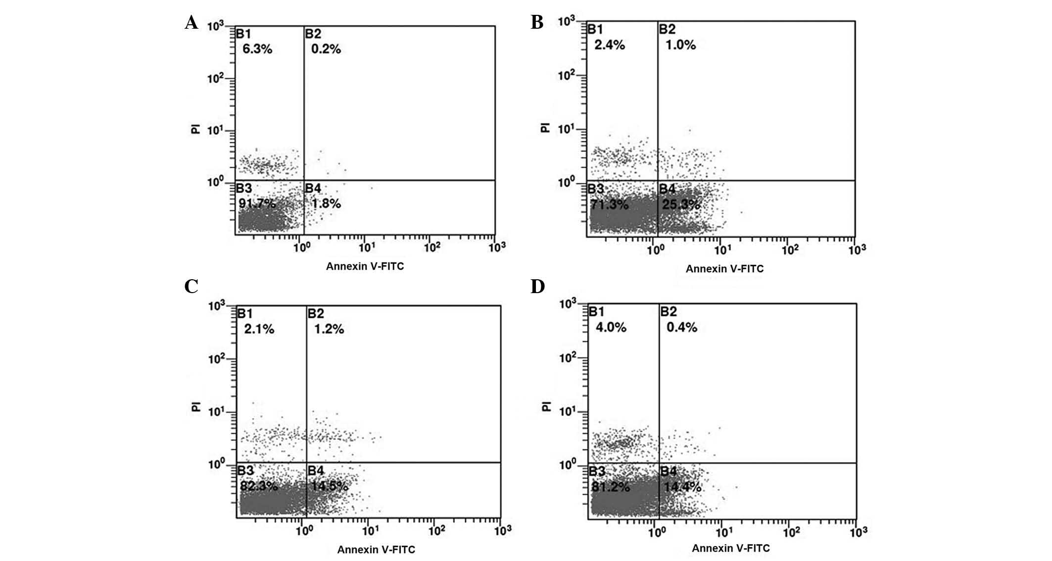 | Figure 7SAN inhibits H9c2 cardiac cell
apoptosis induced by Ang II. Flow cytometry dot plots showing
necrotic cells (Annexin V−/PI+) in the upper left, late apoptotic
cells (Annexin V+/PI+) in the upper right, early apoptotic cells
(Annexin V+/PI−) in the lower right and viable cells (Annexin
V−/PI−) in the lower left. (A) Control group, the percentage of
early apoptotic cells was 1.8%; (B) Stimulated by Ang II (10
µM) only, the percentage of early apoptotic cells was 25.3%;
(C) Stimulated by Ang II (10 µM) and SAN (0.5 µM),
the percentage of early apoptotic cells dropped to 14.5%; (D)
Stimulated by Ang II (10µM) and NAC (1 mM), the percentage
of early apoptotic cells dropped to 14.4%. Values are expressed as
the mean ± standard error of the mean for three independent
experiments. SAN, sanguinarine; Ang, angiotensin; NAC,
N-acetylcysteine; FITC, fluorescein isothiocyanate; PI,
propidium iodide. |
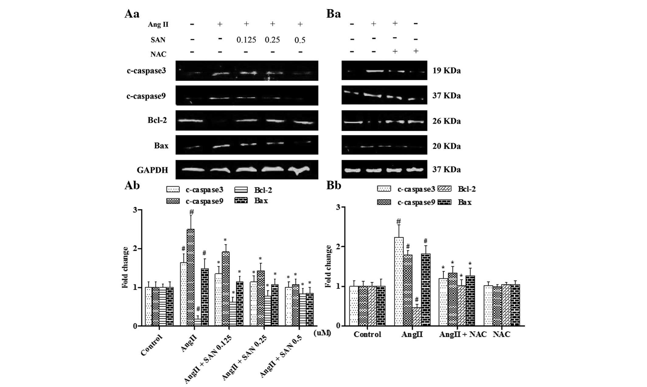
SAN ameliorates the expression of
apoptosis family proteins
The lysate of cells of the experimental groups was
assessed regarding the expression of c-caspase 3, c-caspase 9 and
Bcl-2 family proteins. The expression of c-caspase3 and c-caspase 9
was increased in the Ang II group, while the expression of the
anti-apoptotic protein Bcl-2 was significantly decreased.
Furthermore, the expression levels of pro-apoptotic protein Bax
were increased. These results are in line with the finding that Ang
II induced H9c2 cardiac cell apoptosis. Treatment with SAN or NAC
was able to significantly reduce the Ang II-induced expression of
c-caspase 3 and -9 as well as Bax, and to decrease the expression
of Bcl-1 (Fig. 8). The results
therefore indicated that SAN is able to block apoptosis of cardiac
cells by interfering with the mitochondrial-mediated apoptosis
signaling pathway.
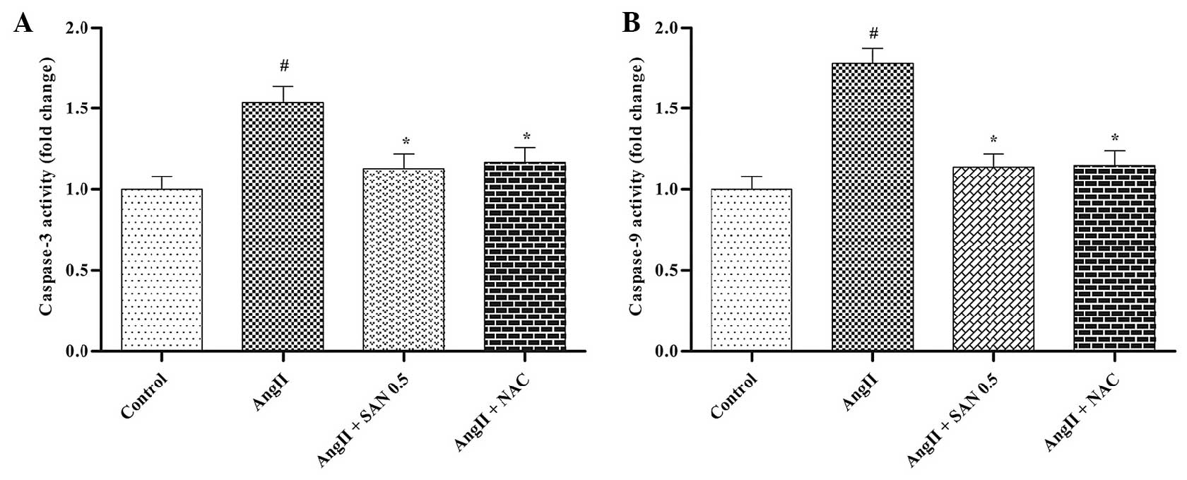
SAN inhibits caspase-3 and caspase-9
activation induced by Ang II
Caspase-3 and caspase-9 activity were significantly
increased in Ang II-treated H9c2 cardiac cells, while SAN and NAC
significantly inhibited Ang II-induced caspase activation (Fig. 9). This result further confirmed the
mechanism of action of SAN as an inhibitor of the
mitochondrial-mediated apoptotic pathway to inhibit apoptosis of
cardiac cells.
Discussion
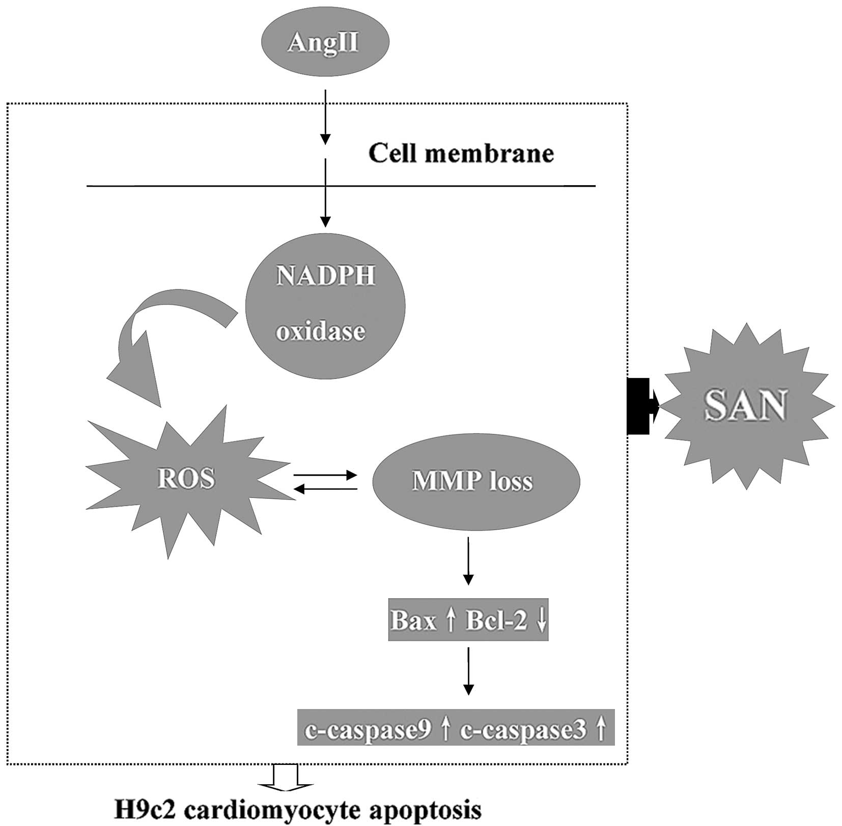
The present study demonstrated that Ang II increased
ROS generation, initiating a signaling cascade contributing to MMP
loss and resulting in an up-regulation of apoptosis of H9c2 cardiac
cells. Of note, SAN ameliorated ROS generation and MMP loss in H9c2
cardiac cells induced by Ang II, and decreased caspase 3 and -9
protein expression and activity, as well as enhancing the Bcl-2/Bax
ratio. In addition, the present study demonstrated that nearly all
of these pro-apoptotic factors of Ang II were eliminated by NAC as
a positive control treatment. These results indicated that SAN
inhibits H9c2 cardiac cell apoptosis caused by Ang II, which is
likely to be due to restoration of ROS-mediated decreases of the
MMP (Fig. 10).
Increasing evidence suggests that Ang II treatment
significantly increases NADPH oxidase activity via the Ang II
receptor, type 1, and subsequently leads to ROS generation, which
can cause damage to mitochondria and lipid peroxidation (17,18).
A study by Chu et al (19)
showed that Ang II was able to stimulate intracellular
Ca2+ accumulation, which altered the MMP and caused a
release of cytochrome C from the mitochondria to the cytoplasm and
subsequent apoptotic cascades in neonatal rat ventricular myocytes.
Chang et al (20)
demonstrated that caspase 3 and -9 were increased in H9c2 cardiac
cells stimulated by Ang II, which was confirmed by the present
study. Therefore, it was hypothesized that Ang II causes
mitochondrial damage and MMP loss via ROS generation and subsequent
activation of apoptosis, and the present study was designed to
investigate whether SAN was able to decrease ROS generation in H9c2
cardiac cells and ameliorate MMP loss and apoptosis caused by Ang
II.
Numerous studies have focused on SAN as an
anti-oxidant and anti-inflammatory drug (21–23).
Ahmad et al (14) showed
that low-dose SAN (1 µM) treatment of A431 cells resulted in
a significantly decreased cell viability and an enhanced apoptotic
index, while this treatment had no effect on NHEK normal
keratinocyte cells, which exclusively showed necrotic staining at
the high doses of 2–5 µM. In the present study, in order to
avoid H9c2 cardiac cell necrosis, a maximum SAN concentration of
0.5 µM was used, and after treatment for 12 h, the viability
of H9c2 cardiac cells treated with different concentrations of SAN
with or without Ang II was lower than that in the control group,
while always remaining >85%; this indicated that SAN had no
significant toxicity to H9c2 cardiac cells.
The chemical reactivity of SAN is based on the
nucleophilic character of its iminium moiety, which may participate
in oxidant scavenging and/or enzyme inhibition (24). SAN was shown to inhibit phorbol
myristate-induced oxidative burst (25), and the most important enzyme in
oxidative burst is the NADPH oxidase complex (26). SAN may have exerted its
anti-oxidative function by impairing the activity of the NADPH
enzyme, which is supported by a study by Qin et al (26), which demonstrated that SAN is an
enzyme inhibitor rather than an ROS scavenger. NADPH oxidases are
transmembrane enzymes designated to produce super-oxide by
transferring an electron from NADPH to molecular oxygen. NOX2 is a
major NADPH oxidase isoform expressed in cardiac cells (27,28),
and it is well established that several actions of NOX2 NADPH in
cardiac remodeling are activated through activation by Ang II, and
alongside tumor necrosis factor alpha, it constitutes the main
source of ROS generation (29,30).
NOX2-derived ROS has a major role in the regulation of cardiac
hypertrophy, apoptosis, fibrosis and mitochondrial dysfunction
(26,31,32).
As NOX2 NADPH oxidase has an important role in ROS generation
induced by Ang II, it was speculated that SAN impaired the
generation of ROS through decreasing the expression of NOX2 NADPH
oxidase. These hypotheses were confirmed by the results of the
present study, showing that SAN (0.5 µM) inhibited NOX2
NADPH oxidase activity in a time-dependent manner and that ROS
generation induced by Ang II was also inhibited by SAN in a
dose-dependent manner.
In the present study, ROS were significantly
elevated and the MMP was declined in the Ang II-treated group. A
high level of ROS production may cause mitochondrial oxidative
attack as well as accumulating damage to mitochondrial DNA and
proteins, which further stimulates ROS generation (ROS-induced ROS
release) (33). These interactions
cause mutual damage and lead to H9c2 cardiac cell apoptosis, which,
however, was demonstrated to be inhibited by pre-treating the H9c2
cardiac cells with SAN. NOX2 levels induced by Ang II were also
inhibited by SAN, and Ang II-induced H9c2 cardiac cell apoptosis as
well as c-caspase 3 and -9 expression were significantly reduced by
SAN. Furthermore, the present study investigated the possible
molecular pathway underlying the anti-apoptotic effect of SAN. The
results showed that the expression of Bcl-2 was decreased, while
Bax increased following treatment with Ang II, which was rescued by
treatment with SAN. In addition, Ang II enhanced the activation of
caspase 9 and caspase 3, while pre-treatment of H9c2 cardiac cells
with SAN blocked these effects. Pre-treatment with NAC as a
positive reference exerted similar effects to those of SAN on
protein expression, changes in the MMP and the apoptotic index in
response to Ang II.
In conclusion, the present study provided novel
insight into the cardioprotective effect of SAN as well as the
underlying molecular mechanisms. SAN inhibits ROS generation, MMP
loss and apoptosis of H9c2 cardiac cells induced by Ang II,
possibly via inhibiting NOX2 and the mitochondrial-mediated
apoptotic pathway. Although the precise mechanism remains to be
fully elucidated, the present study may contribute to the selection
of SAN as a candidate drug for the treatment or prevention of
cardiovascular diseases.
Acknowledgments
This study was supported by the Fundamental Research
Funds for the Central Universities of China (2012302020211).
References
|
1
|
Wencker D, Chandra M, Nguyen K, et al: A
mechanistic role for cardiac myocyte apoptosis in heart failure. J
Clin Invest. 111:1497–1504. 2003. View
Article : Google Scholar : PubMed/NCBI
|
|
2
|
Mihl C, Dassen WR and Kuipers H: Cardiac
remodelling: concentric versus eccentric hypertrophy in strength
and endurance athletes. Neth Heart J. 16:129–133. 2008. View Article : Google Scholar : PubMed/NCBI
|
|
3
|
Qipshidze-Kelm N, Piell KM, Solinger JC
and Cole MP: Co-treatment with conjugated linoleic acid and nitrite
protects against myocardial infarction. Redox Biol. 2:1–7. 2013.
View Article : Google Scholar : PubMed/NCBI
|
|
4
|
Whaley-Connell A, Johnson MS and Sowers
JR: Aldosterone: role in the cardiometabolic syndrome and resistant
hypertension. Prog Cardiovasc Dis. 52:401–409. 2010. View Article : Google Scholar : PubMed/NCBI
|
|
5
|
Aroor AR, Demarco VG, Jia G, et al: The
role of tissue renin-angiotensin-aldosterone system in the
development of endothelial dysfunction and arterial stiffness.
Front Endocrinol (Lausanne). 4:1612013.
|
|
6
|
Sayer G and Bhat G: The
renin-angiotensin-aldosterone system and heart failure. Cardiol
Clin. 32:21–32. 2014. View Article : Google Scholar
|
|
7
|
Xuan CL, Yao FR, Guo LR, et al: Comparison
of extracts from cooked and raw lentil in antagonizing angiotensin
II-induced hypertension and cardiac hypertrophy. Eur Rev Med
Pharmacol Sci. 17:2644–2653. 2013.PubMed/NCBI
|
|
8
|
Bendall JK, Cave AC, Heymes C, Gall N and
Shah AM: Pivotal role of a gp91(phox)-containing NADPH oxidase in
angiotensin II-induced cardiac hypertrophy in mice. Circulation.
105:293–296. 2002. View Article : Google Scholar : PubMed/NCBI
|
|
9
|
Byrne JA, Grieve DJ, Bendall JK, et al:
Contrasting roles of NADPH oxidase isoforms in pressure-overload
versus angiotensin II-induced cardiac hypertrophy. Circ Res.
93:802–805. 2003. View Article : Google Scholar : PubMed/NCBI
|
|
10
|
Nakagami H, Takemoto M and Liao JK: NADPH
oxidase-derived superoxide anion mediates angiotensin II-induced
cardiac hypertrophy. J Mol Cell Cardiol. 35:851–859. 2003.
View Article : Google Scholar : PubMed/NCBI
|
|
11
|
Adams JW, Pagel AL, Means CK, Oksenberg D,
Armstrong RC and Brown JH: Cardiomyocyte apoptosis induced by
Galphaq signaling is mediated by permeability transition pore
formation and activation of the mitochondrial death pathway. Circ
Res. 87:1180–1187. 2000. View Article : Google Scholar : PubMed/NCBI
|
|
12
|
Dong XZ, Zhang M, Wang K, et al:
Sanguinarine inhibits vascular endothelial growth factor release by
generation of reactive oxygen species in MCF-7 human mammary
adenocarcinoma cells. Biomed Res Int. 2013:5176982013. View Article : Google Scholar : PubMed/NCBI
|
|
13
|
Chaturvedi MM, Kumar A, Darnay BG, Chainy
GB, Agarwal S and Aggarwal BB: Sanguinarine (pseudochelerythrine)
is a potent inhibitor of NF-kappaB activation, IkappaBalpha
phosphorylation and degradation. J Biol Chem. 272:30129–30134.
1997. View Article : Google Scholar : PubMed/NCBI
|
|
14
|
Ahmad N, Gupta S, Husain MM, Heiskanen KM
and Mukhtar H: Differential antiproliferative and apoptotic
response of sanguinarine for cancer cells versus normal cells. Clin
Cancer Res. 6:1524–1528. 2000.PubMed/NCBI
|
|
15
|
Burgeiro A, Bento AC, Gajate C, Oliveira
PJ and Mollinedo F: Rapid human melanoma cell death induced by
sanguinarine through oxidative stress. Eur J Pharmacol.
705:109–118. 2013. View Article : Google Scholar : PubMed/NCBI
|
|
16
|
Deng W, Fang Y, Liu Y, et al: Sanguinarine
protects against pressure overloadinduced cardiac remodeling via
inhibition of nuclear factor-kappaB activation. Mol Med Rep.
10:211–216. 2014.PubMed/NCBI
|
|
17
|
Choi WY, Jin CY, Han MH, et al:
Sanguinarine sensitizes human gastric adenocarcinoma AGS cells to
TRAIL-mediated apoptosis via down-regulation of AKT and activation
of caspase-3. Anticancer Res. 29:4457–4465. 2009.PubMed/NCBI
|
|
18
|
Liu JJ, Li DL, Zhou J, et al:
Acetylcholine prevents angiotensin II-induced oxidative stress and
apoptosis in H9c2 cells. Apoptosis. 16:94–103. 2011. View Article : Google Scholar
|
|
19
|
Chu CH, Lo JF, Hu WS, et al: Histone
acetylation is essential for ANG-II-induced IGF-IIR gene expression
in H9c2 cardiomyoblast cells and pathologically hypertensive rat
heart. J Cell Physiol. 227:259–268. 2012. View Article : Google Scholar
|
|
20
|
Chang YM, Tsai CT, Wang CC, et al:
Alpinate oxyphyllae fructus (Alpinia Oxyphylla Miq) extracts
inhibit angiotensin-II induced cardiac apoptosis in H9c2
cardiomyoblast cells. Biosci Biotechnol Biochem. 77:229–234. 2013.
View Article : Google Scholar : PubMed/NCBI
|
|
21
|
Kumar A, Husain F, Das M and Khanna SK: An
out-break of epidemic dropsy in the Barabanki District of Uttar
Pradesh, India: a limited trial for the scope of antioxidants in
the management of symptoms. Biomed Environ Sci. 5:251–256.
1992.PubMed/NCBI
|
|
22
|
Vavrecková C, Ulrichová J, Hajdúch M,
Grambal F, Weigl E and Simánek V: Effect of quaternary
benzo[c]phenanthridine alkaloids sanguinarine, chelerythrine and
fagaronine on some mammalian cells. Acta Univ Palacki Olomuc Fac
Med. 138:7–10. 1994.
|
|
23
|
Chaturvedi MM, Kumar A, Darnay BG, Chainy
GB, Agarwal S and Aggarwal BB: Sanguinarine (pseudochelerythrine)
is a potent inhibitor of NF-kappaB activation, IkappaBalpha
phosphorylation, and degradation. J Biol Chem. 272:30129–30134.
1997. View Article : Google Scholar : PubMed/NCBI
|
|
24
|
Ulrichova J, Dvorák Z, Vicar J, et al:
Cytotoxicity of natural compounds in hepatocyte cell culture
models. The case of quaternary benzo[c]phenanthridine alkaloids.
Toxicol Lett. 125:125–132. 2001. View Article : Google Scholar
|
|
25
|
Varga Z, Czompa A, Kakuk G and Antus S:
Inhibition of the superoxide anion release and hydrogen peroxide
formation in PMNLs by flavonolignans. Phytother Res. 15:608–612.
2001. View
Article : Google Scholar : PubMed/NCBI
|
|
26
|
Qin F, Patel R, Yan C and Liu W: NADPH
oxidase is involved in angiotensin II-induced apoptosis in H9C2
cardiac muscle cells: effects of apocynin. Free Radic Biol Med.
40:236–246. 2006. View Article : Google Scholar : PubMed/NCBI
|
|
27
|
Kim YM, Guzik TJ, Zhang YH, et al: A
myocardial Nox2 containing NAD(P)H oxidase contributes to oxidative
stress in human atrial fibrillation. Circ Res. 97:629–636. 2005.
View Article : Google Scholar : PubMed/NCBI
|
|
28
|
Niccoli G, Celestini A, Calvieri C, et al:
Patients with micro-vascular obstruction after primary percutaneous
coronary intervention show a gp91phox (NOX2) mediated persistent
oxidative stress after reperfusion. Eur Heart J Acute Cardiovasc
Care. 2:379–388. 2013. View Article : Google Scholar : PubMed/NCBI
|
|
29
|
Wang G, Anrather J, Glass MJ, et al: Nox2,
Ca2+ and protein kinase C play a role in angiotensin II-induced
free radical production in nucleus tractus solitarius.
Hypertension. 48:482–489. 2006. View Article : Google Scholar : PubMed/NCBI
|
|
30
|
Zhang J, Chandrashekar K, Lu Y, et al:
Enhanced expression and activity of Nox2 and Nox4 in the macula
densa in ANG II-induced hypertensive mice. Am J Physiol Renal
Physiol. 306:F344–F350. 2014. View Article : Google Scholar :
|
|
31
|
Kuroda J and Sadoshima J: NADPH oxidase
and cardiac failure. J Cardiovasc Transl Res. 3:314–320. 2010.
View Article : Google Scholar : PubMed/NCBI
|
|
32
|
Nabeebaccus A, Zhang M and Shah AM: NADPH
oxidases and cardiac remodelling. Heart Fail Rev. 16:5–12. 2011.
View Article : Google Scholar
|
|
33
|
Ide T, Tsutsui H, Kinugawa S, et al:
Mitochondrial electron transport complex I is a potential source of
oxygen free radicals in the failing myocardium. Circ Res.
85:357–363. 1999. View Article : Google Scholar : PubMed/NCBI
|