Introduction
Colorectal cancer is one of the leading causes
cancer-associated mortality and morbidity in several countries
(1). Colorectal cancer, a highly
prevalent cancer in males and females, includes colon cancer and
rectal cancer, due to their adjacent anatomical locations. Colon
cancer is the second most commonly diagnosed type of cancer in
females and the third in males worldwide (2). Carethers et al demonstrated a
higher prevalence for microsatellite instability (MSI) among
cancers patients of African American ethnicity, and the MSI
prevalence in these cancer patients was observed to be half that of
Caucasian patients without changes in CD8+ T cell
infiltration, which may contribute towards the higher mortality
rates in the former group from colon cancer (3). Several tumor biomarkers, including
carcinoembryonic antigen (CEA), carbohydrate antigen (CA) 19.9 and
CA 12.5, have been detected in a number of patients with colon
cancer (4). The levels of CEA and
CA19.9 are often increased in advanced colon cancer (5) and have been considered as an early
biological signal of colon cancer recurrence (6). Increasing current knowledge of cancer
cell proliferation, migration and invasion are central to
understanding tumor progression and metastasis. The local tumor
environment may provide specific environmental cues to alter the
behavior of the cells and promote metastasis.
For the last two decades, the liver has been the
most frequent site of colon cancer metastasis (7), which affects the efficacy of
treatment and the prognosis of patients. Therefore, it is important
that the pathogenesis and treatment of colon cancer is
addressed.
Human trophoblast cell-surface marker (TROP-2) is a
cell surface glycoprotein, which was originally identified in human
placental trophoblasts and has been reported to be highly expressed
in various types of human carcinoma, including oral (8), lung (9), pancreatic (10) and gastric carcinoma (11). By contrast, the expression of
TROP-2 in adult somatic tissues is minimal or absent (12). TROP-2, as a type I transmembrane
protein, has been cloned from human (13) and mouse cells (14), and the overexpression of TROP-2
cell surface protein was correlated with aggressive behavior and
poor prognosis in certain types of epithelial carcinoma by Coldren
et al (15). It has been
reported that TROP-2 is important in determining the fate of tumor
growth, and the expression of TROP-2 may be necessary for
anchorage-independent growth, invasiveness and tumorigenesis in
cancer cells (16). Notably,
previous studies have suggested that overexpression of TROP-2 is
associated with increased tumor aggressiveness and metastasis, and
decreased patient survival rate (17). Therefore, there is a increasing
interest in TROP-2 as a potential therapeutic target for solid
types of cancer (18). For
example, hRS7, a humanized anti-TROP-2 antibody, is currently under
clinical trial as a drug for patients with advanced epithelial
cancer (19).
The functional role of TROP-2 in cancer remains to
be fully elucidated. Several studies have demonstrated that the
TROP-2 gene encodes a tumor-associated calcium signal transducer,
which is involved in the regulation of cell-cell adhesion, and an
antibody against TROP-2 has been observed to alter intracellular
calcium levels (20,21). Other studies have suggested that
TROP-2 is highly expressed in certain developing tissues, it may be
important in morphogenesis (18,22,23).
Notably, evidence has revealed that humans born with homozygous
inactivating mutations in TROP-2 exhibited only limited pathology
(24). The biological function of
TROP-2 is focused on promoting self-renewal and hyperplasia in the
prostate, which has been attributed to the accumulation of the
intracellular domain of TROP-2 in the nucleus, following its
cleavage through regulating intramembrane proteolysis (25). Additionally, reports suggest that
prostate basal cells overexpressing TROP-2 possess stem cell
capacities, including tissue regeneration, self-renewal and
multilineage differentiation (25,26).
To the best of our knowledge, the expression and
biological function of TROP-2 in the tumorigenesis and invasiveness
of colon cancer has not been investigated. Therefore, the purpose
of the present study was to investigate the expression of TROP-2 in
colon cancer tissues, and to assess its biological and clinical
significance.
Patients and methods
Patients
Cancer tissues and tumor-adjacent tissues, the
latter sited 5 cm from the cancer tissue edge and confirmed as
normal tissue by a pathologist, were obtained from 82 patients
diagnosed with colon cancer, who had not received radiation,
chemotherapy or immunotherapy prior to surgery. Normal tissue
specimens were collected from 30 patients with non-neoplastic colon
mucosa. All specimens were obtained from the Department of General
Surgery, Hebei United University Affiliated Hospital (Hebei,
China), between January 2012 and December 2013. Eligible cases were
between the ages of 35 and 90 years (59.8±5.7), included 53 male
cases and 30 female cases, and had pathologically confirmed
invasive adenocarcinoma of the colon. Additional clinicopathologic
features of the colon cancer are listed in Table 1. Under sterile conditions, tissues
samples of 0.5 cm diameter were obtained and shock-frozen in liquid
nitrogen. The present study was approved by the Ethics Committee of
Hebei Medical University (Shijiazhuang, China), and written
informed consent was obtained from all patients enrolled.
 | Table IAssociation between the expression of
TROP-2 in and clinicopathological features in patients with colon
cancer. |
Table I
Association between the expression of
TROP-2 in and clinicopathological features in patients with colon
cancer.
| Factor | n | TROP-2(+) | TROP-2(−) | P-value |
|---|
| Gender | | | | |
| Male | 52 | 48 | 4 | 0.512 |
| Female | 30 | 27 | 3 | |
| Age (years) | | | | |
| ≤59 | 39 | 34 | 5 | 0.684 |
| >59 | 43 | 41 | 2 | |
| Differentiation | | | | |
| High | 24 | 21 | 3 | 0.857 |
| Middle | 33 | 30 | 3 | |
| Low | 25 | 24 | 1 | |
| Lymph node
metastases | | | | |
| Yes | 54 | 51 | 3 | 0.018 |
| No | 28 | 24 | 4 | |
| Dukes stage | | | | |
| A+B | 38 | 33 | 5 | 0.002 |
| C+D | 44 | 42 | 2 | |
Western blot analysis
Total proteins were extracted from the tissues with
radioimmunoprecipitation assay buffer (Cell Signaling Technology,
Inc., Danvers, MA, USA) with the addition of Halt Protease
Inhibitor Cocktail (Cell Signaling Technology, Inc.), and protein
concentration was determined using a bicinchoninic acid reagent
(Solarbio, Beijing, China) method. The samples were then subjected
to 10% sodium dodecyl sulfate polyacrylamide gel electrophoresis
(SDS-PAGE). The separated proteins on the gel were transferred onto
polyvinylidene difluoride membranes (Roche Diagnostics, Mannheim,
Germany). The blots were blocked with 5% fat-free dry milk for 1 h
at room temperature. Subsequently, the blots were incubated
overnight at 4°C with the following primary antibodies: Rabbit
anti-human TROP-2 polyclonal antibody (cat. no., sc-80406;
dilution, 1:400; Santa Cruz Biotechnology, Inc., Santa Cruz, CA,
USA) and rabbit anti-human β-actin monoclonal antibody (cat. no.,
sc-130657; dilution, 1:600; Santa Cruz Biotechnology, Inc.). The
blots were then incubated with horseradish peroxidase-conjugated
anti-rabbit immunoglobulin (IgG; 1:5,000; Cell Signaling
Technology, Inc.) for 1 h at room temperature. The immunoblot on
the membrane was visible following development using an enhanced
chemiluminescence detection system (ChemiDoc XRS; Bio-Rad
Laboratories, Inc., Hercules, CA, USA), and the densitometric
signals were quantified using an imaging program. The
immunoreactive bands of all the proteins expressed were normalized
to the intensity of corresponding bands for β-actin. The densities
of the western blotting bands were quantified using National
Institutes of Health ImageJ software, version 1.41 (Bethesda, MD,
USA).
Immunofluorescence analyses
All specimens were fixed in 4% paraformaldehyde for
24 h, following 30% sucrose solution in 0.1 M PBS (pH 7.4 was added
until the cells were observed to sink to the bottom, when they were
then embedded in OCT. Frozen sections (10 μm) were sliced
using a frozen slicer, treated with 0.4% Triton-100 for 15 min, and
blocked in normal donkey serum for 1 h. The frozen sections were
then incubated with rabbit anti-human TROP-2 polyclonal antibody
(1:50; Santa Cruz Biotechnology, Inc.), overnight at 4°C. The next
day, the sections were incubated with a mixture of
fluorescein-conjugated horse anti-rabbit IgG (cat. no., SA00003-8;
dilution, 1:1,000; Cell Signaling Technology, Inc) for 2 h at 37°C
in the dark. The nuclei were stained using
4,6-diamino-2-phenylindole (DAPI; Wuhan Boster Biological
Engineering, Wuhan, China). Images were captured under a laser
scanning confocal microscope (Olympus FV1000: Olympus Corporation,
Tokyo, Japan). Primary antibodies were replaced with PBS in the
negative control group.
Statistical analysis
All experiments were repeated three times and
similar results were obtained. Statistical analysis was performed
using SPSS 16.0 statistical software (SPSS, Inc., Chicago, IL,
USA). Data are expressed as the mean ± standard error of the mean.
Statistical analysis was performed using one-way analysis of
variance, Student's t-test and a χ2 test. P<0.05 was
considered to indicate a statistically significant difference.
Results
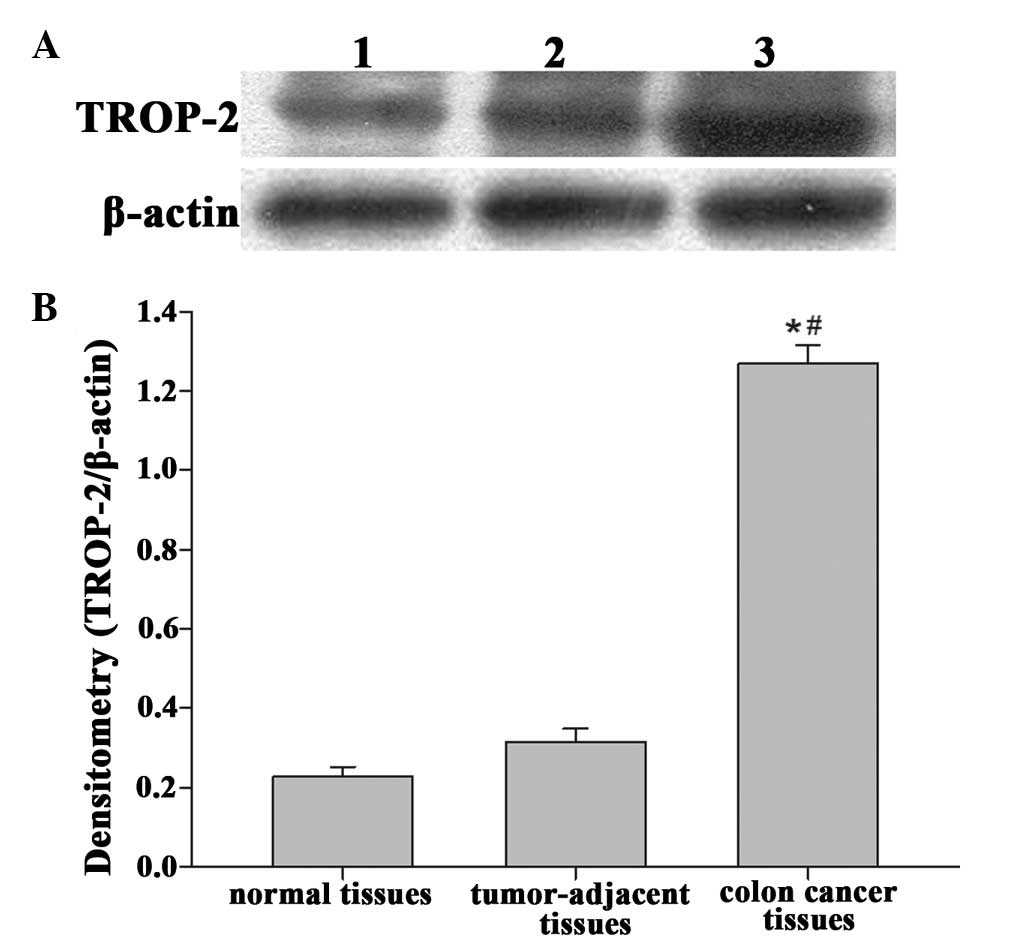
Expression of TROP-2 in colon cancer,
tumor-adjacent and normal tissues
The expression levels of TROP-2 in colon cancer
tissues, tumor-adjacent tissues and normal tissues were examined
using western blot analysis (Fig.
1). Notably, in contrast to the colon cancer tissues, the
normal tissues exhibited decreased protein expression of Trop-2.
The immunoreactivity for TROP-2 in the tumor-adjacent tissues was
higher than in the normal tissues, however, this difference was not
statistically significant(P>0.05). However, the expression of
TROP-2 in colon cancer tissues was significantly higher than that
in the tumor-adjacent tissues and normal tissues(P<0.05). These
result demonstrated that TROP-2 was highly expressed in colon
cancer and may offer potential as an important biomarker for early
diagnosis in colon cancer.
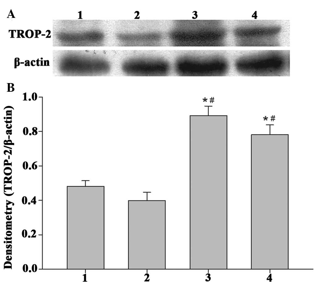
Expression of TROP-2 in colon cancer
tissues with/without lymph node metastases
As shown in Fig. 2,
positive expression of TROP-2 was observed in the colon cancer with
lymph node metastasis group, which was significantly higher,
compared with that in the non-lymph node metastasis group
(P<0.05). This indicated that the protein expression of TROP-2
was associated with lymph node metastasis in colon cancer.
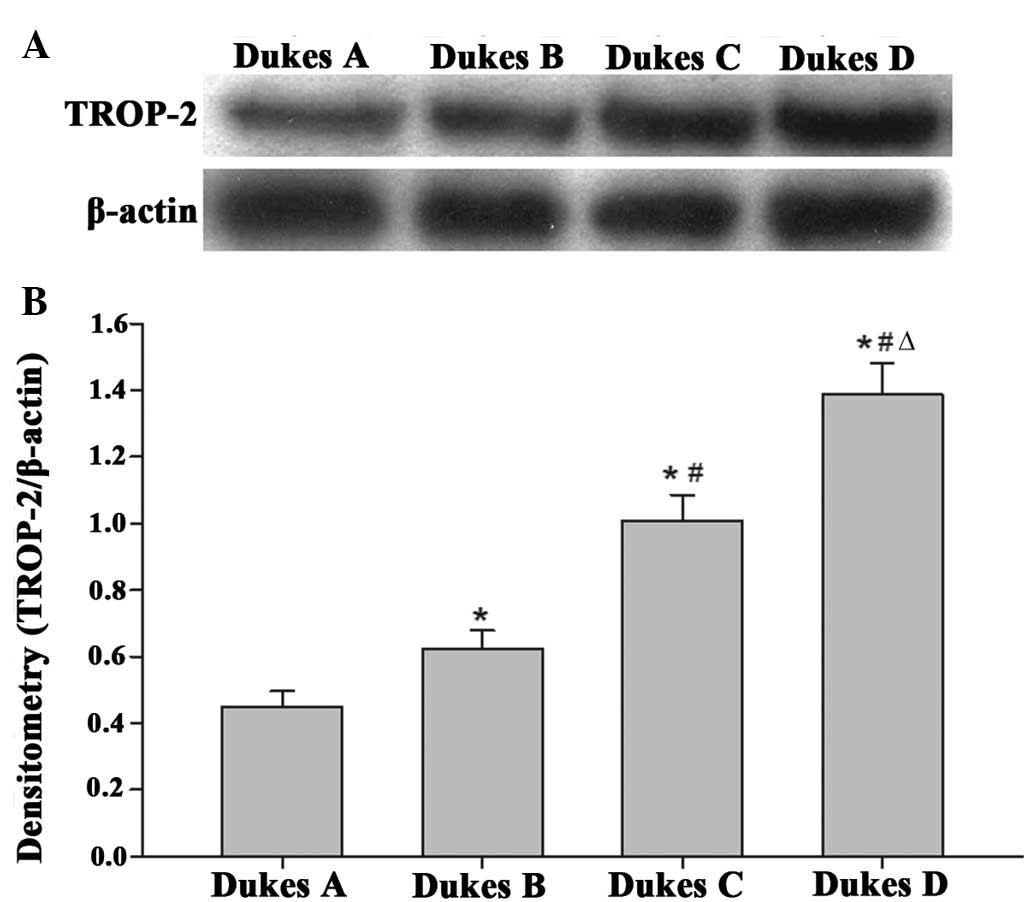
Expression of TROP-2 in colon cancer
tissues of different Dukes stages
The expression of Trop-2 in the tissue samples of
the Dukes A, Dukes B, Dukes C and Dukes D stages were analyzed
using western blot analysis. As shown in Fig. 3, the expression of, TROP-2
expression was upregulated in the Dukes A stage cancer tissues, and
the level of expression increased with the increasing Dukes stage.
These differences were statistically significant (P<0.05) and
these findings indicated that TROP-2 was associated with the
invasion and metastasis of colon cancer.
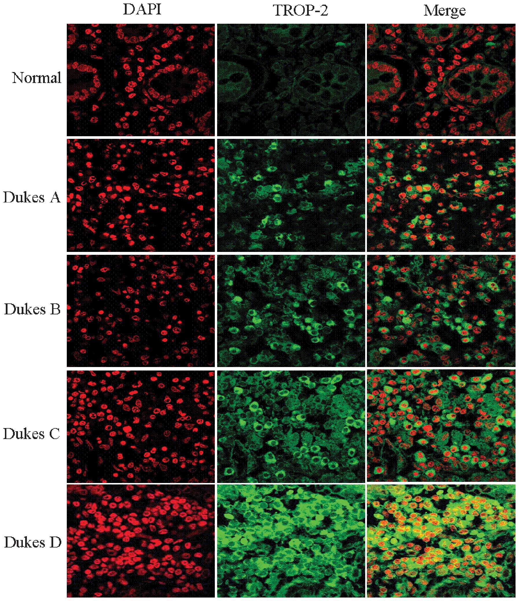
Confirmation of the protein expression of
TROP-2 using immunofluorescence analyses
Further experiments were performed to evaluate the
localization of the TROP-2 protein in colon cancer tissues. TROP-2
protein in cancer tissues of different Dukes stages was detected
using immunofluorescence staining. As shown in Fig. 4, TROP-2 protein was stained with
rabbit anti-TROP-2 antibody and green fluorescent protein-labeled
secondary antibody. TROP-2 protein was located in the cell membrane
surface of the colon cancer cells. The TROP-2-immunoreactive
structures appeared green and circular, and the nuclei were stained
with DAPI, observed as red fluorescence. The cells, which appeared
green in the centre and surrounded by red subsequent to merging,
were observed under a laser scanning confocal microscope. The
results also demonstrated that the expression of TROP-2 was
upregulated with increasing Dukes stage.
Correlations between the expression of
TROP-2 and clinical pathological features of colon cancer
The association between the expression of TROP-2 and
the clinicopathological features of colon cancer patients were
analyzed using a χ2 test. As shown in Table I, the high protein expression level
of TROP-2 in colon cancer was associated with lymph node metastasis
and Dukes classification (P=0.018 and P=0.002, respectively). By
contrast, no statistically signifi-cant correlation was observed
with gender, age or the degree of differentiation. These results
indicated that the protein expression of TROP-2 protein is
important in the development and metastasis of colon cancer.
Discussion
Colon cancer remains a major public health concern
worldwide, and presents a considerable disease burden. Despite
increasing knowledge of the cellular and molecular signaling
pathways underlying colon cancer, therapeutic outcomes remain only
moderately successful, with poor efficacy and marked variation in
therapeutic outcomes among patients. At present, surgical resection
remains the only curative treatment option, however, this involves
costly and invasive procedures with certain considerable
limitations. Despite efforts to determine its molecular mechanism,
the precise mechanisms underlying the malignant outcomes of colon
cancer remain to be elucidated. There are certain risk factors
associated with colon cancer, including inflammatory syndromes,
hereditary factors and environmental factors (27). Biologically active molecules, which
are involved in aggressiveness and metastasis are important for the
mechanism of tumor dissemination (28). Therefore, examining gene and
protein changes associated with the invasion and metastasis of
colon cancer is important for identifying the molecular targets for
clinical treatment.
In the present study, the protein expression of
TROP-2 was analyzed in patients with colon cancer. A significant
upregulation in the protein expression of TROP-2 was observed in
colon cancer, relative to the corresponding normal tissues and
tumor-adjacent tissues. However, whether the overexpres-sion of
TROP-2 is actively involved in tumorigenesis, and whether targeting
TROP-2 is of therapeutic use remains to be elucidated. The present
study investigated the expression of TROP-2 in colon cancer
with/without lymph node metastases and in tissues exhibiting
different Dukes stages. In addition, the correlation between the
expression of TROP-2 and clinical factors were examined to
determine its clinicopathological significance in colon cancer. The
findings revealed that over-expression of TROP-2 protein in colon
cancer was significantly associated with lymph node metastasis and
Dukes classification, suggesting the overexpression of TROP-2 in
colon cancer tissue could induces mucosal instability, premalignant
polyps and malignant transformation. TROP-2 was observed to be
necessary for the tumorigenesis and invasiveness of colon cancer,
and these effects may be reduced effectively with an antibody
against the extracellular domain of TROP-2. Notably, these findings
led to the hypothesis that antibodies against TROP-2 protein may
inhibit the tumorigenesis of colon cancer, presenting a novel
anticancer target. TROP-2 has also been suggested as a target for
anticancer immunotherapy in other types of epithelial carcinoma
(28). Anti-TROP-2 monoclonal or
polyclonal antibodies have been observed to destroy uterine serous
papillar carcinoma cells through Antibody-dependent cell-mediated
cytotoxicity in vitro (19). Therefore, in contrast to previous
studies, which focused predominantly on the oncogenic effects of
Trop-2, the results of the present study demonstrated that TROP-2
is functionally important in tumorigenesis, and present established
biological markers of colon cancer. Given its accessibility to
antibodies as a cell surface protein and its selective expression
in tumor cells, TROP-2 may be a potential therapeutic target for
colon cancer. Additionally, the restricted expression of TROP-2 in
normal tissues suggested that anti-TROP-2 therapeutics may be of
limited cytotoxicity.
In conclusion, the results of the present study
indicated for the first time, to the best of our knowledge, that
TROP-2 protein is overexpressed in colon cancer tissues.
Furthermore, correlation exists between the expression of Trop-2
and the pathogenesis of colon cancer. The TROP-2 cell surface
protein may regulate the growth, invasion and metastasis of tumor
cells. These findings provide novel insight into the role of TROP-2
in human colon cancer, as a novel, widespread stimulator of colon
cancer growth and a unique biomarker. Therefore, targeting the
overexpression of TROP-2 using immunotherapeutic strategies may
offer an attractive and potentially effective approach in the
treatment of patients with colon cancer.
Acknowledgments
The present study was supported by a grant from the
Science and Technology Development Project of Tangshan City (grant
no. 10140201A-3). The authors would like to thank Dr Jian-Hui Cai
for their critical evaluation of the manuscript, the members of the
Hebei United University Laboratory for providing technical
assistance, and our colleagues for their assistance during the
course of the study.
Abbreviations:
|
CC
|
colon cancer
|
|
CRC
|
colorectal cancer
|
|
TROP-2
|
trophoblast cell-surface marker
|
|
CEA
|
carcinoembryonic antigen
|
|
CA
|
carbohydrate antigen
|
References
|
1
|
Chiang EP, Tsai SY, Kuo YH, et al: Caffeic
Acid Derivatives Inhibit the Growth of Colon Cancer: Involvement of
the PI3-K/Akt and AMPK Signaling Pathways. PloS One. 9:e996312014.
View Article : Google Scholar : PubMed/NCBI
|
|
2
|
Boncheva V, Bonney SA, Brooks SE, et al:
New targets for the immunotherapy of colon cancer- does reactive
disease hold the answer? Cancer Gene Ther. 20. pp. 157–168. 2013,
View Article : Google Scholar
|
|
3
|
Carethers JM, Murali B, Yang B, et al:
Influence of Race on Microsatellite Instability and CD8+ T Cell
Infiltration in Colon Cancer. PloS One. 9:e1004612014. View Article : Google Scholar : PubMed/NCBI
|
|
4
|
Selcukbiricik F, Bilici A, Tural D,
Erdamar S, Soyluk O, Buyukunal E, Demirelli F and Serdengecti S:
Are high initial CEA and CA 19-9 levels associated with the
presence of K-ras mutation in patients with metastatic colorectal
cancer? Tumour Biol. 34:2233–2239. 2013. View Article : Google Scholar : PubMed/NCBI
|
|
5
|
Mayer RJ, Garnick MB, Steele GD Jr and
Zamcheck N: Carcinoembryonic antigen (CEA) as a monitor of
chemotherapy in disseminated colorectal cancer. Cancer.
42:1428–1433. 1978. View Article : Google Scholar : PubMed/NCBI
|
|
6
|
Petrioli R, Licchetta A, Roviello G, et
al: CEA and CA19.9 as early predictors of progression in
advanced/metastatic colorectal cancer patients receiving
oxaliplatin- based chemotherapy and bevacizumab. Cancer Invest.
30:65–71. 2012. View Article : Google Scholar : PubMed/NCBI
|
|
7
|
Van den Eynden GG, Majeed AW, Illemann M,
et al: The multifaceted role of the microenvironment in liver
metastasis: biology and clinical implications. Cancer Res.
73:2031–2043. 2013. View Article : Google Scholar : PubMed/NCBI
|
|
8
|
Fong D, Spizzo G, Gostner JM, et al:
TROP2: a novel prognostic marker in squamous cell carcinoma of the
oral cavity. Modern Pathol. 21:186–191. 2008.
|
|
9
|
Pak MG, Shin DH, Lee CH and Lee MK:
Significance of EpCAM and TROP2 expression in non-small cell lung
cancer. World J Surg Oncol. 10:532012. View Article : Google Scholar : PubMed/NCBI
|
|
10
|
Fong D, Moser P, Krammel C, et al: High
expression of TROP2 correlates with poor prognosis in pancreatic
cancer. Br J Cancer. 99:1290–1295. 2008. View Article : Google Scholar : PubMed/NCBI
|
|
11
|
Mühlmann G, Spizzo G, Gostner J, et al:
TROP2 expression as prognostic marker for gastric carcinoma. J Clin
Pathol. 62:152–158. 2009. View Article : Google Scholar
|
|
12
|
Zhang L, Zhou W, Velculescu VE, et al:
Gene expression profiles in normal and cancer cells. Science.
276:1268–1272. 1997. View Article : Google Scholar : PubMed/NCBI
|
|
13
|
Fornaro M, Dell'Arciprete R, Stella M, et
al: Cloning of the gene encoding Trop- 2, a cell- surface
glycoprotein expressed by human carcinomas. Int J Cancer.
62:610–618. 1995. View Article : Google Scholar : PubMed/NCBI
|
|
14
|
El Sewedy T, Fornaro M and Alberti S:
Cloning of the murine TROP2 gene: conservation of a PIP2- binding
sequence in the cytoplasmic domain of TROP- 2. Int J Cancer.
75:324–330. 1998. View Article : Google Scholar : PubMed/NCBI
|
|
15
|
Coldren CD, Helfrich BA, Witta SE, et al:
Baseline gene expression predicts sensitivity to gefitinib in non-
small cell lung cancer cell lines. Mol Cancer Res. 4:521–528. 2006.
View Article : Google Scholar : PubMed/NCBI
|
|
16
|
Bignotti E, Todeschini P, Calza S, et al:
Trop- 2 overexpression as an independent marker for poor overall
survival in ovarian carcinoma patients. Eur J Cancer. 46:944–953.
2010. View Article : Google Scholar : PubMed/NCBI
|
|
17
|
Ambrogi F, Fornili M, Boracchi P, et al:
Trop-2 is a determinant of breast cancer survival. PloS One.
9:e969932014. View Article : Google Scholar : PubMed/NCBI
|
|
18
|
Cubas R, Li M, Chen C and Yao Q: Trop2: a
possible therapeutic target for late stage epithelial carcinomas.
Biochim Biophys Acta. 1796:309–314. 2009.PubMed/NCBI
|
|
19
|
Varughese J, Cocco E, Bellone S, et al:
Uterine serous papillary carcinomas overexpress human trophoblast-
cell- surface marker (Trop- 2) and are highly sensitive to
immunotherapy with hRS7, a humanized anti- Trop- 2 monoclonal
antibody. Cancer. 117:3163–3172. 2011. View Article : Google Scholar : PubMed/NCBI
|
|
20
|
Ripani E, Sacchetti A, Corda D and Alberti
S: Human Trop- 2 is a tumor- associated calcium signal transducer.
IntJ Cancer. 76:671–676. 1998. View Article : Google Scholar
|
|
21
|
Trerotola M, Li J, Alberti S and Languino
LR: Trop- 2 inhibits prostate cancer cell adhesion to fibronectin
through the beta1 integrin- RACK1 axis. J Cell Physiol.
227:3670–3677. 2012. View Article : Google Scholar : PubMed/NCBI
|
|
22
|
Trerotola M, Cantanelli P, Guerra E, et
al: Upregulation of Trop- 2 quantitatively stimulates human cancer
growth. Oncogene. 32:222–233. 2013. View Article : Google Scholar
|
|
23
|
Huang HY: Role of TROP2 in cancer and as
potential therapeutic target. Zhonghua Bing Li Xue Za Zhi.
42:860–863. 2013.Article in Chinese.
|
|
24
|
Lu J, Izvolsky KI, Qian J and Cardoso WV:
Identification of FGF10 targets in the embryonic lung epithelium
during bud morphogenesis. J Biol Chem. 280:4834–4841. 2005.
View Article : Google Scholar
|
|
25
|
Stoyanova T, Goldstein AS, Cai H, Drake
JM, Huang J and Witte ON: Regulated proteolysis of Trop2 drives
epithelial hyperplasia and stem cell self- renewal via beta-
catenin signaling. Genes Dev. 26:2271–2285. 2012. View Article : Google Scholar : PubMed/NCBI
|
|
26
|
Goldstein AS, Lawson DA, Cheng D, Sun W,
Garraway IP and Witte ON: Trop2 identifies a subpopulation of
murine and human prostate basal cells with stem cell
characteristics. Proc the Natl Acad Sci USA. 105:20882–20887. 2008.
View Article : Google Scholar
|
|
27
|
Antonic V, Stojadinovic A, Kester KE,
Weina PJ, Brücher BL, Protic M, Avital I and Izadjoo M:
Significance of infectious agents in colorectal cancer development.
J Cancer. 4:227–240. 2013. View
Article : Google Scholar : PubMed/NCBI
|
|
28
|
Messick CA, Church J, Casey G and Kalady
MF: Identification of the methylator (serrated) colorectal cancer
phenotype through precursor serrated polyps. Dis Colon Rectum.
52:1535–1541. 2009. View Article : Google Scholar : PubMed/NCBI
|