Introduction
Neutrophil elastase (NE) is a neutrophil-specific
serine protease, which is able to degrade virulence factors and
kill bacteria. NE knockout mice are susceptible to bacterial and
fungal infections (1,2). It has also been suggested that NE may
be involved in the development of obesity and obesity-related
complications (3). Since NE has
various functions, numerous studies have focused on its effects on
inflammatory diseases, such as acute lung injury and obesity
(3–5). Previous studies have demonstrated
that NE contributes to the progression of various malignancies, and
promotes cell proliferation and metastasis during the occurrence
and development of certain solid tumors (6,7).
NE is predominantly produced in promyelocytes. Lane
and Ley (8) demonstrated that
importing the fusion protein, PML-RARα, into early myeloid cells
that express high levels of NE resulted in a marked increase in the
risk of acute promyelocytic leukemia (APL); therefore, NE
expression may aid in determining susceptibility of hematopoietic
cells to APL (9). To date, there
have been few studies regarding the effects of NE on leukemia;
therefore, determining the effect of NE on leukemia may provide
evidence for the development of novel target drugs for leukemia
therapy.
NE inhibitors, both endogenous and synthetic, are
able to inhibit NE activity and regulate the release of
inflammatory cytokines and chemokines (10). Endogenous inhibitors include
α1-proteinase inhibitor, a secretory leukocyte protease inhibitor
and α2-macroglobulin, whereas the synthetic inhibitors include
GW311616A and Sivelestat (11,12).
GW311616A is a potent, long-acting intracellular inhibitor of human
NE. As compared with other inhibitors, it is orally bioavailable,
long-lasting and is associated with lower clearance levels
(13). Currently, studies
regarding NE inhibitors have focused on acute lung injury and
chronic obstructive pulmonary disorder (14,15).
GW311616A is an NE inhibitor; however, its role in human leukemia
has yet to be elucidated. Further studies regarding the
proliferation and apoptosis of cells following treatment with NE
inhibitors, such as GW311616A, may provide novel information
regarding leukemia, and identify a novel direction for future
research on leukemia treatment.
In our preliminary experiment, NE expression was
detected in five cases of APL (unpublished data), and NE expression
was higher in patients with APL, as compared with healthy controls,
thus confirming its important role. In order to clarify the role of
NE in the process and development of leukemia, two different
leukemia cell lines were selected: K562 and U937. The K562 cells
express NE at low levels, whereas U937 cells contain abundant NE
(8,9).
To explore the possibility of NE as a target for
diagnosis and treatment, NE was upregulated in K562 cells by
recombinant adenovirus, and silenced in U937 cells by small
interfering (si)RNA and treatment with a specific inhibitor of NE,
GW311616A. The exact effects of NE were then determined on the
various cell lines, including alterations in cell proliferation,
apoptosis, expression of apoptotic proteins, and activation of
related signaling pathways.
Materials and methods
Cell culture and construction of
adenovirus (Ad)-NE
The K562 and U937 leukemia cells (Institutes for
Biological Sciences, Shanghai, China) were cultured in RPMI 1640
(Gibco Life Technologies, Carlsbad, CA, USA) supplemented with 10%
fetal bovine serum (Gibco Life Technologies) at 37°C in an
environment containing 5% CO2. The medium was refreshed
daily. An Ad-Easy system (American Type Culture Collection,
Rockville, MA, USA) was used to construct the recombinant
adenovirus (16).
Cell infection by adenovirus
K562 cells (2×10⁵/ml) were cultured in
24-well plates, and were infected with 5 μl recombinant
adenovirus Ad-NE (containing NE) and Ad-KZ (empty vector),
conjugated to GFP. Fluorescence microscopy was used to observe
green fluorescence 48 h post-infection. The cells were divided into
three groups: Infection group (K562/Ad-NE), empty vector group
(K562/Ad-KZ) and untreated K562 cell group.
RNA interference
Cell density was 60–80% confluent on the day of
transfection. The U937 cells (5×10⁵ logarithmic growth
phase cells) were seeded into 6-well plates. For cell transfection,
5 μl siRNA and 5 μl Lipofectamine® 2000
(Invitrogen Life Technologies, Carlsbad, CA, USA) were diluted in
100 μl Opti-MEM (Invitrogen Life Technologies) separately.
The siRNA and Lipofectamine® 2000 were then gently mixed
and incubated for 25 min at room temperature. The
siRNA-Lipofectamine® 2000 complexes were subsequently
added to each well and mixed by gentle agitation. All siRNA were
purchase from RiboBio (Guangzhou, China) and the sequences of the
NE-specific siRNAs were as follows: siRNA101,
5′-CCGUAAACUUGCUCAACGAdTdT-3′ and 3′- dTdTGGCAUUUGAACGAGUUGCU-5′;
siRNA102, 5′-CCGGUGGCACAGUUUGUAAdTdT-3′ and
3′-dTdTGGCCACCGUGUCAAACAUU-5′; siRNA103,
5′-GAUCGACUCUAUCAUCCAAdTdT-3′ and 3′-dTdTCU-AGCUGAGAUAGUAGGUU-5′.
Following a 48 h incubation at 37°C, red fluorescence protein (RFP)
was visualized by fluorescence microscopy. Transfection efficiency
was determined by calculating the number of cells expressing RFP.
The cells were divided into the following experimental groups:
Untreated group, negative control (NC) group, siRNA-101 group,
siRNA-102 group and siRNA-103 group.
NE activity assay
The U937 and K562 cells were seeded into a 6-well
plate (6×10⁴ cells/well), and 150 μmol/l
GW311616A (Sigma-Aldrich, St. Louis, MO, USA) was subsequently
added to the cells. After 48 h, the protein was extracted using
radioimmunoprecipitation lysis buffer (Zhongshan Goldenbridge
Biotechnology Co., Ltd., Beijing, China) and concentration was
measured by a bicinchoninic acid assay (BCA). Purified human
leukocyte elastase (Sigma-Aldrich) was used as a standard, and was
diluted in pure water to provide the following final
concentrations: 100, 200, 400, 800, 1,600 and 3,200 pmol/l. Another
three wells contained the protein samples. A total of 100 μl
substrate solution (Sigma-Aldrich) was added to the samples and
incubated at 37°C for 1 h. The optical density (OD) was measured at
405 nm in an ELISA analyzer. According to the OD value and a linear
regression equation of the standards, the activity of NE in the
samples was calculated.
Western blot analysis
The cells (5×10⁶) were collected in
Eppendorf tubes, washed with ice-cold phosphate-buffered saline
(PBS), and lysed in radioimmunoprecipitation solution containing a
protease inhibitor cocktail (Roche, Los Angeles, CA, USA). Protein
concentration was determined using the BCA method. A total of 30–50
μg protein was separated by 10% sodium dodecyl
sulfate-polyacrylamide gel electrophoresis, and was then
transferred to a polyvinylidene difluoride membrane (EMD Millipore,
Billerica, MA, USA). The membrane was blocked with 5% skimmed milk
(EMD Millipore) for 2 h at room temperature, and was then incubated
with the following primary antibodies overnight at 4°C: Mouse
anti-human NE monoclonal antibody (diluted 1:2,000; Abcam,
Cambridge, MA, USA); and rabbit anti-human polyclonal B-cell
lymphoma 2 (Bcl-2), rabbit anti-human polyclonal Bcl-2-associated X
protein (Bax) and rabbit anti-human AKT polyclonal antibodies
(diluted 1:1,000; Santa Cruz Biotechnology, Inc., Dallas, TX, USA).
The membranes were then incubated with goat anti-rabbit and goat
anti-mouse secondary antibodies (1:2,000 dilution; Zhongshan
Goldenbridge Biotechnology Co., Ltd., Beijing, China) for 1 h at
37°C. After washing with Tris-buffered saline containing Tween-20
(TBST), the immunoreactive complexes were visualized using an
enhanced chemiluminescence system (Bio-Rad Laboratories, Inc.,
Hercules, CA, USA). β-actin was used as an internal positive
control.
Cell Counting kit (CCK)-8 proliferation
assay
The cells were plated into 96-well plates
(5×10³/well). A total of 10 μl CCK-8 was added to
the cells, in order to quantify cell proliferation 1 to 4 days
post-infection. Following a 2 h incubation, the absorbance of each
well was measured at 450 nm, using a spectrophotometer (Bio-Rad
Laboratories, Inc.). A cell growth curve was generated with
absorbance as the ordinate and time as the abscissa. The experiment
was repeated three times.
Flow cytometric assay
The cells were routinely collected and centrifuged
at 500 × g for 5 min at room temperature. After the cells had been
washed twice with PBS, a staining mixture was prepared containing 5
μl Annexin-V-fluorescein isothiocyanate fluorescent dye and
5 μl propidium iodide (Sigma-Aldrich). The rate of cell
apoptosis was analyzed using a FACsorter (BD Biosciences, San Jose,
CA, USA) following a 15 min incubation at room temperature.
Furthermore, 5×10⁵ cells were collected, centrifuged at
500 × g for 5min and washed with pre-cooled PBS twice. The
supernatant was discarded and precooled 70% ethanol (1 ml) was
added and the samples were fixed overnight at 4°C. Following
fixation, the samples were centrifuged at 500 × g for 5 min and
washed with 1 ml PBS twice. The samples were resuspended cells in
100 μl 1 mg/ml RNase solution in a 37°C water bath for 30
min. Following incubation, 150 μl 50 μg/ml propidium
iodide staining solution was added and incubated for 30 min in dark
at room temperature. The cell cycle distribution was detected using
a FACsorter. Experiments were repeated three times.
Statistical analysis
Statistical analyses were performed using SPSS 17.0
software (SPSS Inc., Chicago, IL, USA). Experimental results are
presented as the mean ± standard deviation. An independent samples
t-test was used to compare the results of two groups. P<0.05 was
considered to indicate a statistically significant difference. Each
experiment was repeated at least three times.
Results
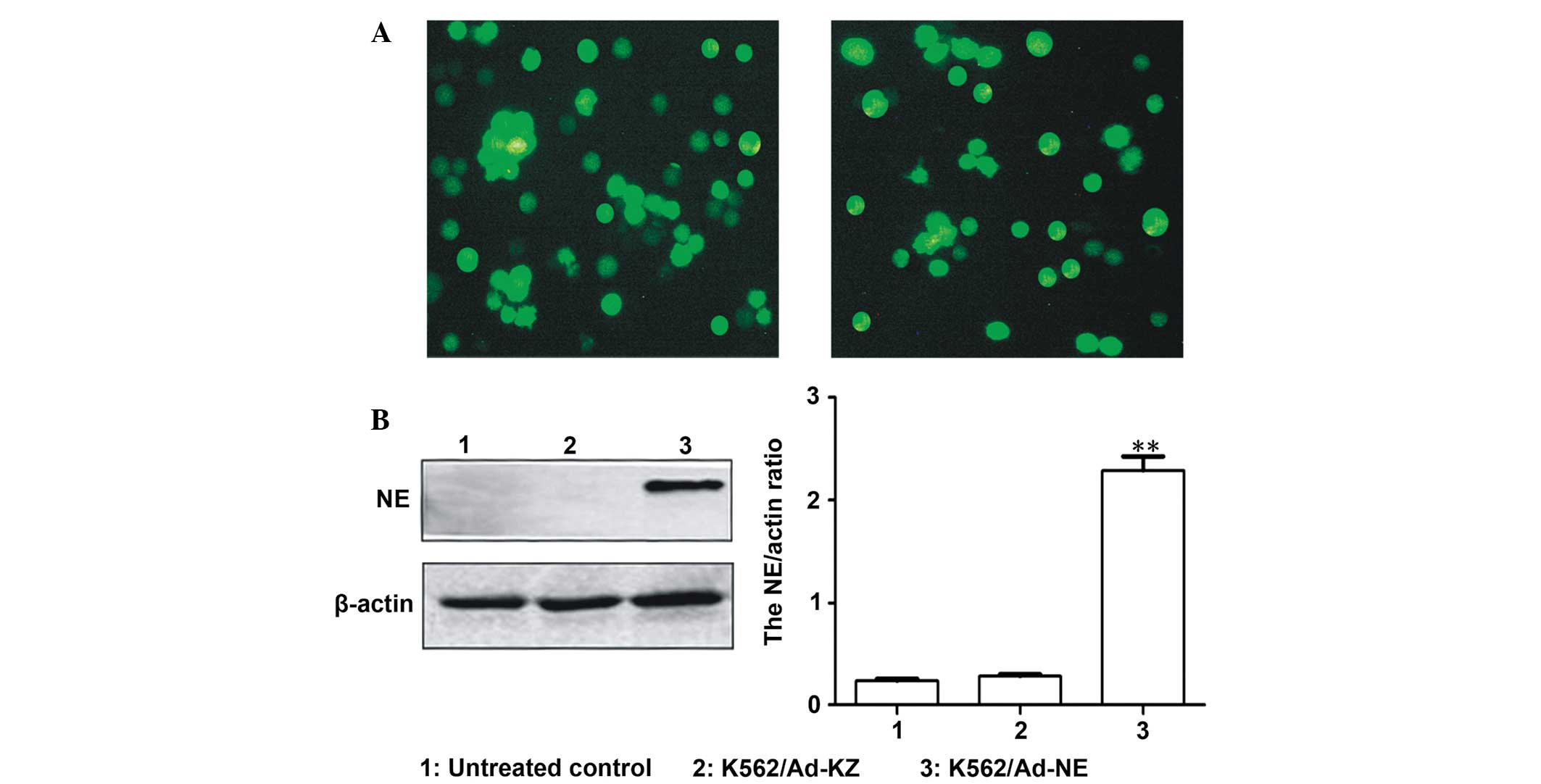
NE protein expression levels in K562
cells
To upregulate NE gene expression, the K562 cells
were infected with recombinant adenovirus Ad-NE or Ad-KZ with
enhanced green fluorescent protein (GFP). After 48 h, the
percentage of GFP-positive cells in the Ad-NE and Ad-KZ infection
groups was 70 and 60%, respectively (Fig. 1A). These results suggest that the
K562 cells were successfully infected with the recombinant
adenoviruses, as determined by expression of the GFP reporter gene.
The protein expression levels of NE were detected following
infection of the cells with Ad-NE, by western blotting. The
expression levels of NE were significantly higher in the K562/Ad-NE
group, as compared with the control groups (P<0.001, Fig. 1B).
Effects of Ad-NE on proliferation,
apoptosis and cell cycle distribution in K562 cells
To determine whether upregulation of NE had an
effect on cell growth and apoptosis in vitro, CCK-8 and flow
cytometric analysis were performed to detect the proliferation and
apoptosis of the K562 cells, respectively. The CCK-8 assay showed
that cell growth was markedly enhanced in the Ad-NE group in a
time-dependent manner, as compared with the untreated control and
Ad-KZ groups (P<0.05, Fig. 2A).
Flow cytometric analysis demonstrated there was no significant
difference in cell cycle distribution; however, the rate of
apoptosis was reduced. The apoptotic rate in the Ad-NE group was
11.23±0.56%, whereas the apoptotic rate in the Ad-KZ and blank
groups was 28.94±0.44 and 27.68±0.49%, respectively, thus
indicating that NE was able to inhibit apoptosis of K562 cells
(P<0.05, Fig. 2B).
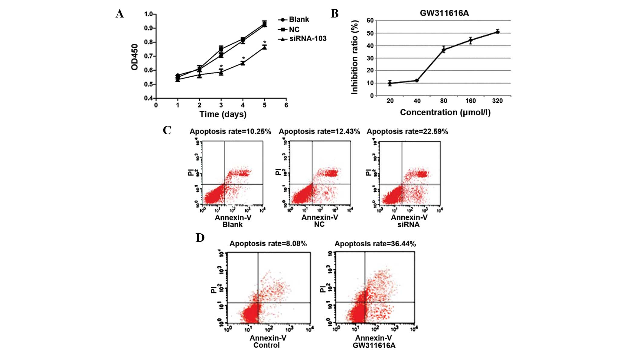
Efficiency of siRNA and GW311616A on NE
expression in various cell lines
In order to determine the function of NE in
leukemia, the present study knocked down its endogenous expression
by RNA interference. The percentage of RFP-positive cells in the
U937/siRNA group was 85% 48 h post-transfection (Fig. 3A). The effects of RNA interference
were also detected by western blotting. The protein expression
levels of NE were decreased in the three RNA interference groups,
as compared with the untreated and NC groups, and the interference
efficiency was most marked in the U937/siRNA-103 group (P<0.001,
Fig. 3B). NE activity was tested
before and after treatment with GW311616A in two cell lines, and
GW311616A was shown to markedly suppress NE activity (Fig. 3C).
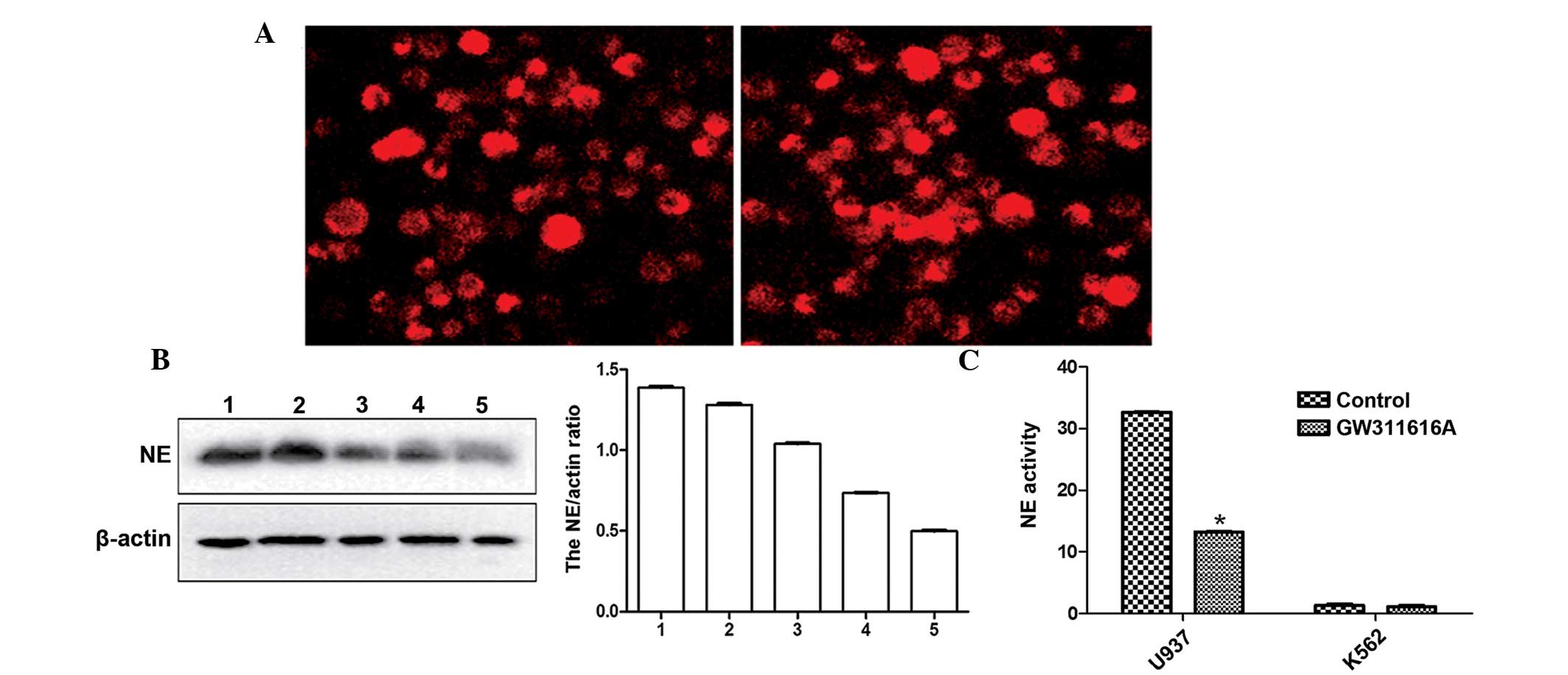 | Figure 3Detection of neutrophil elastase (NE)
following downregulation by small interfering (si)RNA and GW311616A
in two cell lines. (A) Red fluorescent protein expression was
detected in the siRNA-transfected U937 human leukemia cells by
fluorescence microscopy (magnification, ×200), indicating
successful expression of the siRNA in the U937 cells. (B) Western
blotting was performed to measure the protein expression levels of
NE in U937 siRNA-transfected cells. The protein expression levels
of NE in the 5th group were significantly decreased in the
U937/siRNA-103 group, as compared with control groups.
Representative results of western blot and densitometric analysis
for quantitative evaluation are shown. Data are presented as the
mean ± standard deviation of triplicate experiments.
***P<0.001, vs. untreated control. Lane 1, U937
cells; lane 2, U937/si negative control (NC) cells; lane 3,
U937/siRNA-101 cells; lane 4, U937/siRNA-102 cells; and lane 5,
U937/siRNA-103 cells). (C) Effects of GW311616A on NE activity in
U937 and K562 human leukemia cells. NE activity was strongly
surpressed by GW311616A. *P<0.05, vs. control. |
Cell proliferation and apoptosis in U937
cells following knockdown of NE expression
In order to accurately evaluate the effect of NE
knockdown, the growth and apoptosis of siRNA-infected and
GW311616A-treated U937 cells was detected. Following downregulation
of NE expression, cell proliferation was inhibited in a
time-dependent manner (Figs. 4A and
B). Flow cytometric analysis was performed to measure the rate
of apoptosis. The rate of apoptosis was markedly higher in the
siRNA-103 group (22.59±0.51%), as compared with the NC siRNA group
(12.43±0.62%) and the untreated group (10.25±0.45%) 48 h
post-transfection (P<0.05, Fig.
4C).
In the GW311616A-treated group, the rate of
apoptosis was enhanced, as compared with the control group
(P<0.05, Fig. 4D).
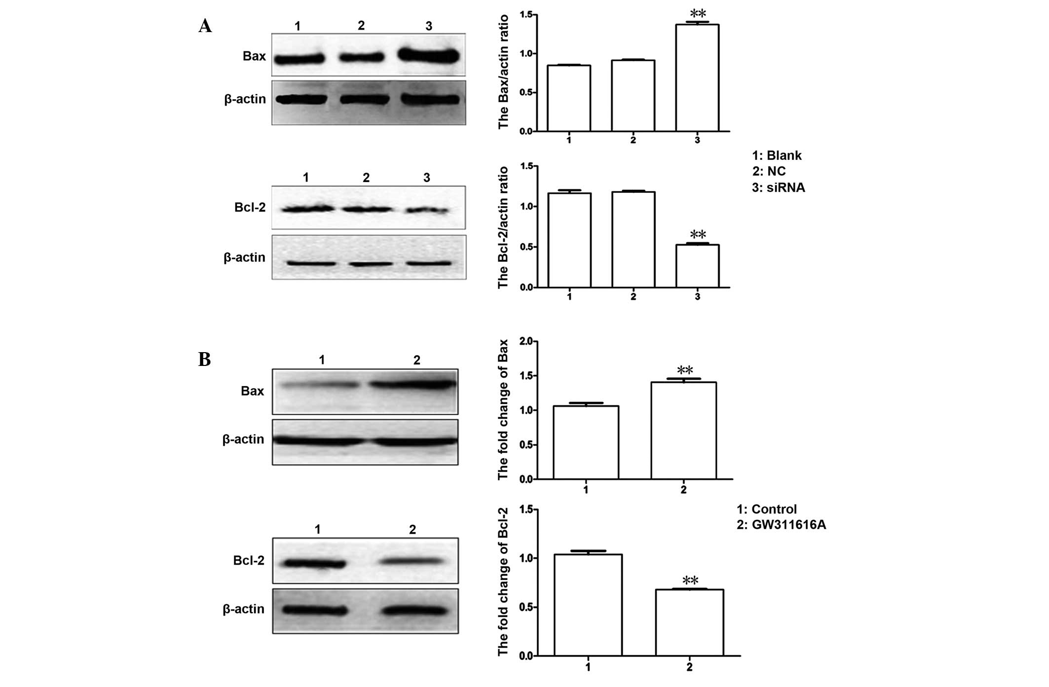
Expression levels of apoptosis-associated
proteins Bax and Bcl-2
To further investigate the effects of NE on
apoptosis in leukemia, the expression levels of relevant
apoptosis-associated proteins Bax and Bcl-2 were detected following
NE knockdown in U937 cells. Downregulation of NE with siRNA-103 was
able to increase the protein expression levels of Bax and decrease
the expression of Bcl-2, as compared with the control group
(Fig. 5A). Following treatment
with GW311616A, similar results were observed (Fig. 5B).
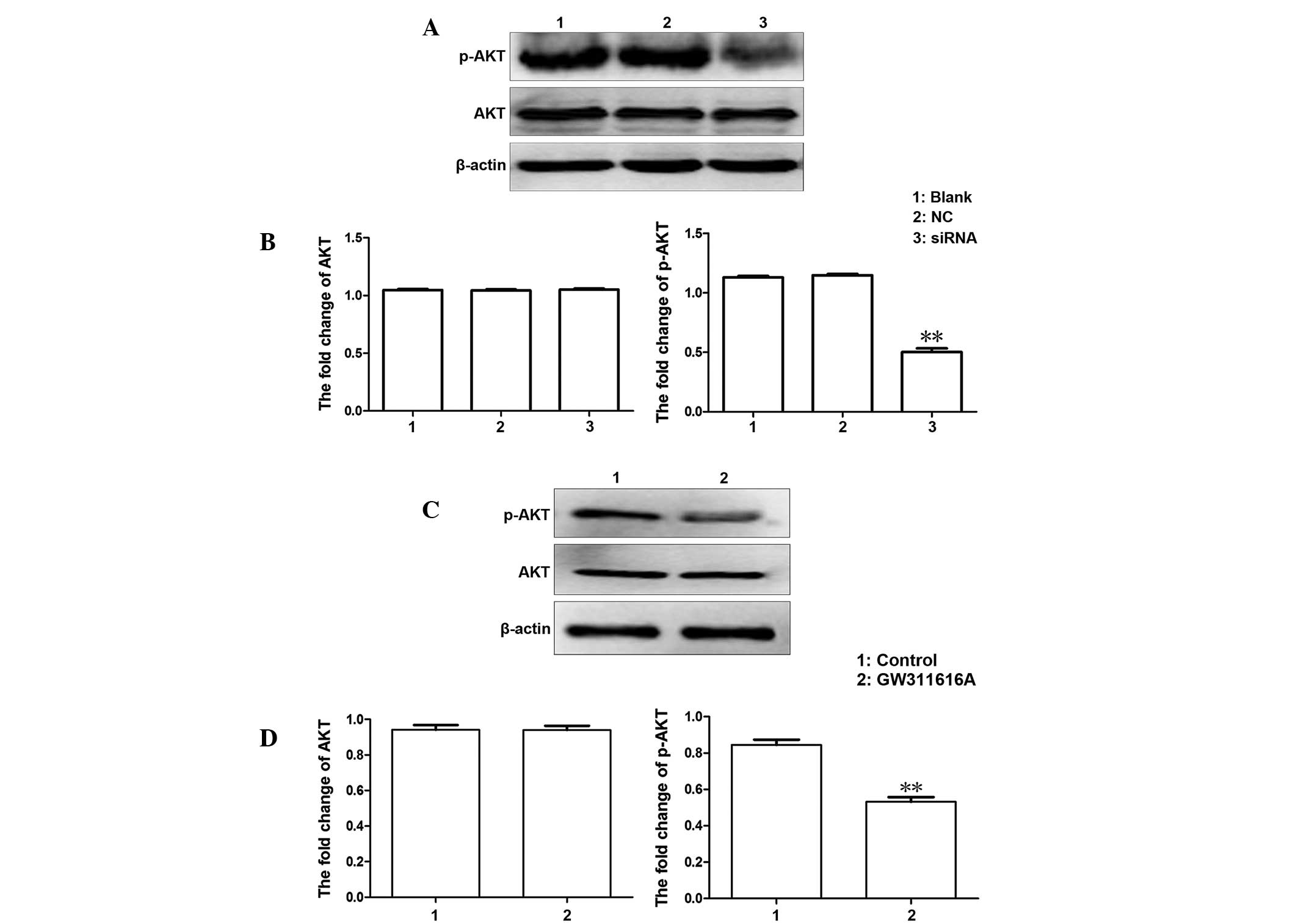
Associated signaling pathway
In order to determine the underlying mechanism of
the effects of siRNA and GW311616A on the proliferation and
apoptosis of leukemia cells, the expression levels of AKT and
phosphorylated-AKT in the PI3K/AKT signaling pathway were detected.
The expression levels of phosphorylated AKT were decreased, whereas
the expression levels of total AKT were similar in each group,
which suggested that the siRNA inhibited the activation of the AKT
signaling pathway (Fig. 6).
Discussion
In recent years, numerous studies have focused on
leukemia, particular aims have been exploration of its therapeutic
targets and drug interventions, and identification of novel
approaches for molecular diagnosis.
NE, encoded by Ela-2, is a type of serine protease
(17), which is predominantly
expressed in promyelocytes and packaged in azurophilic granules
(9). Numerous studies have
reported on its involvement in the progression of certain tumors,
such as breast cancer, non-small cell lung cancer and colorectal
cancer (3,18,19).
In addition, through the generation of elastin fragments, NE may
potently stimulate cancer cell invasiveness and angiogenesis
(20). A previous study identified
natural compounds and synthetic agents that are able to antagonize
NE activity, which may be of value as therapeutic agents for
suppressing cancer cell growth in certain types of cancer (8). The effects of NE in leukemia were
initially investigated by Lane et al (8,9),
whose studies demonstrated the importance of NE in the occurrence
and development of APL. Since NE was identified as having an
important role in the occurrence of APL, it is of great importance
to determine the underlying mechanisms of the effects of NE in
leukemia cells, and to detect its effects on leukemia cell lines
that express abundant of NE. These findings may be beneficial for
the identification of specific inhibitors to treat leukemia through
inhibition of NE activity.
In order to explore the exact effects of NE and to
clarify its underlying mechanism, the present study upregulated NE
in K562 cells, which express little NE; and downregulated NE in
U937 cells, which contain various levels of NE. Following up- or
downregulation of NE, the proliferation and apoptosis of the two
cell lines was measured, and the results demonstrated that NE had a
proliferation-inducing and anti-apoptotic effect in leukemia cells.
The cell cycle of the Ad-NE infected cells was arrested in S-phase,
indicating that NE may be a potential therapeutic target.
It is well-known that apoptosis is characterized by
a series of biochemical events, including condensation of the
nuclei, DNA fragmentation, and ultimately cell death (21,22).
The Bcl-2 protein family contains key regulators of apoptosis,
including apoptosis-inducing factors (e.g. Bax) and anti-apoptotic
factors (e.g. Bcl-2) (23–25). A slight change in the dynamic
balance of anti-apoptotic to pro-apoptotic proteins may result in
either inhibition or promotion of cell death (26–28).
The results of the present study demonstrated that
silencing NE by siRNA in U937 cells resulted in an upregulation of
the expression of Bax, whereas Bcl-2 expression was downregulated.
Further experiments demonstrated that these findings may be
achieved through regulation of the PI3K/AKT pathway, which has an
important role in orchestrating various cellular processes,
including proliferation, differentiation and apoptosis (29–31).
The expression levels of phosphorylated-AKT were decreased
following the downregulation of NE using siRNA, whereas the
expression levels of total AKT were similar in each group, thus
indicating that the activation of phosphorylated-AKT in the
PI3K/AKT pathway was inhibited by siRNA, which induced
apoptosis-associated protein Bax and Bcl-2 expression changes.
These results demonstrated the positive effects of low level
expression of NE.
To further understand the effects of NE, and to
identify therapeutic targets for leukemia, a specific inhibitor of
NE, GW311616A was selected. GW311616A is a potent, intracellular,
orally bioavailable, and long-acting inhibitor of human NE
(2,32). In the present study, the inhibitory
efficiency of GW311616A was apparent, and its ability to induce
apoptosis in a concentration-dependent manner was observed. In the
GW311616A-treated group, cell apoptosis was enhanced (P<0.05).
These results demonstrated the therapeutic effects of GW311616A.
Furthermore, the expression levels of apoptosis-associated proteins
Bax and Bcl-2, phosphorylated-AKT and total AKT were detected, the
results of which were the same as in the siRNA-treated groups; thus
suggesting that GW311616A has a potential therapeutic effect.
In the present study, the protein expression levels
of apoptosis-inducing Bax were reduced and the expression levels of
anti-apoptotic proteins were increased following downregulation of
NE, by siRNA and treatment with GW311616A. These results indicated
that NE may be a therapeutic target for leukemia, and the
inhibitory drug GW311616A may be used as a potential treatment.
However, it will be beneficial to continue studying the effects of
NE inhibition in various leukemia cell lines, including HL60
cells.
The present study demonstrated that upregulating NE
was able to promote the growth of leukemia cells and decrease the
proportion of apoptotic cells. Conversely, downregulation of NE by
siRNA and GW311616A treatment inhibited proliferation and induced
apoptosis in leukemia cells. This novel information regarding the
involvement of NE in leukemia cell proliferation and apoptosis may
be used to identify a potential biomarker or treatment target for
leukemia, and the NE-specific inhibitor GW311616A may be a
promising treatment target option.
Acknowledgments
The present study was supported by a grant from the
National Natural Science Foundation of China (grant no. 81171658)
and the Natural Science Foundation Project of CQ CSTC (grant no.
2011BA5037).
References
|
1
|
Papayannopoulos V, Metzler KD, Hakkim A
and Zychlinsky A: Neutrophil elastase and myeloperoxidase regulate
the formation of neutrophil extracellular traps. J Cell Biol.
191:677–691. 2010. View Article : Google Scholar : PubMed/NCBI
|
|
2
|
Korkmaz B, Moreau T and Gauthier F:
Neutrophil elastase, proteinase 3 and cathepsin G: Physicochemical
properties, activity and physiopathological functions. Biochimie.
90:227–242. 2008. View Article : Google Scholar
|
|
3
|
Mansuy-Aubert V, Zhou QL, Xie X, Gong Z,
Huang JY, Khan AR, Aubert G, Candelaria K, Thomas S, Shin DJ, et
al: Imbalance between neutrophil elastase and its inhibitor
α1-antitrypsin in obesity alters insulin sensitivity, inflammation,
and energy expenditure. Cell Metab. 17:534–548. 2013. View Article : Google Scholar : PubMed/NCBI
|
|
4
|
Kawabata K, Hagio T and Matsuoka S: The
role of neutrophil elastase in acute lung injury. Eur J Pharmacol.
451:1–10. 2002. View Article : Google Scholar : PubMed/NCBI
|
|
5
|
Tamakuma S, Ogawa M, Aikawa N, Kubota T,
Hirasawa H, Ishizaka A, Taenaka N, Hamada C, Matsuoka S and Abiru
T: Relationship between neutrophil elastase and acute lung injury
in humans. Pulm Pharmacol Ther. 17:271–279. 2004. View Article : Google Scholar : PubMed/NCBI
|
|
6
|
Sato T, Takahashi S, Mizumoto T, Harao M,
Akizuki M, Takasugi M, Fukutomi T and Yamashita J: Neutrophil
elastase and cancer. Surg Oncol. 15:217–222. 2006. View Article : Google Scholar
|
|
7
|
Ho AS, Chen CH, Cheng CC, Wang CC, Lin HC,
Luo TY, Lien GS and Chang J: Neutrophil elastase as a diagnostic
marker and therapeutic target in colorectal cancers. Oncotarget.
30:473–480. 2014.
|
|
8
|
Lane AA and Ley TJ: Neutrophil elastase
cleaves PML-RARalpha and is important for the development of acute
promyelocytic leukemia in mice. Cell. 115:305–318. 2003. View Article : Google Scholar : PubMed/NCBI
|
|
9
|
Lane AA and Ley TJ: Neutrophil elastase is
important for PML-retinoic acid receptor alpha activities in early
myeloid cells. Mol Cell Biol. 25:23–33. 2005. View Article : Google Scholar :
|
|
10
|
Lee SK, Son BS, Hwang JJ, et al: The use
of neutrophil elastase inhibitor in the treatment of acute lung
injury after pneumonectomy. J Cardiothorac Surg. 8(69)2013.
View Article : Google Scholar
|
|
11
|
Lee WL and Downey GP: Leukocyte elastase:
Physiological functions and role in acute lung injury. Am J Respir
Crit Care Med. 164:896–904. 2001. View Article : Google Scholar : PubMed/NCBI
|
|
12
|
Hoshi K, Kurosawa S and Kato M:
Sivelestat, a neutrophil elastase inhibitor, reduces mortality rate
of critically ill patients. Tohoku J Exp Med. 207:143–148. 2005.
View Article : Google Scholar : PubMed/NCBI
|
|
13
|
Macdonald SJ, Dowle MD, Harrison LA, Shah
P, Johnson MR, Inglis GG, Clarke GD, Smith RA, Humphreys D, Molloy
CR, et al: The discovery of a potent, intracellular, orally
bioavailable, long duration inhibitor of human neutrophil elastase
- GW311616A a development candidate. Bioorg Med Chem Lett.
11:895–898. 2001. View Article : Google Scholar : PubMed/NCBI
|
|
14
|
Ohbayashi H: Neutrophil elastase
inhibitors as treatment for COPD. Expert Opin Investig Drugs.
11:965–980. 2002. View Article : Google Scholar : PubMed/NCBI
|
|
15
|
Miyoshi S, Hamada H, Ito R, Katayama H,
Irifune K, Suwaki T, Nakanishi N, Kanematsu T, Dote K, Aibiki M, et
al: Usefulness of a selective neutrophil elastase inhibitor,
sivelestat, in acute lung injury patients with sepsis. Drug Des
Devel Ther. 7:305–316. 2013. View Article : Google Scholar : PubMed/NCBI
|
|
16
|
Luo J, Deng ZL, Luo X, et al: A protocol
for rapid generation of recombinant adenoviruses using the AdEasy
system. Nature Protocols. 2:1236–1247. 2007. View Article : Google Scholar : PubMed/NCBI
|
|
17
|
Horwitz M, Benson KF, Person RE, Aprikyan
AG and Dale DC: Mutations in ELA2, encoding neutrophil elastase,
define a 21-day biological clock in cyclic haematopoiesis. Nat
Genet. 23:433–436. 1999. View
Article : Google Scholar : PubMed/NCBI
|
|
18
|
Akizuki M, Fukutomi T, Takasugi M,
Takahasi S, Sato T, Harao M, Mizumoto T and Yamashita J: Prognostic
significance of immunoreactive neutrophil elastase in human breast
cancer: Long-term follow-up results in 313 patients. Neoplasia.
9:260–264. 2007. View Article : Google Scholar : PubMed/NCBI
|
|
19
|
Wislez M, Antoine M, Rabbe N, Gounant V,
Poulot V, Lavolé A, Fleury-Feith J and Cadranel J: Neutrophils
promote aerogenous spread of lung adenocarcinoma with
bronchioloalveolar carcinoma features. Clin Cancer Res.
13:3518–3527. 2007. View Article : Google Scholar : PubMed/NCBI
|
|
20
|
Moroy G, Alix AJ, Sapi J, Hornebeck W and
Bourguet E: Neutrophil elastase as a target in lung cancer.
Anticancer Agents Med Chem. 12:565–579. 2012. View Article : Google Scholar : PubMed/NCBI
|
|
21
|
Zhang X, Zhong L, Liu BZ, Gao YJ, Gao YM
and Hu XX: Effect of GINS2 on proliferation and apoptosis in
leukemic cell line. Int J Med Sci. 10:1795–1804. 2013. View Article : Google Scholar : PubMed/NCBI
|
|
22
|
Buggins AG and Pepper CJ: The role of
Bcl-2 family proteins in chronic lymphocytic leukaemia. Leuk Res.
34:837–842. 2010. View Article : Google Scholar : PubMed/NCBI
|
|
23
|
Li Y, Yin S, Nie D, Xie S, Ma LM, Wang X,
Wu Y and Xiao J: MK886 inhibits the proliferation of HL-60 leukemia
cells by suppressing the expression of mPGES-1 and reducing
prostaglandin E2 synthesis. Int J Hematol. 94:472–478. 2011.
View Article : Google Scholar : PubMed/NCBI
|
|
24
|
Korsmeyer SJ: BCL-2 gene family and the
regulation of programmed cell death. Cancer Res. 59(7 Suppl):
1693s–1700s. 1999.PubMed/NCBI
|
|
25
|
Ola MS, Nawaz M and Ahsan H: Role of Bcl-2
family proteins and caspases in the regulation of apoptosis. Mol
Cell Biochem. 351:41–58. 2011. View Article : Google Scholar : PubMed/NCBI
|
|
26
|
Chipuk JE, Moldoveanu T, Llambi F, Parsons
MJ and Green DR: The BCL-2 family reunion. Mol Cell. 37:299–310.
2010. View Article : Google Scholar : PubMed/NCBI
|
|
27
|
Willis S, Day CL, Hinds MG and Huang DC:
The Bcl-2-regulated apoptotic pathway. J Cell Sci. 116:4053–4056.
2003. View Article : Google Scholar : PubMed/NCBI
|
|
28
|
Juin P, Geneste O, Raimbaud E and Hickman
JA: Shooting at survivors: Bcl-2 family members as drug targets for
cancer. Biochim Biophys Acta. 1644:251–260. 2004. View Article : Google Scholar : PubMed/NCBI
|
|
29
|
Osaki M, Oshimura M and Ito H: PI3K-Akt
pathway: Its functions and alterations in human cancer. Apoptosis.
9:667–676. 2004. View Article : Google Scholar : PubMed/NCBI
|
|
30
|
Chang F, Lee JT, Navolanic PM, Steelman
LS, Shelton JG, Blalock WL, Franklin RA and McCrubey JA:
Involvement of PI3K/Akt pathway in cell cycle progression,
apoptosis, and neoplastic transformation: A target for cancer
chemotherapy. Leukemia. 17:590–603. 2003. View Article : Google Scholar : PubMed/NCBI
|
|
31
|
Fresno Vara JA, Casado E, de Castro J,
Cejas P, Belda-Iniesta C and González-Barón M: PI3K/Akt signalling
pathway and cancer. Cancer Treat Rev. 30:193–204. 2004. View Article : Google Scholar : PubMed/NCBI
|
|
32
|
Ma PP, Zhu D, Liu BZ, Zhong L, Zhu XY,
Wang H, Zhang X, Gao YM and Hu XX: Neutrophil elastase inhibitor on
proliferation and apoptosis of U937 cells. Zhonghua Xue Ye Xue Za
Zhi. 34:507–511. 2013.In Chinese. PubMed/NCBI
|