Introduction
Glioma is a collective group of brain tumors, which
can occur in adults and children. According to pathological
evaluation of the tumor, glioma are further classified to low-grade
and high-grade glioma. Low-grade glioma progresses slowly, and the
patients have a median survival rate of 11–17 years (1,2).
However, patients with high-grade glioma, including anaplastic
astrocytomas and glioblastoma multiforme, have a poor prognosis and
a substantially lower 3-year relative survival rate despite the
advances in understanding of tumor pathology and the development of
cancer therapy (3,4). At present, the standard treatment for
glioma includes surgical resection, followed by radiation therapy
and chemotherapy. Temozolomide, an alkylating drug capable of
methylating DNA and causing DNA damage, is the most commonly used
chemotherapeutic agent in glioblastoma multiforme and relapsed
anaplastic astrocytomas (5,6).
However, chemoresistance occurs frequently and recurrence is
common, therefore the 5-year survival rate remains low in these
patients (3). Novel and effective
chemotherapeutic and targeted therapeutic strategies are urgently
required and are an area of continued investigation.
Dysregulated gene expression and aberrant signal
transduction have been attributed to the pathogenesis and
tumorigenesis of various types of human malignancies. Insulin-like
growth factor (IGF) and its receptor IGF-IR are important in growth
and development. Circulating IGF-1 and IGF-2 bind to the cell
surface receptor IGF-IR and activate the downstream
phosphoinositide 3-kinase (PI3K)/AKT and mitogen-activated protein
kinase (MAPK) signaling pathways, both of which have well defined
proliferative, anti-apoptotic and oncogenic transformative roles
(7,8). Increased levels of circulating IGF-1
and IGF-2 have been detected in patients with colorectal cancer,
prostate cancer, breast cancer and ovarian cancer (9–12).
In glioma, deregulated IGF signaling has been associated with
progression of the disease state (13). The inhibition of IGF signaling by
small interfering RNA, specific kinase inhibitors or antibodies,
suppresses tumor cell growth and survival (14–16).
Furthermore, IGF signaling is involved in cellular migration and
invasion, with the observation that the tumor cells at the margin
of infiltration exhibit higher expression levels of IGF-IR compared
with low-grade tumor cells (13,17).
IGF signaling serves as an effective therapeutic target for glioma,
as a result of the central role IGF/IGF-IR has in tumor survival,
migration and metastasis.
GSK1838705A, also termed
1H-pyrrolo[2,3-d]pyrimidine, is a novel, small molecule kinase
inhibitor of IGF-IR and anaplastic lymphoma kinase (ALK) (18,19).
GSK1838705A has been demonstrated to inhibit the proliferation of
cells derived from a variety of types of human tumor, including
multiple myeloma, neuroblastoma and certain subtypes of non-small
cell lung cancer (18). The
present study aimed to investigate the inhibitory effect of
GSK1838705A on glioma tumor cell proliferation and migration, and
the growth of tumor xenografts in vivo. The results may
support the use of GSK1838705A as a potential antitumor drug for
the treatment of human glioma.
Materials and methods
Reagents, cells and mouse model
GSK1838705A was purchased from Selleck Chemicals LLC
(Houston, TX, USA). The human U87MG glioma cell line was purchased
from American Type Culture Collection (Rockville, MD, USA). The
U87MG cells were maintained in Eagle's minimal essential medium
(EMEM), supplemented with 10% heat-inactivated fetal bovine serum
(FBS) and 100 U/ml penicillin-streptomycin in a humidified
incubator at 37°C with 5% CO2. Athymic nude mice were
purchased from Slack Company (Shanghai, China) and were handled in
compliance with the Experimental Animal Care and Use Committee
guidelines of Xinxiang Medical University (Xinxiang, Henan, China).
A total of 18 female mice (age, 7–8 weeks; weight, 24–28 g) were
housed in a specific-pathogen-free facility maintained between 20
and 25°C with a 12 h light/12 h dark cycle, and were allowed ad
libitum access to food and water. The study was approved by the
ethics committee of Xinxiang Medical University (Xinxiang,
China).
Cell viability assay
The cells (1×10⁵) were seeded into
96-well plates in triplicate and were treated with dimethyl
sulfoxide (DMSO) or different concentrations of GSK1838705A for 24,
48 or 72 h. Cell viability at the end of each treatment was
measured using a CellTiter-Glo Assay kit (Promega Corporation,
Madison, WI, USA), according to the manufacturer's
instructions.
Flow cytometric analysis
The cells (1×10⁶) were treated with DMSO
or different concentrations of GSK1838705A. Following treatment for
48 h, the cells were harvested, fixed and stained with propidium
iodide using a CycleTEST plus DNA reagent kit (Becton Dickinson,
Franklin lakes, NJ, USA), according to the manufacturer's
instructions. Following staining, the cells were collected and
processed using a FACSCalibur (BD Biosciences, San Jose, CA, USA).
The DNA content was analyzed using CellQuest Pro flow cytometry
analytical software (version 5.1; Becton Dickinson).
Nuclear staining
Exponentially growing cells (5×10⁵/ml)
were seeded onto polylysine coated glass coverslips overnight and
treated with DMSO or different concentrations of GSK1838705A for 48
h. The cells were subsequently fixed with 4% paraformaldehyde for
10 min and stained with Hoechst (Sigma-Aldrich, St Louis, MO, USA)
for a further 10 min. Images were captured using a fluorescent
microscope (BX51; Olympus America Inc., Melville, NY, USA).
Transwell assay
A total of 1×10⁵ cells in EMEM,
supplemented with 1% FBS, were seeded into the upper compartment of
a 24-transwell Boyden chamber (Costar, Bedford, MA, USA).
Subsequently, DMSO or different concentrations of GSK1838705A were
added to the cells. EMEM (650 µl), supplemented with 10%
FBS, was loaded into the lower chamber for use as a
chemoattractant. Following incubation for 12 h at 37°C, the cells
were fixed with 70% ethanol and stained with 0.1% crystal violet
(Sigma-Aldrich). The non-migrating cells, which remained on the
upper surface were removed by gentle scraping with a cotton swab
and images of the migrated cells on the lower side were captured
using an Olympus BH2 microscope (Olympus, Tokyo, Japan), following
which the cells were lysed with 10% acetic acid. The absorbance was
measured at 595 nm using a Beckman DU-640 spectrophotometer
(Beckman Coulter, Pasadena, CA, USA).
In vivo efficacy investigations using a
mouse xenograft model
Exponentially growing U87MG cells were injected into
the axillary region of nude mice (2×10⁶ cells/mouse).
When the tumors reached ~70 mm3 in size, the mice were
randomly allocated into a control and a treatment group, each
containing six animals. The mice in the control group were
administered orally with formulating vehicle, consisting of 20%
sulfobutyl ether β-cyclodextrin (ISP; pH 3.5; Sigma-Aldrich) and
the mice in the treatment group were administered orally with 4 or
8 mg/kg GSK1838705A once daily. The mice were weighed and the tumor
size was measured every other day for 11 days. The tumor volume was
calculated using the following formula: Tumor volume
(mm3) = (width × width × length) / 2. At the end of the
treatment period, the mice were sacrificed via carbon dioxide
inhalation by trained personnel, and the tumors were harvested for
analysis.
Immunohistochemical analysis of tumor
samples
The tumor samples were cut into 5 mm3
tissue sections and were immediately fixed in 10% neutral buffered
formalin (Sigma-Aldrich), followed by transfer into 70% ethanol.
Following fixation, the samples were embedded into paraffin blocks
and sectioned at a thickness of 5 mm. A terminal deoxynucleotidyl
transferase dUTP nick end labeling (TUNEL) assay was performed
using a FragEL kit (Calbiochem, Darmstadt, Germany), according to
manufacturer's instructions. The nuclei were stained with Hoechst
for 20 min and images were captured using an Olympus fluorescent
microscope.
Statistical analysis
Student's t-test and analysis of variance were
performed using StatView software version 5.0 (SAS Institute, Cary,
NC, USA). The data are expressed as the mean ± standard deviation
of triplicate measurements. P<0.05 was considered to indicate a
statistically significant difference.
Results
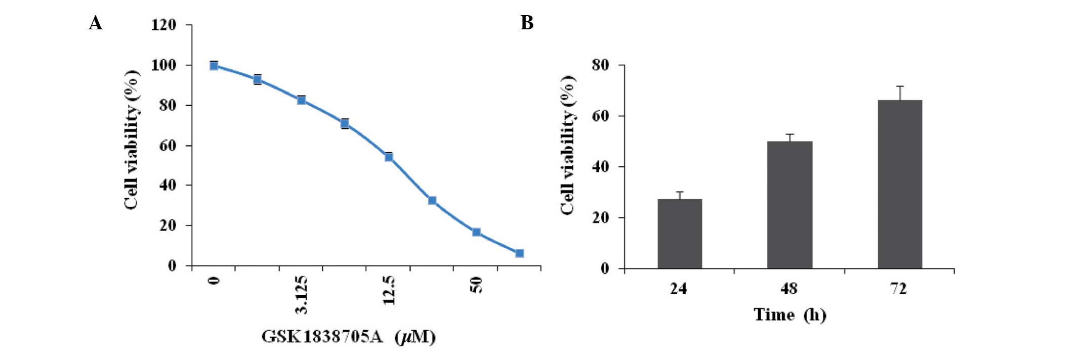
GSK1838705A decreases glioma cell
viability
Cellular proliferation is regulated by the
cooperative action of various growth stimulatory and inhibitory
signals. IGF signaling favors cell survival by activating
downstream signaling transduction cascades, including the Ras,
MAPK, PI3K and AKT pathways (8,20).
To determine whether the suppression of IGF signaling affected
cells proliferation in the present study, the U87MG glioma cells
were treated with different concentrations of GSK1838705A for 72 h
and the cell viability was measured. A dose-dependent inhibition of
glioma cell viability was observed following treatment with
GSK1838705A (Fig. 1A). To
determine the onset of drug action, the U87MG cells were treated
with GSK1838705A (20 µM) for 24, 48 or 72 h, followed by the
measurement of cell viability. The results indicated an early onset
for the inhibitory effect of GSK1838705A, which was observed 24 h
following treatment (Fig. 1B).
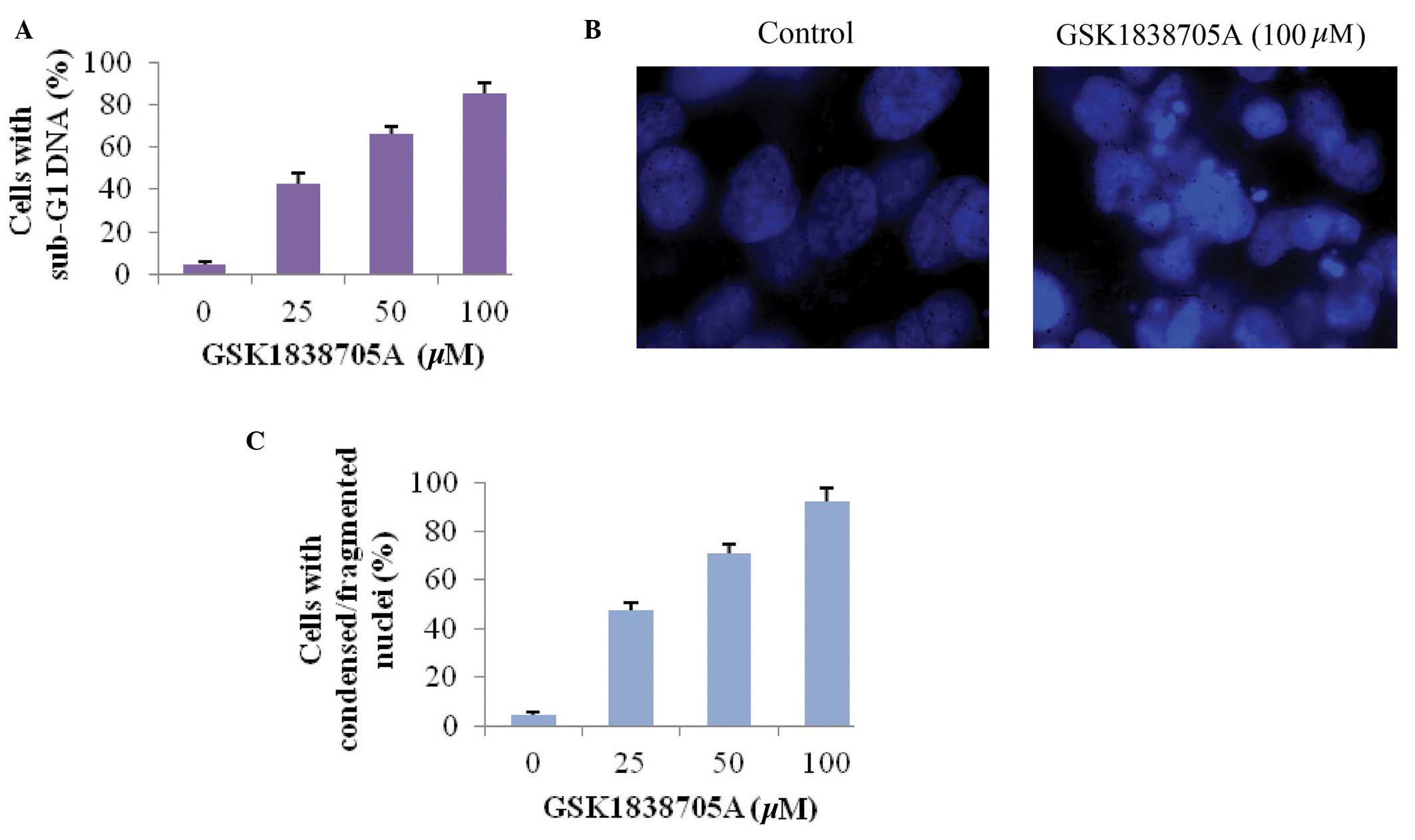
GSK1838705A induces the apoptosis of
glioma cells
Tumor cells often evolve mechanisms to evade or
antagonize programmed cell death, and IGF is a potent pro-survival
factor, which increases cell proliferation, therefore activation of
IGF signaling provides cells with a growth advantage (8). The present study investigated whether
inhibiting IGF signaling via treatment of glioma cells with
GSK1838705A induced apoptosis in the glioma cell. The sub-G1 DNA
content in cells was measured as an indicator of late stage
apoptosis (21). Consistent with
the results obtained in the measurement of cell viability,
GSK1838705A caused an increase in sub-G1 DNA content in a
dose-dependent manner, which confirmed that the inhibition of IGF
signaling promoted apoptosis in the affected cells (Fig. 2A). Apoptosis-induced condensation
and fragmentation of DNA was visualized following nuclear staining.
Compared with the untreated cells, the cells treated with
GSK1838705A exhibited marked apoptosis, as shown in Fig. 2B. Similar to the results shown in
Fig. 2A, the percentage of cells
with condensed/fragmented DNA increased proportionally with
increasing concentrations of GSK1838705A (Fig. 2C).
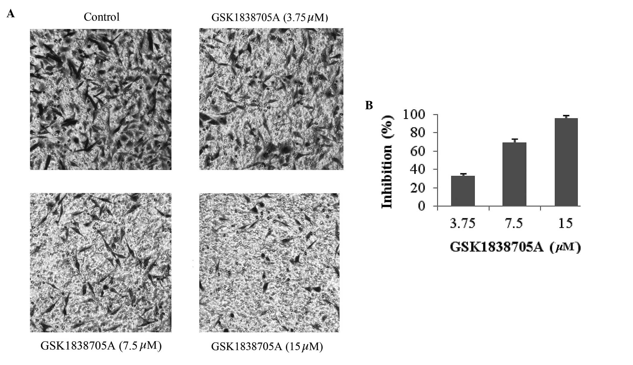
GSK1838705A inhibits the migration of
glioma cells
Malignant tumor cells, including gliomas, are
capable of migrating and invading to a secondary site through the
processes of angiogenesis and metastasis, in which IGF signaling is
important (8,13). Therefore, IGF/IGF-IR is an
attractive target in cancer therapeutics. The present study further
investigated the effect of IGF inhibition on glioma cell migration.
U87MG cells, in the presence or absence of GSK1838705A, were
induced to migrate in a Transwell assay. As shown in Fig. 3A and B, an inhibitory effect of
GSK1838705A on cellular migration was observed after 8 h of
treatment at concentrations as low as 3.75 µM and reached
its peak effect at 15 µM. The reduced number of migrating
cells was not a result of apoptosis due to GSK1838705A treatment
itself, as the duration of treatment was significantly shorter and
the concentrations of GSK1838705A were significantly lower compared
with those used to examine the cell viability and apoptosis
(Figs. 1 and 2).
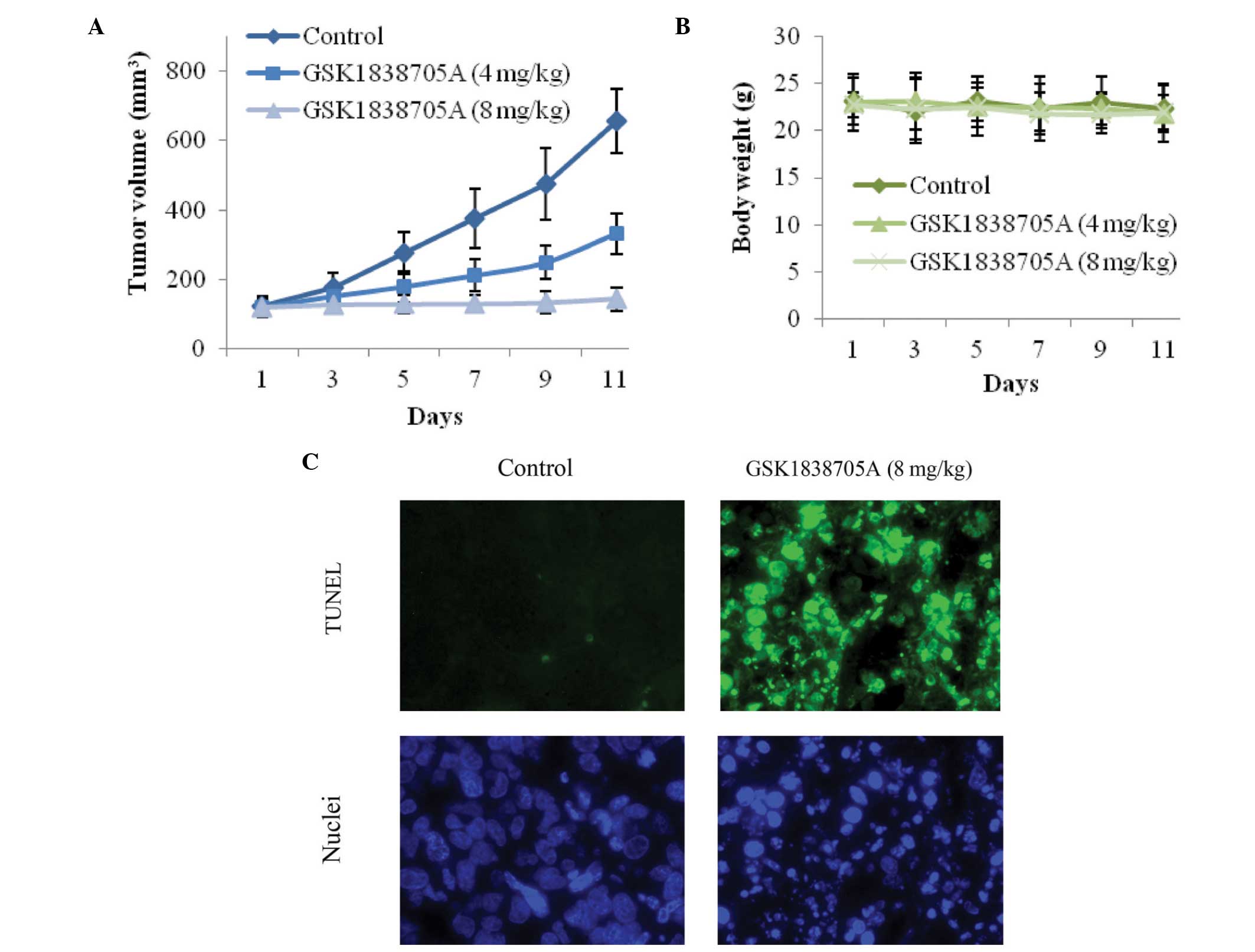
GSK1838705A suppresses tumor cell growth
in vivo
Subsequent to the results obtained from the in
vitro investigations, the present study investigated the
antitumor efficacy of GSK1838705A in vivo, in which U87MG
cells were injected into athymic nude mice. Following successful
inoculation, GSK1838705A (4 or 8 mg/kg) was administered once daily
and the tumor volumes were measured every other day. GSK1838705A
significantly inhibited the growth of the tumor mass. Treatment
with GSK1838705A at 4 or 8 mg/kg resulted in reductions of ~45 and
85% in tumor volume, respectively, 11 days after the first
administration (Fig. 4A). The
antitumor efficacy of GSK1838705A was consistent with the
concentration used. No significant weight loss was observed in
either treatment group during the course of treatment, indicating
that the concentrations of GSK1838705A used were well tolerated by
the recipient mice, and no significant cytotoxicity accompanied the
GSK1838705A treatment (Fig. 4B).
Consistent with the results of the in vitro investigations,
GSK1838705A induced significant apoptosis in the tumor cells.
Following treatment of tumors with 8 mg/kg GSK1838705A, significant
DNA fragmentation was detected using a TUNEL assay and nuclear
staining (Fig. 4C). Taken
together, these results provided clear evidence indicating that
GSK1838705A effectively suppressed the growth of the tumor in
vivo by inducing apoptosis of the tumor cells.
Discussion
Dysregulated signaling pathways have been implicated
in the tumorigenesis and angiogenesis of a wide variety of human
malignancies. The critical elements involved in signal
transduction, including surface receptors, kinases, adaptor
proteins and various signaling molecules, have been identified with
advances in biological research. The IGF signaling pathway
represents an example of the transformation of a functional growth
regulator into a tumor promoter when disturbance occurs (8). Since IGF has a common mode of action
in a wide range of tumor types, IGF signaling is an attractive
therapeutic target for cancer therapy. In human glioma,
overexpression of IGF-1, IGF-2 and IGF-IR has been identified, and
the paracrine stimulatory loop promotes tumor growth and invasion
(22). By selecting biological
inhibitors with a specific targeting spectrum, effective
therapeutic agents can be directed to a targeted population with
higher sensitivity. The high-grade gliomas, glioblastoma in
particular, are aggressive and the clinical outcomes are
particularly poor. Molecular examinations of glioblastoma cells
have revealed frequent loss of the phosphatase and tensin homologue
(PTEN), a tumor suppressor gene, which results in increased
activation of the PI3K/AKT signaling pathway (23,24).
Using the PTEN-deficient human U87MG glioma cell line in the
present study enabled manipulation of the activity of AKT through
alteration of the upstream IGF signaling (25). The present study demonstrated the
use of GSK1838705A, a novel kinase inhibitor of IGF-IR, as a
therapeutic possibility for the treatment of human glioma.
Previous studies in different tumor cell lines have
revealed the importance of GSK1838705A in the inhibition of
cellular proliferation (18,19).
The results from the present study provided additional evidence to
demonstrate the efficacy of GSK1838705A in inhibiting the growth of
glioma tumor cells. Treatment with GSK1838705A decreased the
survival and induced the apoptosis of U87MG glioma cells in a
dose-dependent manner. Previous investigation has also demonstrated
the role of IGF signaling in the assembly of vascular networks,
which are required for angiogenesis and metastasis. IGF-1 was
reported to stimulate the expression of vascular endothelial growth
factor (VEGF) through the AKT pathway in a thyroid carcinoma model
(26). The perivascular glioma
cells exhibit higher expression levels of IGF-IR than the cells in
other tumor zones (13,17). In the present study,
GSK1838705A-treated U87MG cells exhibited reduced migratory
activity in response to chemoattractants, therefore, a beneficial
effect of GSK1838705A on the suppression of angiogenesis and tumor
invasion is expected. Furthermore, administration of GSK1838705A
significantly inhibited the growth of U87MG tumor cells in
vivo. The present study demonstrated for the first time, to the
best of our knowledge, the antitumor activity of GSK1838705A in
glioma cells.
Tumorigenesis involves multiple steps and receives
contribution from different gene products and signaling pathways.
Tumor cells usually exhibit a high degree of molecular
heterogeneity, which leads to different sensitivities to targeted
chemotherapies and the emergence of chemoresistance. Future
treatment approaches to combat life-threatening malignancies,
including glioma, are most likely to be combinational strategies,
in which multi-target based chemotherapies against various
disease-associated pathways increase the effects of conventional
cytotoxic drugs and radiation therapy. Therefore, in glioma
therapy, future investigations are required to identify the
effective combination of targeted therapies. The inhibition of IGF
signaling by GSK1838705A may be combined with suppression of other
growth factor pathways, including the epidermal growth factor (EGF)
or VEGF pathways, to assess the antitumor efficacy. It is
hypothesized that innovative targeted therapies are likely to offer
significant clinical benefit and lead to the development of
personalized medicine.
References
|
1
|
Ohgaki H and Kleihues P: Population-based
studies on incidence, survival rates and genetic alterations in
astrocytic and oligodendroglial gliomas. J Neuropathol Exp Neurol.
64:479–489. 2005.PubMed/NCBI
|
|
2
|
Smoll NR, Gautschi OP, Schatlo B, Schaller
K and Weber DC: Relative survival of patients with supratentorial
low-grade gliomas. Neuro Oncol. 14:1062–1069. 2012. View Article : Google Scholar : PubMed/NCBI
|
|
3
|
Spinelli GP, Miele E, Lo Russo G, Miscusi
M, Codacci-Pisanelli G, Petrozza V, Papa A, Frati L, Della Rocca C,
Gulino A and Tomao S: Chemotherapy and target therapy in the
management of adult high-grade gliomas. Curr Cancer Drug Targets.
12:1016–1031. 2012. View Article : Google Scholar : PubMed/NCBI
|
|
4
|
Wang Y and Jiang T: Understanding high
grade glioma: Molecular mechanism, therapy and comprehensive
management. Cancer Lett. 331:139–146. 2013. View Article : Google Scholar : PubMed/NCBI
|
|
5
|
Newlands ES, Stevens MF, Wedge SR,
Wheelhouse RT and Brock C: Temozolomide: A review of its discovery,
chemical properties, pre-clinical development and clinical trials.
Cancer Treat Rev. 23:35–61. 1997. View Article : Google Scholar : PubMed/NCBI
|
|
6
|
Stupp R, Hegi ME, Mason WP, van den Bent
MJ, Taphoorn MJ, Janzer RC, Ludwin SK, Allgeier A, Fisher B,
Belanger K, et al: Effects of radiotherapy with concomitant and
adjuvant temozolomide versus radiotherapy alone on survival in
glioblastoma in a randomised phase III study: 5-year analysis of
the EORTC-NCIC trial. Lancet Oncol. 10:459–466. 2009. View Article : Google Scholar : PubMed/NCBI
|
|
7
|
Cao Z, Liu LZ, Dixon DA, Zheng JZ,
Chandran B and Jiang BH: Insulin-like growth factor-I induces
cyclooxygenase-2 expression via PI3K, MAPK and PKC signaling
pathways in human ovarian cancer cells. Cell Signal. 19:1542–1553.
2007. View Article : Google Scholar : PubMed/NCBI
|
|
8
|
Weroha SJ and Haluska P: The insulin-like
growth factor system in cancer. Endocrinol Metab Clin North Am.
41:335–350. vi2012. View Article : Google Scholar : PubMed/NCBI
|
|
9
|
Jenkins PJ, Frajese V, Jones AM,
Camacho-Hubner C, Lowe DG, Fairclough PD, Chew SL, Grossman AB,
Monson JP, Besser GM, et al: Insulin-like growth factor I and the
development of colorectal neoplasia in acromegaly. J Clin
Endocrinol Metab. 85:3218–3221. 2000.PubMed/NCBI
|
|
10
|
Lukanova A, Lundin E, Toniolo P, Micheli
A, Akhmedkhanov A, Rinaldi S, Muti P, Lenner P, Biessy C, Krogh V,
et al: Circulating levels of insulin-like growth factor-I and risk
of ovarian cancer. Int J Cancer. 101:549–554. 2002. View Article : Google Scholar : PubMed/NCBI
|
|
11
|
Roddam AW, Allen NE, Appleby P, Key TJ,
Ferrucci L, Carter HB, Metter EJ, Chen C, Weiss NS, Fitzpatrick A,
et al: Insulin-like growth factors, their binding proteins and
prostate cancer risk: Analysis of individual patient data from 12
prospective studies. Ann Intern Med. 149:461–471. W83–W88. 2008.
View Article : Google Scholar
|
|
12
|
Endogenous Hormones and Breast Cancer
Collaborative Group; Key TJ, Appleby PN, Reeves GK and Roddam AW:
Insulin-like growth factor 1 (IGF1), IGF binding protein 3 (IGFBP3)
and breast cancer risk: Pooled individual data analysis of 17
prospective studies. Lancet Oncol. 11:530–542. 2010. View Article : Google Scholar : PubMed/NCBI
|
|
13
|
Hirano H, Lopes MB, Laws ER Jr, Asakura T,
Goto M, Carpenter JE, Karns LR and VandenBerg SR: Insulin-like
growth factor-1 content and pattern of expression correlates with
histopathologic grade in diffusely infiltrating astrocytomas. Neuro
Oncol. 1:109–119. 1999.
|
|
14
|
Resnicoff M, Sell C, Rubini M, Coppola D,
Ambrose D, Baserga R and Rubin R: Rat glioblastoma cells expressing
an antisense RNA to the insulin-like growth factor-1 (IGF-1)
receptor are nontumorigenic and induce regression of wild-type
tumors. Cancer Res. 54:2218–2222. 1994.PubMed/NCBI
|
|
15
|
Yin S, Girnita A, Strömberg T, Khan Z,
Andersson S, Zheng H, Ericsson C, Axelson M, Nistér M, Larsson O,
et al: Targeting the insulin-like growth factor-1 receptor by
picropodophyllin as a treatment option for glioblastoma. Neuro
Oncol. 12:19–27. 2010. View Article : Google Scholar : PubMed/NCBI
|
|
16
|
Gariboldi MB, Ravizza R and Monti E: The
IGFR1 inhibitor NVP-AEW541 disrupts a pro-survival and
pro-angiogenic IGF-STAT3-HIF1 pathway in human glioblastoma cells.
Biochem Pharmacol. 80:455–462. 2010. View Article : Google Scholar : PubMed/NCBI
|
|
17
|
Schlenska-Lange A, Knüpfer H, Lange TJ,
Kiess W and Knüpfer M: Cell proliferation and migration in
glioblastoma multiforme cell lines are influenced by insulin-like
growth factor I in vitro. Anticancer Res. 28:1055–1060.
2008.PubMed/NCBI
|
|
18
|
Sabbatini P, Korenchuk S, Rowand JL, Groy
A, Liu Q, Leperi D, Atkins C, Dumble M, Yang J, Anderson K, et al:
GSK1838705A inhibits the insulin-like growth factor-1 receptor and
anaplastic lymphoma kinase and shows antitumor activity in
experimental models of human cancers. Mol Cancer Ther. 8:2811–2820.
2009. View Article : Google Scholar : PubMed/NCBI
|
|
19
|
Bao NR, Lu M, Bin FW, Chang ZY, Meng J,
Zhou LW, Guo T and Zhao JN: Systematic screen with kinases
inhibitors reveals kinases play distinct roles in growth of
osteoprogenitor cells. Int J Clin Exp Pathol. 6:2082–2091.
2013.PubMed/NCBI
|
|
20
|
Anisimov VN and Bartke A: The key role of
growth hormone-insulin-IGF-1 signaling in aging and cancer. Crit
Rev Oncol Hematol. 87:201–223. 2013. View Article : Google Scholar : PubMed/NCBI
|
|
21
|
Nicoletti I, Migliorati G, Pagliacci MC,
Grignani F and Riccardi C: A rapid and simple method for measuring
thymocyte apoptosis by propidium iodide staining and flow
cytometry. J Immunol Methods. 139:271–279. 1991. View Article : Google Scholar : PubMed/NCBI
|
|
22
|
Trojan J, Cloix JF, Ardourel MY, Chatel M
and Anthony DD: Insulin-like growth factor type I biology and
targeting in malignant gliomas. Neuroscience. 145:795–811. 2007.
View Article : Google Scholar : PubMed/NCBI
|
|
23
|
Freeman DJ, Li AG, Wei G, Li HH, Kertesz
N, Lesche R, Whale AD, Martinez-Diaz H, Rozengurt N, Cardiff RD, et
al: PTEN tumor suppressor regulates p53 protein levels and activity
through phosphatase-dependent and -independent mechanisms. Cancer
Cell. 3:117–130. 2003. View Article : Google Scholar : PubMed/NCBI
|
|
24
|
Choe G, Horvath S, Cloughesy TF, Crosby K,
Seligson D, Palotie A, Inge L, Smith BL, Sawyers CL and Mischel PS:
Analysis of the phosphatidylinositol 3′-kinase signaling pathway in
glioblastoma patients in vivo. Cancer Res. 63:2742–2746.
2003.PubMed/NCBI
|
|
25
|
Ishii N, Maier D, Merlo A, Tada M,
Sawamura Y, Diserens AC and Van Meir EG: Frequent co-alterations of
TP53, p16/CDKN2A, p14ARF, PTEN tumor suppressor genes in human
glioma cell lines. Brain Pathol. 9:469–479. 1999. View Article : Google Scholar : PubMed/NCBI
|
|
26
|
Poulaki V, Mitsiades CS, McMullan C,
Sykoutri D, Fanourakis G, Kotoula V, Tseleni-Balafouta S, Koutras
DA and Mitsiades N: Regulation of vascular endothelial growth
factor expression by insulin-like growth factor I in thyroid
carcinomas. J Clin Endocrinol Metab. 88:5392–5398. 2003. View Article : Google Scholar : PubMed/NCBI
|