Introduction
Cadmium is a hazardous environmental pollutant with
economic value, however no known biological function. It is an
industrial toxicant, which has been classified as a type I
carcinogen (1,2). It is a highly reactive metal and
complexes with ligands to form different compounds, which affect
numerous biological molecules and organs (3). A significant quantity of cadmium is
introduced into the environment through anthropogenic activities,
including copper and nickel smelting, electroplating, galvanizing,
nickel-cadmium battery production, welding, phosphate fertilizers,
sewage sludge and cigarette smoke (3,4). The
toxicokinetics of cadmium depend on the form of cadmium, the dose,
the time of exposure and the accumulation in the affected organ
(5,6). Cadmium can be observed in all organs
of the body; however, the majority accumulates in the lungs, liver
and kidney (7,8). Although the level of cadmium exposure
to humans is low, it can accumulate and remain in the system for
15–30 years (9–11), which is responsible for the
toxicity in various organs. Cadmium molecular toxicity has been
associated with various diseases, including tumor formation.
Our previous study investigated cadmium toxicity and
the protective effect of different antioxidant or chelating
compounds, which reduce the toxic effect of cadmium in liver cells
(12–15). Inhalation is one of the predominant
causes of cadmium exposure in humans and it has been reported to
cause chronic inflammation and is responsible for various lung
diseases (16–19). Cytokines are the predominant
mediators of inflammation. Interleukin (IL)-1α is an important
pro-inflammatory cytokine, which regulates the expression levels of
other cytokines and chemokines (20–22).
Conversely, cells respond to various toxic insults by secreting
different anti-inflammatory cytokines, including IL-10, for defense
and repair mechanisms (23).
The aim of the present study was to measure the
expression levels of the IL-1α and IL-10 cytokines and to determine
the viability of normal and cancerous human lung cells treated with
various concentrations of cadmium chloride (CdCl2) for
different incubation periods in order to elucidate the mechanism of
cadmium toxicity. The IL-1α and IL-10 cytokines were selected as
they were significantly upregulated following treatment with 75
µM CdCl2 after 24 h in our previous study using
human A549 cancer cells (24).
Materials and methods
Chemicals
The F12 K medium, penicillin/streptomycin antibiotic
solution (100×), fetal bovine serum (FBS), trypsin-EDTA solution
(1X), amphotericin B (1,000×), phosphate-buffered saline without
calcium and magnesium, CdCl2, 25% glutaraldehyde and
crystal violet were purchased from Sigma-Aldrich (St. Louis, MO,
USA). The human IL-10 (cat. no. ELH-IL10–001) and human IL-1α (cat.
no. ELH-IL1alpha-001) enzyme-linked immunosorbent assay (ELISA)
kits were purchased from Ray Biotech, Inc. (Norcross, GA, USA).
Maintenance of the cell lines
The human MRC-9 normal lung (cat. no. CCL-212) and
human A549 lung cancer (cat. no. CCL-185) cell lines were purchased
from the American Type Culture Collection (Manassas, VA, USA). The
supplied frozen cells were cultured, according to the
manufacturer's instructions. The cells were grown in 10 ml minimum
essential medium (American Type Culture Collection) (MRC-9) or F12K
(A549) medium, containing 100 U/ml penicillin, 100 µg/ml
streptomycin, 0.025 µg/ml amphotericin B and 10% FBS in T-75
cm2 tissue culture flasks at 37°C in a 5% CO2
incubator (Nuaire Co., Plymouth, MN, USA).
Crystal violet viability test
The viability test was performed as previously
reported (25). Briefly, to
investigate the effect of CdCl2 on the viability of the
cells, ~1×10⁵ A549 lung cancer cells or 5×10⁴
normal MRC-9 lung cells were plated into each well of a 24-well
tissue culture plate and allowed to stabilize overnight in a 5%
CO2 incubator at 37°C. The cells were treated with 0,
25, 50, 75, 100, 125, 150 or 200 µM CdCl2 in a
final volume of 1 ml in triplicate wells and were subsequently
incubated for 24 h at 37°C in a 5% CO2 incubator.
Following incubation, the viability of the cells was measured. The
median lethal dose (LD50) value was calculated from the
LD50 graph where the two lines meet, according to a
previous study (26).
Preparation of cell extracts
A total of ~3.9×10⁶ A549 lung cancer
cells or 1.95×10⁶ normal MRC-9 lung cells were plated
into T-75 cm2 flasks in complete medium. Each cell line
was treated with 0, 50, 100 or 150 µM CdCl2 in
triplicate flasks for various durations (0, 6, 12, 18 or 24 h) and
were incubated at 37°C at 5% CO2. At the end of each
treatment period, the cells were trypsinized and centrifuged at
1,350 × g for 5 min. The cells were lysed in 1 ml of 50 mM
potassium phosphate (pH 7.0) lysis buffer, containing 0.1% Triton
X-100, by homogenization in a vial on ice three times for 10 sec
using a polytron homogenizer (Pro Scientific, Inc., Oxford, CT,
USA). The homogenate was subsequently transferred to an eppendorf
tube and centrifuged at 12,740 × g for 10 min at 4°C to remove the
lysed cell membrane debris. The supernatants were transferred into
fresh tubes and the cell lysates were stored at −20°C for cytokine
analysis.
Protein estimation
The protein concentration of the cell lysates were
determined using a Bicinchoninic Acid Protein Assay kit (Pierce
Biotechnology, Inc., Rockford, IL, USA) with bovine serum albumin
as a standard protein. The protein standards and working reagents
were prepared, according to the manufacturer's instructions. In
triplicate eppendorf tubes, 25 µl standard or lysate sample
were added. The working reagent (500 µl) was added to each
tube, incubated at 37°C for 30 min and was subsequently measured at
562 nm in a Beckman spectrophotometer (Beckman Coulter, Inc.,
Fullerton, CA, USA). The concentration of the lysate samples were
determined using the standard curve.
ELISA
The human IL-1α and IL-10 ELISA kits were purchased
from Ray Biotech Inc. and used for the quantitative measurement of
the expression levels of the cytokines in human lung cells. The
ELISA was performed according to the manufacturer's instructions.
The intensity of the color in the 96-well plate was measured at 450
nm using a plate reader (EL 800; Bio-Tek Instruments, Inc.,
Winooski, VT, USA).
Statistical analysis
The viability and cytokine assay results are
presented as the mean ± standard deviation (n=3). All
CdCl2-treated cell data are presented as a percentage
value compared with the untreated control cells (100%). The data
were analyzed for significance by Dunnet's multiple comparison
test, using GraphPad Prism software version 3.0 (GraphPad Software
Inc., San Diego, CA, USA). P<0.05 was considered to indicate a
statistically significant difference.
Results
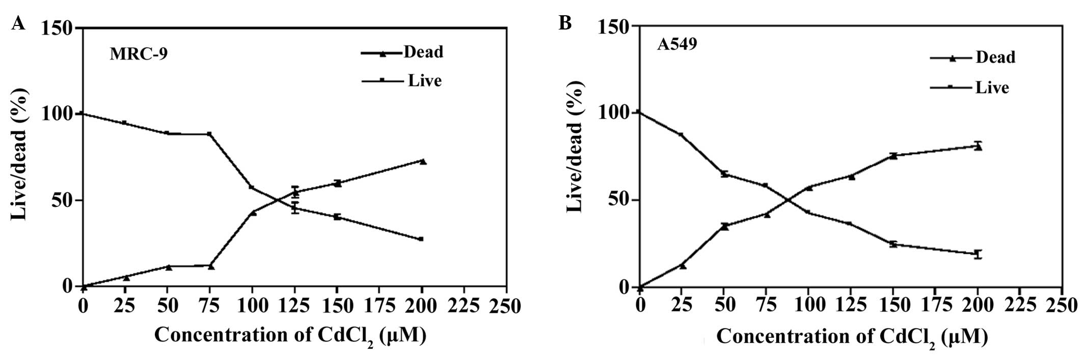
Effect of CdCl2 on the cell
viability of human MRC-9 normal lung and A549 cancer cells
The viability of the normal and cancerous lung cells
exposed to various CdCl2 concentrations was demonstrated
to be dose-dependent (Fig. 1). The
viability of the normal lung MRC-9 cells was reduced by 6, 11, 11,
44, 55, 60 and 75% (Fig. 1A),
while the viability of the A594 lung cancer cells was decreased by
13, 36, 42, 58, 65, 76 and 81% (Fig.
1B) following 24 h treatment with 25, 50, 75, 100, 125, 150 and
200 µM CdCl2, respectively. The significant toxic
effect of CdCl2 was observed at 25 µM
CdCl2 in the A549 lung cancer cells, while the MRC-9
normal lung cells exhibited a significant decrease in viability at
50 µM CdCl2. The LD50 of
CdCl2 was revealed to be 87.5 µM in the A549
cells and 112.5 µM in the MRC-9 cells. These results
demonstrated the toxic effect of CdCl2 on normal human
lung cells and lung cancer cells.
Effect of CdCl2 concentration
and treatment duration on the expression levels of IL-1α and IL-10
cytokines in human MRC-9 normal lung cells
The human MRC-9 normal lung cells were treated with
different concentrations of CdCl2 (0, 50, 100 or 150
µM) for various durations (6, 12, 18 or 24 h), and the
expression levels of IL-1α and IL-10 cytokines were measured. The
cells treated with 50 µM CdCl2 for 6 h
demonstrated the maximum expression levels of each cytokine
(Fig. 2). The cytokine levels
decreased with increasing concentration and duration (Fig. 2). The results clearly demonstrated
that normal MRC-9 lung cells responded to the toxic effect of
CdCl2 with high expression levels of the cytokines in
the lysate at early time points and high concentrations and longer
exposure durations of CdCl2 demonstrated toxic effects
on the expression levels of the cytokines.
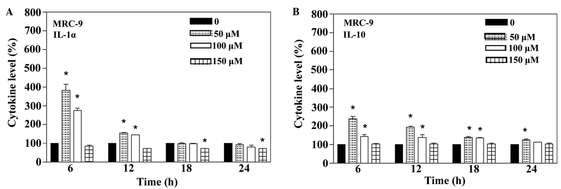 | Figure 2Expression levels of the cytokines,
IL-1α and IL-10, in the CdCl2-treated human MRC-9 normal
lung cells. The cells were treated with 0, 50, 100 or 150 µM
CdCl2 for 6, 12, 18 or 24 h and the cytokine expression
levels were measured by Ray Biotech, Inc. (Norcross, GA, USA) IL-1α
or IL-10 specific ELISA kits. The expression levels of (A) IL-1α
and (B) IL-10 were determined following treatment
(*P<0.05, compared with the control). IL,
interleukin. |
Effect of the concentration of
CdCl2 and duration on the expression level of IL-1α and
IL-10 cytokines in human A549 lung cancer cells
As shown in Fig. 3,
the expression levels of IL-1α and IL-10 cytokines in the A549 lung
cancer cells treated with 0, 50, 100 or 150 µM
CdCl2 concentrations for 6, 12, 18 or 24 h were
detected. The lung cancer cells treated with 50 µM
CdCl2 for 12 h demonstrated the maximum expression of
each cytokine, and the expression levels decreased as the duration
of exposure and concentration of CdCl2 increased
(Fig. 3). The results clearly
demonstrated the response of the A549 lung cancer cells to the
toxic effect of CdCl2, with high expression of cytokines
at high concentrations and longer exposure durations of
CdCl2, demonstrating the toxic effect on the expression
of cytokines.
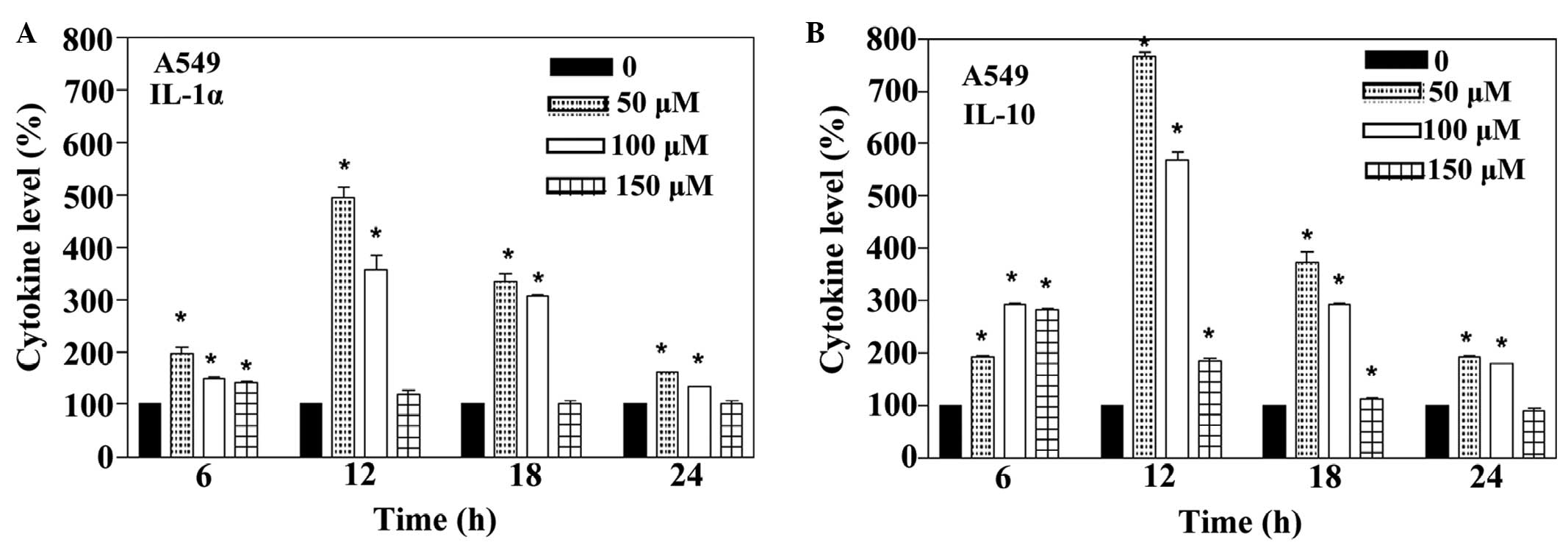 | Figure 3Expression levels of the cytokines,
IL-1α and IL-10, in the CdCl2-treated human A549 lung
cancer cells. The cells were treated with 0, 50, 100 or 150
µM CdCl2 for 6, 12, 18 or 24 h and the expression
levels of the cytokines were measured by Ray Biotech, Inc.
(Norcross, GA, USA) IL-1α or IL-10 specific ELISA kits. The
expression levels of (A) IL-1α and (B) IL-10 were determined
following the treatment (*P<0.05, compared with the
control). IL, interleukin. |
Discussion
The widespread industrial usage of cadmium presents
a health risk directly and indirectly to humans and other living
organisms. Industrial waste fumes, burning of fossil fuels and
cigarette smoke are the predominant direct sources of cadmium
exposure to humans. It is estimated that 90% of the inhaled cadmium
particles are absorbed by lung tissue and cause pulmonary damage,
emphysema and lung cancer (16–19).
Organisms respond to xeno-biotics via inflammation and it is
initiated through various signaling molecules, including cytokines,
in the cells. To date, few investigations into the expression
levels of the cytokines, IL-1α (pro-inflammatory) and IL-10
(anti-inflammatory), have been reported (27). Therefore, elucidating the
expression levels of IL-1α and IL-10, and the cell viability at
various CdCl2 concentrations and incubation durations in
human MRC-9 normal lung and A549 lung cancer cells may provide an
understanding of how lung cells response to cadmium toxicity.
The results indicated that A549 lung cancer cells
were observed to be more sensitive to CdCl2 compared
with the MRC-9 normal lung cells. The difference in their
sensitivities was reflected in their LD50 values. The
LD50 of CdCl2 for the normal MRC-9 lung cells
was 112.5 µM, whereas the LD50 of the lung cancer
cells was 87.5 µM (Fig. 1).
A previous study demonstrated that carcinomas contain significantly
less metallothionein compared with their corresponding normal cells
(28). This may be one of the
contributing factors for the higher sensitivity of cancer cells to
cadmium toxicity, however, further investigation is required to
prove this hypothesis.
Normal lung cells and the lung cancer cells revealed
high expression levels of the IL-1α and IL-10 cytokines following
treatment with CdCl2 (Figs.
1 and 2). In a previous study
(29), a significant increase in
IL-1α and IL-10 cytokines was observed in the tested cells, when
human-derived bronchial epithelium was exposed to tobacco smoke
components. Furthermore, other previous studies have revealed that
cells release pro-inflammatory cytokines during inflammation, to
activate the cytokine network and the secretion of
anti-inflammatory cytokines (30,31).
The higher expression of cytokines, which lead to inflammation, may
be responsible for the later cytotoxic effects in the cells, which
lead to a decrease in viability, as observed in the present study
(Fig. 1). This is supported by a
previous study (32) demonstrating
that high levels of pro-inflammatory cytokines due to cadmium
exposure cause pathological conditions in a biological system.
When the cytokine levels were compared in the normal
lung cells, a higher expression of pro-inflammatory cytokine,
IL-1α, was observed compared with the anti-inflammatory cytokine,
IL-10, at 6 h exposure (Fig. 2). A
high level of IL-1α cytokine in the cells exposed to xeno-biotics
has been reported to inhibit the expression of metallothionein
protein (33). In that previous
study, IL-1α inhibited the mRNA expression of metallothionein in
endometrial stromal cells and amniotic cells treated with
CdCl2. Based on the above report, the present study
hypothesized that increased levels of IL-1α may reduce the protein
expression of metallothionein, which in turn leads to the increase
in unbound CdCl2 in the cell, which may now damage
biological molecules, including proteins, as shown by the decreased
expression of IL-1α and IL-10 observed in the present study
(Fig. 2).
The expression pattern of the IL-1α and IL-10
cytokines from the A549 cancer lung cells was different compared
with the normal lung cells. The maximum levels of the IL-1α and
IL-10 cytokines were observed at a later period (12 h, Fig. 3) in the lung cancer cells compared
with the normal cells (6 h, Fig.
2). In addition, it was also observed that each cytokine was
highly expressed in cancer cells compared with the normal cells
treated with CdCl2 (Figs.
2 and 3). A similar
observation was observed when the cytokine levels of patients with
cancer were compared with the cytokine levels of normal individuals
(34). The delayed expression and
higher expression levels of the cytokines observed in cancer cells
may be as a result of the malfunction of the cell regulatory
mechanisms commonly observed in cancer cells.
Lung cancer cells demonstrated higher expression of
the anti-inflammatory cytokine, IL-10, compared with the
pro-inflammatory cytokine, IL-1α (Fig.
3). This was consistent with our previously reported findings
(24) and another previous study
(35). IL-10 acts as
anti-inflammatory cytokine and is also responsible for cell death.
IL-10 cytokine has been demonstrated to decrease the translocation
of nuclear factor-κB, which is important in increasing apoptotic
markers, which later leads to apoptotic cell death (31). Therefore, the higher levels of
IL-10 cytokine in the A549 lung cancer cells may be one of the
reasons for the higher cytotoxicity caused by CdCl2
(Fig. 1).
In the present study, higher cadmium concentrations
(100 and 150 µM) decreased the expression levels of the
cytokines in the normal and cancer cells (Figs. 2 and 3). Higher concentrations of cadmium
induced higher levels of reactive oxygen species, which degrade
macromolecules, including proteins and DNA (12,13,36).
The low expression levels of the IL-1α and IL-10 cytokines in each
cell line may also be linked to the toxic effect of unbound cadmium
in the cells, as a result of the lack of metallothionein protein.
This result is consistent with a previous study, which reported
inhibition of the expression of IL-1α in rat hepatocytes following
treatment with high concentrations of CdCl2 (32). In addition, longer exposure
durations may also be a factor in causing higher toxicity, as
decreased expression of cytokines were observed with increased
incubation durations in the present study (Figs. 2 and 3). The longer exposure duration led to
the accumulation of cadmium inside of the cell, which in turn
causes decreased cytokine levels leading to cell death.
In conclusion, the viability result revealed that
human A549 lung cancer cells exhibited higher sensitivity to
CdCl2 compared with the normal MRC-9 lung cells.
Furthermore, the cells demonstrated a differential expression of
the cytokines in response to CdCl2. The maximum cytokine
levels were observed in the normal MRC-9 lung cells at an early
incubation time (6 h) compared with the lung cancer cells (12 h),
demonstrating an early immune response of normal lung cells. The
present study clearly demonstrated the effect of CdCl2
on the expression levels of cytokines in lung cells and suggested
that compounds, which activate the cytokines and reduce
inflammation, may prevent cadmium toxicity.
Acknowledgments
This study was supported by the FAMU Title III,
Department of Education (grant no. DOEHBGIPO31B40108–08) and the
RCMI, National Institute of Health (grant nos. G12RR03020 and
G12D007582).
References
|
1
|
International agency for research on
cancer (IARC): Monographs on the Evaluation of the Carcinogenic
Risks to Humans, Beryllium, Cadmium, Mercury and Exposures in the
Glass Manufacturing industry. IARC Scientific Publications; Lyon,
France: pp. 119–238. 1993
|
|
2
|
Goering PL, Waalkes MP and Klaassen CD:
Toxicology of cadmium. Toxicology of metals: Biochemical Aspects.
Goyer RA and Cherias M: Handb Exp Pharmacol. 15:189–214. 1995.
View Article : Google Scholar
|
|
3
|
Agency for Toxic Substances Disease
Registry (ATSDR): Toxicological Profile for Cadmium. Atlanta, GA:
U.S. Department of Health and Human Services, Public Health
Service; pp. 1–512. 2012
|
|
4
|
Joseph P: Mechanisms of cadmium
carcinogenesis. Toxicol Appl Pharmacol. 238:272–279. 2009.
View Article : Google Scholar : PubMed/NCBI
|
|
5
|
Chin TA and Templeton DM: Protective
elevations of glutathione and metallothionein in cadmium-exposed
mesangial cells. Toxicology. 77:145–156. 1993. View Article : Google Scholar : PubMed/NCBI
|
|
6
|
Bridges CC and Zalups RK: Molecular and
ionic mimicry and the transport of toxic metals. Toxicol Appl
Pharmacol. 204:274–308. 2005. View Article : Google Scholar : PubMed/NCBI
|
|
7
|
Habeebu SS, Liu J and Klaassen CD:
Cadmium-induced apoptosis in mouse liver. Toxicol Appl Pharmacol.
149:203–209. 1998. View Article : Google Scholar : PubMed/NCBI
|
|
8
|
Rikans LE and Yamano T: Mechanisms of
cadmium-mediated acute hepatotoxicity. J Biochem Mol Toxicol.
14:110–117. 2000. View Article : Google Scholar : PubMed/NCBI
|
|
9
|
Jin T, Lu J and Nordberg M: Toxicokinetics
and biochemistry of cadmium with special emphasis on the role of
metallothionein. Neurotoxicology. 19:529–535. 1998.PubMed/NCBI
|
|
10
|
Waalkes MP, Anver M and Diwan BA:
Carcinogenic effects of cadmium in the noble (NBL/Cr) rat:
Induction of pituitary, testicular and injection site tumors and
intraepithelial proliferative lesions of the dorsolateral prostate.
Toxicol Sci. 52:154–161. 1999. View Article : Google Scholar
|
|
11
|
Boujelben M, Ghorbel F, Vincent C,
Makni-Ayadi F, Guermazi F, Croute F and El-Feki A: Lipid
peroxidation and HSP72/73 expression in rat following cadmium
chloride administration: Interactions of magnesium supplementation.
Exp Toxicol Pathol. 57:437–443. 2006. View Article : Google Scholar : PubMed/NCBI
|
|
12
|
Ikediobi CO, Badisa VL, Ayuk-Takem LT,
Latinwo LM and West J: Response of antioxidant enzymes and redox
metabolites to cadmium-induced oxidative stress in CRL-1439 normal
rat liver cells. Int J Mol Med. 14:87–92. 2004.PubMed/NCBI
|
|
13
|
Badisa VL, Latinwo LM, Odewumi CO,
Ikediobi CO, Badisa RB, Brooks-Walter A, Lambert AT and Nwoga J:
Cytotoxicity and stress gene microarray analysis in cadmium-exposed
CRL-1439 normal rat liver cells. Int J Mol Med. 22:213–219.
2008.PubMed/NCBI
|
|
14
|
Odewumi CO, Badisa VL, Le UT, Latinwo LM,
Ikediobi CO, Badisa RB and Darling-Reed SF: Protective effects of
N-acetylcysteine against cadmium-induced damage in cultured rat
normal liver cells. Int J Mol Med. 27:243–248. 2011. View Article : Google Scholar
|
|
15
|
Odewumi CO, Buggs R, Badisa VL, Latinwo
LM, Badisa RB, Ikediobi CO, Darling-Reed SF and Owens MA:
Mitigative action of monoisoamy 1-2,3-dimercaptosuccinate (MiADMS)
against cadmium-induced damage in cultured rat normal liver cells.
Toxicol In Vitro. 25:1733–1739. 2011. View Article : Google Scholar : PubMed/NCBI
|
|
16
|
Navarro Silvera SA and Rohan TE: Trace
elements and cancer risk: A review of the epidemiologic evidence.
Cancer Causes Control. 18:7–27. 2007. View Article : Google Scholar
|
|
17
|
Straif K, Benbrahim-Tallaa L, Baan R,
Grosse Y, Secretan B, El Ghissassi F, Bouvard V, Guha N, Freeman C,
Galichet L, et al WHO International Agency for Research on Cancer
Monograph Working Group: A review of human Carcinogens part C:
Metals, arsenic, dusts and fibres. Lancet Oncol. 10:453–454. 2009.
View Article : Google Scholar : PubMed/NCBI
|
|
18
|
Kundu S, Sengupta S and Bhattacharyya A:
EGFR upregulates inflammatory and proliferative responses in human
lung adenocarcinoma cell line (A549), induced by lower dose of
cadmium chloride. Inhal Toxicol. 23:339–348. 2011. View Article : Google Scholar : PubMed/NCBI
|
|
19
|
Rogalska J, Pilat-Marcinkiewicz B and
Brzóska MM: Protective effect of zinc against cadmium
hepatotoxicity depends on this bioelement intake and level of
cadmium exposure: A study in a rat model. Chem Biol Interact.
193:191–203. 2011. View Article : Google Scholar : PubMed/NCBI
|
|
20
|
Shimabukuro DW, Sawa T and Gropper MA:
Injury and repair in lung and airways. Crit. Care Med. 31(8 Suppl):
S524–S531. 2003. View Article : Google Scholar : PubMed/NCBI
|
|
21
|
Grivennikov SI, Kuprash DV, Liu ZG and
Nedospasov SA: Intracellular signals and events activated by
cytokines of the tumor necrosis factor superfamily: From simple
paradigms to complex mechanisms. Int Rev Cytol. 252:129–161. 2006.
View Article : Google Scholar : PubMed/NCBI
|
|
22
|
Barksby HE, Lea SR, Preshaw PM and Taylor
JJ: The expanding family of interleukin-1 cytokines and their role
in destructive inflammatory disorders. Clin Exp Immunol.
149:217–225. 2007. View Article : Google Scholar : PubMed/NCBI
|
|
23
|
Trandem K, Jin Q, Weiss KA, James BR, Zhao
J and Perlman S: Virally expressed interleukin-10 ameliorates acute
encephalomyelitis and chronic demyelination in coronavirus-infected
mice. J Virol. 85:6822–6831. 2011. View Article : Google Scholar : PubMed/NCBI
|
|
24
|
Odewumi CO, Fils-Aime S, Badisa VL,
Latinwo LM, Ruden ML, Ikediobi C and Badisa RB: Chemoprotective
effect of monoisoamyl 2, 3-dimercaptosuccinate (MiADMS) on
cytokines expression in cadmium chloride treated human lung cells.
Environ Toxicol. 30:704–711. 2015. View Article : Google Scholar
|
|
25
|
Badisa RB, Tzakou O, Couladis M and
Pilarinou E: Cytotoxic activities of some Greek Labiatae herbs.
Phytother Res. 17:472–476. 2003. View
Article : Google Scholar : PubMed/NCBI
|
|
26
|
Ipsen J and Feigl P: Bancroft's
Introduction to Biostatistics. Second Edition. Harper and Row; New
York: pp. 1641970
|
|
27
|
Krocova Z, Macela A, Kroca M and
Hernychova L: The immu-nomodulatory effect(s) of lead and cadmium
on the cells of immune system in vitro. Toxicol In Vitro. 14:33–40.
2000. View Article : Google Scholar : PubMed/NCBI
|
|
28
|
Janssen AM, van Duijn W, Kubben FJ,
Griffioen G, Lamers CB, van Krieken JH, van de Velde CJ and
Verspaget HW: Prognostic significance of metallothionein in human
gastrointestinal cancer. Clin Cancer Res. 8:1889–1896.
2002.PubMed/NCBI
|
|
29
|
Balharry D, Sexton K and BéruBé KA: An in
vitro approach to assess the toxicity of inhaled tobacco smoke
components: Nicotine, cadmium, formaldehyde and urethane.
Toxicology. 244:66–76. 2008. View Article : Google Scholar
|
|
30
|
Bergman M, Djaldetti M, Salman H and
Bessler H: Inflammation and colorectal cancer: Does aspirin affect
the interaction between cancer and immune cells? Inflammation.
34:22–28. 2011. View Article : Google Scholar
|
|
31
|
Romoser AA, Chen PL, Berg JM, Seabury C,
Ivanov I, Criscitiello MF and Sayes CM: Quantum dots trigger
immunomodulation of the NFκB pathway in human skin cells. Mol
Immunol. 48:1349–1359. 2011. View Article : Google Scholar : PubMed/NCBI
|
|
32
|
Kayama F, Yoshida T, Elwell MR and Luster
MI: Role of tumor necrosis factor-alpha in cadmium-induced
hepatotoxicity. Toxicol Appl Pharmacol. 131:224–234. 1995.
View Article : Google Scholar : PubMed/NCBI
|
|
33
|
Kawano Y, Furukawa Y, Kawano Y, Abe W,
Hirakawa T and Narahara H: Cadmium chloride induces the expression
of metallothionein mRNA by endometrial stromal cells and
amnion-derived (WISH) cells. Gynecol Obstet Invest. 71:240–244.
2011. View Article : Google Scholar
|
|
34
|
Katsumata N, Eguchi K, Fukuda M, Yamamoto
N, Ohe Y, Oshita F, Tamura T, Shinkai T and Saijo N: Serum levels
of cytokines in patients with untreated primary lung cancer. Clin
Cancer Res. 2:553–559. 1996.PubMed/NCBI
|
|
35
|
Bermúdez-Morales VH, Peralta-Zaragoza O,
Alcocer-González JM, Moreno J and Madrid-Marina V: IL-10 expression
is regulated by HPV E2 protein in cervical cancer cells. Mol Med
Report. 4:369–375. 2011.
|
|
36
|
Thévenod F and Lee WK: Cadmium and
cellular signaling cascades: Interactions between cell death and
survival pathways. Arch Toxicol. 87:1743–1786. 2013. View Article : Google Scholar : PubMed/NCBI
|