Introduction
Regucalcin is a calcium-binding protein which has
been demonstrated to serve a multifunctional role in the regulation
of various types of cells and tissues (1–6). The
regucalcin gene (rgn)), which is located on the X
chromosome, is identified in over 15 species consisting of the
regucalcin family, and is highly conserved in vertebrate species
throughout evolution (7–11). The expression of the regucalcin
gene is regulated by various transcription factors including
activator protein 1, nuclear factor I-A1, regucalcin gene promoter
region-related protein and β-catenin, which are modulated through
intracellular signaling factors associated with the phosphorylation
and dephosphorylation of nuclear proteins in vitro (11). Regucalcin is expressed in the
liver, kidney and various other tissues and is regulated by
hormonal factors including calcium-regulating hormones, insulin,
estrogen and additional steroid hormones (11,12).
Regucalcin is translocated from the cytoplasm to the nucleus in
various types of cells (13).
Regucalcin has been demonstrated to serve a role in the maintenance
of intracellular calcium homeostasis, and it inhibits various
protein kinases and phosphatases, in addition to inhibiting
protein, DNA and RNA syntheses (3–5,13).
Additionally, nuclear regucalcin has been demonstrated to regulate
the gene expression of various proteins (13). Furthermore, regucalcin suppresses
cell proliferation and apoptotic cell death mediated through
various signaling factors in normal kidney NRK52E cells and cloned
rat hepatoma H4II-E cells in vitro (14,15).
Regucalcin has been proposed to serve a physiological role in
maintaining cellular homeostasis and function as a regulatory
protein of intracellular signaling systems (5,6).
Regucalcin has been demonstrated to possess a
pathophysiological role in metabolic disorders (16–18).
In addition, regucalcin has been demonstrated to be involved in
carcinogenesis (19). The
expression of the regucalcin gene and protein have been
demonstrated to be reduced in the tumor tissues of animal models
and human patients in vivo (19,20).
Regucalcin gene expression has been demonstrated to be
downregulated in carcinogenesis, suggesting a potential role of
regucalcin as a suppressor protein in carcinogenesis (19). Overexpression of endogenous
regucalcin has been previously demonstrated to suppress the
enhancement of cell proliferation in cloned rat hepatoma H4-II-E
cells in vitro (21).
The aim of the current study was to investigate
whether exogenous regucalcin possesses a suppressive effect on the
proliferation of human cancer cells in vitro.
Materials and methods
Materials
Dulbecco's modified Eagle's medium (DMEM) with 4.5
g/l glucose, L-glutamine and sodium pyruvate and antibiotics
(penicillin and streptomycin; P/S) were purchased from Invitrogen
Life Technologies (Carlsbad, CA, USA). Fetal bovine serum (FBS) was
purchased from GE Healthcare Life Sciences (Logan, UT, USA).
Gemcitabine was obtained from Hospira, Inc. (Lake Forest, IL, USA),
and diluted in Dulbecco's modified phosphate-buffered saline (PBS).
Tumor necrosis factor-α (TNF-α) was purchased from R&D Systems,
Inc. (Minneapolis, MN, USA). PD98059, staurosporine, Bay K8644,
wortmannin, 5,6-dichloro-1-β-D-ribofuranosylbenzimidazole (DRB) and
all additional reagents were purchased from Sigma-Aldrich (St.
Louis, MO, USA) unless otherwise specified.
Regucalcin
Regucalcin was isolated from rat liver cytosol as
described previously (1). The
livers were perfused with Tris-HCl buffer (pH 7.4), containing 100
mM Tris, 120 mM NaCl and 4 mM KCl, and cooled to 4°C. The livers
were subsequently removed, cut into small pieces, suspended 1:4
(w/v) in Tris-HCl buffer (pH 7.4), and homogenized in a
Potter-Elvehjem homogenizer (Takashima System Ltd., Tokyo, Japan)
with a Teflon pestle (1). The
homogenate was spun at 5,500 × g in a refrigerated centrifuge for
10 min, and the supernatant was spun at 105,000 × g for 60 min at
4°C. The resulting supernatant was purified to electrophoretic
homogeneity by gel filtration using Sephadex G-75 and G-50,
followed by ion-exchange chromatography on
diethylaminoethyl-cellulose (4).
The purity of the isolated regucalcin was analyzed using gel
electrophoresis and western blot analysis, which confirmed that it
did not contain other proteins.
Breast cancer MDA-MB-231-bone metastatic
cells
Breast cancer MDA-MB-231 bone metastatic cells lack
estrogen, progesterone and human epidermal growth factor type 2
receptors, and are therefore considered as triple negative
(22). However, the cells express
epidermal growth factor, transforming growth factor-α and Wnt7B
oncogene, and activation of these receptors and its downstream
signaling events enhances the migration, proliferation, invasion
and progression of the malignant phenotype of these cells. The
MDA-MB-231 bone metastatic cells were provided by Dr Toshi Yoneda
(The University of Texas, San Antonio, TX, USA) (22).
Cell proliferation in MDA-MB-231
cells
Breast cancer MDA-MB-231 cells (1×10⁵/ml
per well) were cultured in a 24-well plate in DMEM containing 10%
FBS and 1% P/S in the presence or absence of regucalcin (0.01, 0.1,
05, 1 or 10 nM) for 1, 2, 3 and 7 days. In separate experiments,
MDA-MB-231 cells (1×10⁵/ml per well) were cultured in
DMEM containing 10% FBS and 1% P/S in the presence of TNF-α (1
ng/ml), Bay K8644 (1 µM), PD98059 (1 µM),
staurosporine (0.1 µM), wortmannin (1 µM) or DRB (1
µM) for 3 days. Following culture, the number of cells was
counted.
Cell death in MDA-MB-231 cells
Breast cancer MDA-MB-231 cells (1×10⁵/ml
per well) were cultured in a 24-well plate in DMEM containing 10%
FBS and 1% P/S in the absence of regucalcin for 7 days, until cells
were confluent. Subsequently, the cells were cultured in the
presence or absence of regucalcin (0.1, 1 or 10 nM) with or without
gemcitabine (10, 50, 100, 250, and 1,000 nM) for 7 days. Following
culture, the number of cells was counted.
Cell counting
Following trypsinization of each culture dish in
0.2% trypsin with 0.02% EDTA in
Ca2+/Mg2+-free PBS for 2 min at 37°C, the
detached cells were collected by centrifugation at 12 × g for 5 min
at 4°C (Eppendorf 5810 R). The cells were resuspended in PBS
solution and stained with eosin. Cell numbers were counted under a
microscope (Olympus MTV-3; Olympus Corporation, Tokyo, Japan) using
a hemocytometer (Brightline; Sigma-Aldrich). For each dish, the
cells were counted twice from which the average was calculated.
Cell numbers are presented as the number/well of plate.
Statistical analysis
Data are presented as the mean ± standard deviation.
Statistical significance was determined using GraphPad InStat
software, version 3 (GraphPad Software, Inc., La Jolla, CA, USA).
Multiple comparisons were conducted using a one-way analysis of
variance and a Tukey-Kramer multiple comparisons post-test for
parametric data. P<0.05 was considered to indicate a
statistically significant difference.
Results
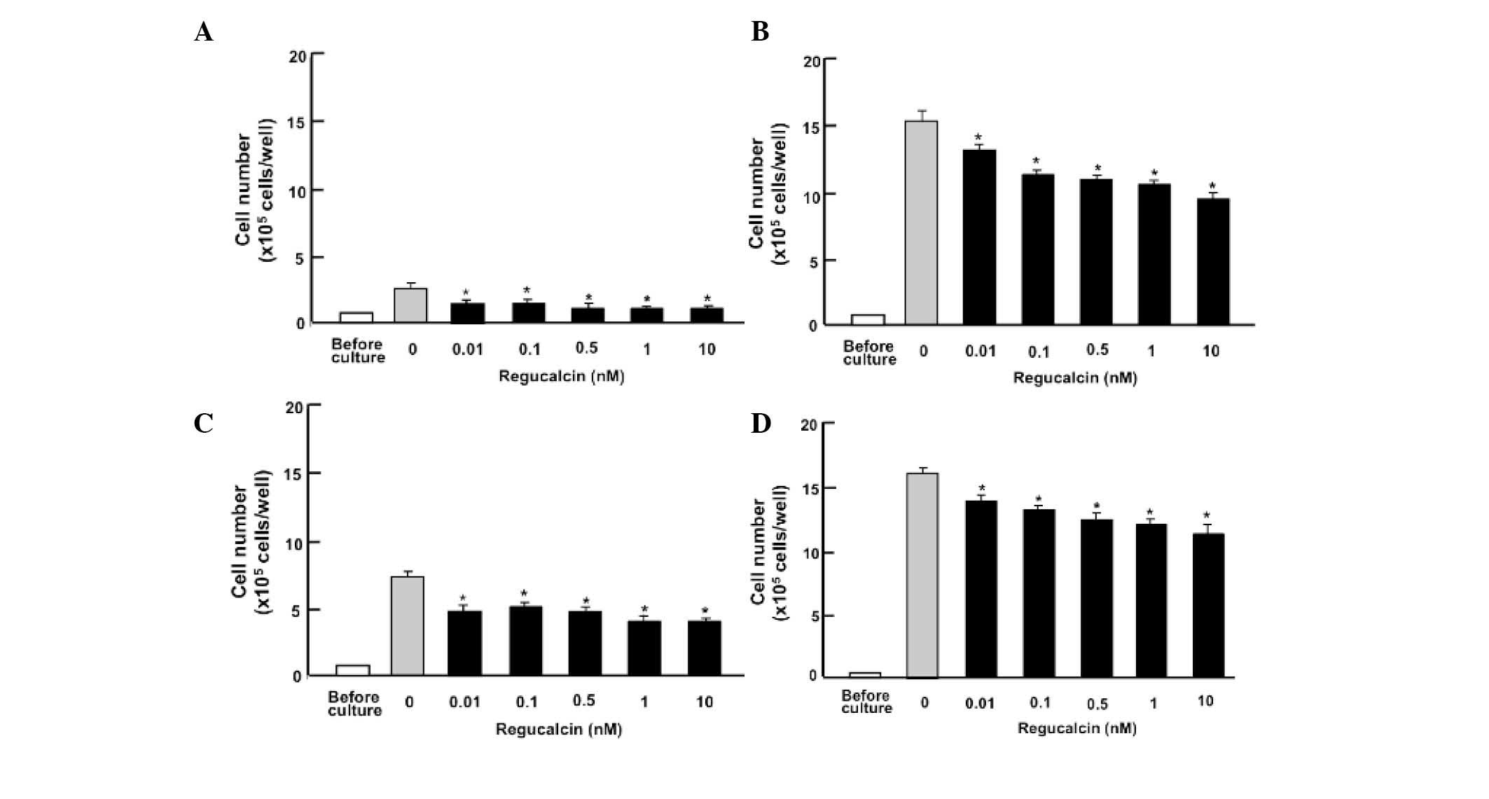
The effect of exogenous regucalcin on the
proliferation of breast cancer MDA-MB-231 bone metastatic cells
in vitro is presented in Fig.
1. MDA-MB-231 cells were cultured in the presence of exogenous
regucalcin (0.1–10 nM) for 1–7 days. The number of cells increased
with the increasing duration of culture. The addition of exogenous
regucalcin reduced the increase in cell number, indicating that
cell proliferation was suppressed by the physiological
concentrations of serum regucalcin (23).
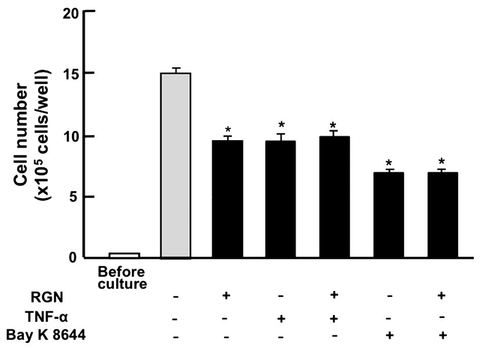
The suppressive effects of regucalcin (1 nM) on cell
proliferation in MDA-MB-231 cells were not enhanced in the presence
of TNF-α (1 ng/ml), an enhancer of nuclear factor-κB (NF-κB)
signaling (24) or Bay K8644 (1
µM), an agonist of Ca2+ influx in cells (25), which resulted in significantly
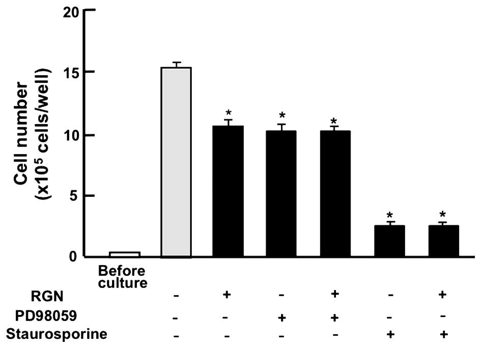
reduced cell numbers when applied alone (Fig. 2). In addition, the effects of
exogenous regucalcin in reducing cell proliferation were not
enhanced in the presence of PD98059 (1 µM), a
mitogen-activated protein kinase (MAPK) inhibitor (26) or staurosporine (0.1 µM), an
inhibitor of protein kinase C (27), which caused a significant reduction
in cell numbers (Fig. 3).
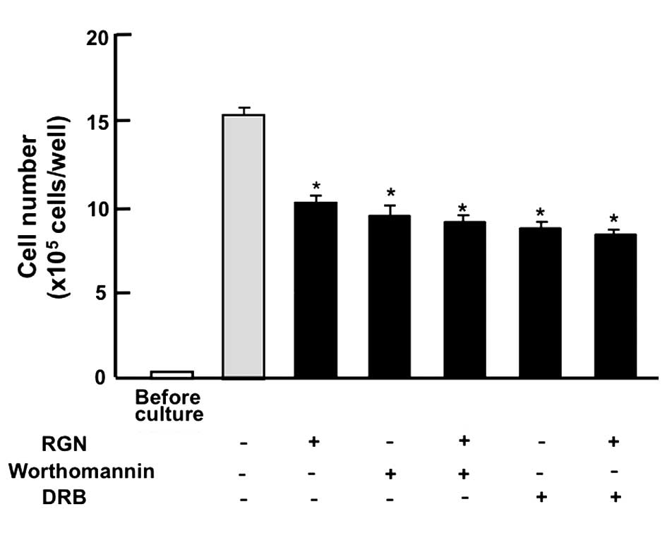
Furthermore, the suppressive effects of regucalcin on cell
proliferation were not enhanced in the presence of wortmannin (1
µM), an inhibitor of phosphatidylinositol 3-kinase (PI3K)
(28) or DRB (1 µM), an
inhibitor of transcriptional activity via RNA polymerase II
inhibition (29) (Fig. 4).
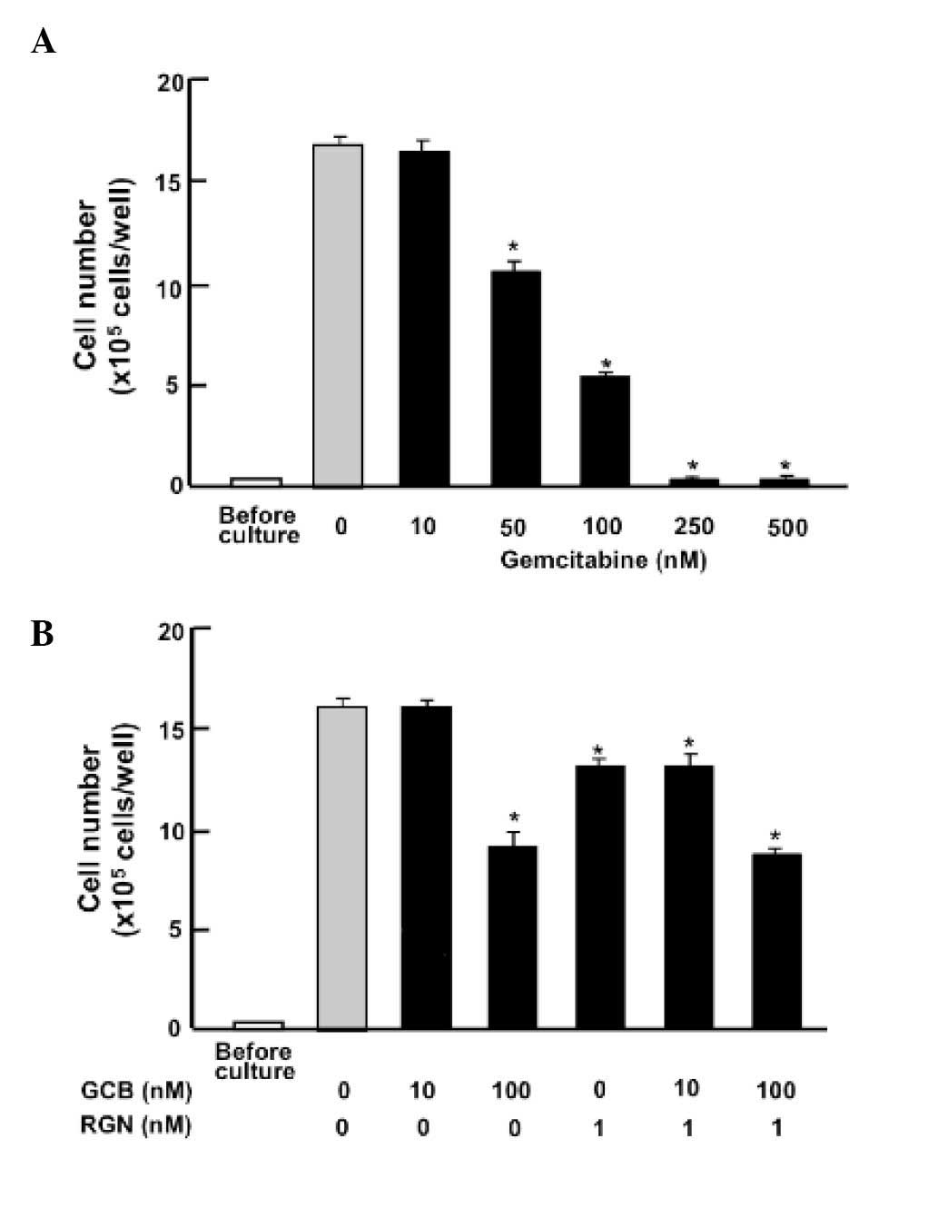
The suppressive effects of regucalcin on the
proliferation of MDA-MB-231 cells were investigated in the presence
of gemcitabine, an antitumor agent, which induces nuclear DNA
damage (30). The addition of
gemcitabine (50–500 nM) to MDA-MB-231 cultures reduced cell
proliferation (Fig. 5A). This
effect was not altered by the application of regucalcin (1 nM) with
gemcitabine (Fig. 5B). The
addition of regucalcin (1 nM) significantly reduced cell numbers in
the presence of gemcitabine (10 nM).
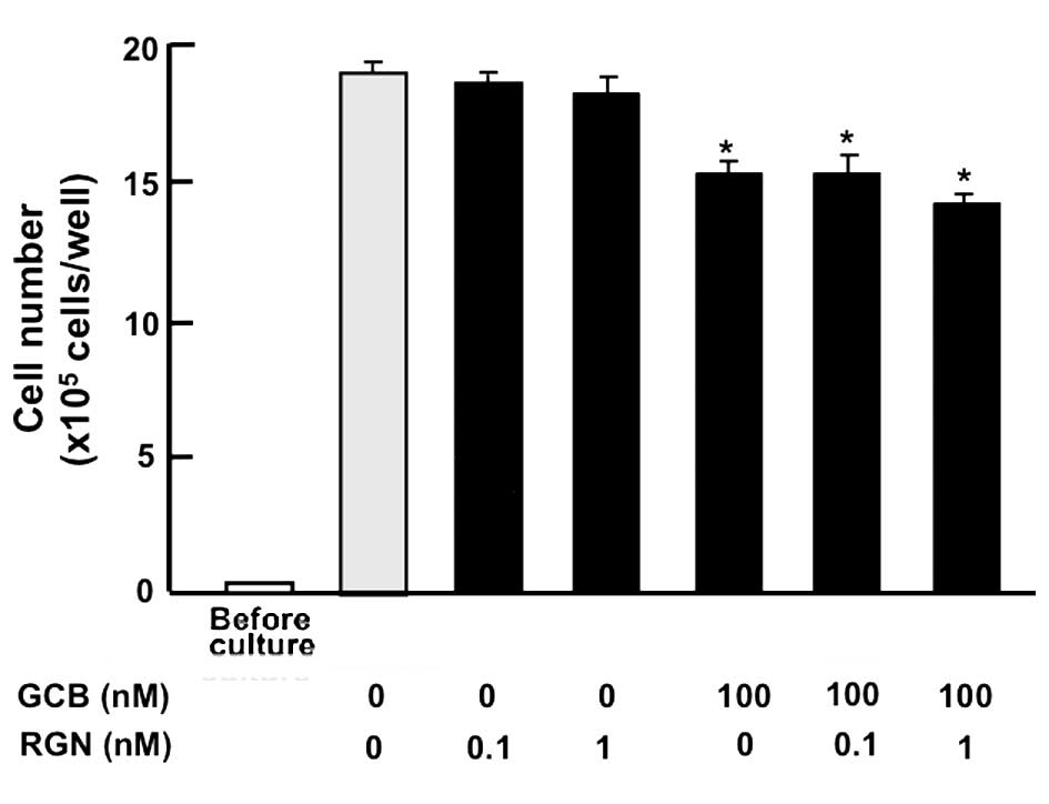
The effect of regucalcin on cell death in breast
cancer MDA-MB-231 cells is presented in Fig. 6. Cells were cultured for 7 days
until confluent, and subsequently cultured for 7 days in the
presence of regucalcin (0.1 or 1 nM) with or without gemcitabine
(100 nM). The addition of regucalcin was not observed to
significantly affect the cell number, however culture with
gemcitabine reduced cell number. Therefore, it is suggested that
regucalcin does not induce cell death.
Discussion
Regucalcin has been demonstrated to serve a
multifunctional role in the regulation of cell function by
suppressing various signaling pathways in various types of cells
and tissues (4–6). Previous studies have demonstrated
that regucalcin serves a potential role as a suppressor of cell
proliferation and carcinogenesis (14,18,19).
Regucalcin gene expression was observed to be downregulated in the
tumor tissues of human patients (20) and in human cancer cells (18,31).
The current study demonstrated that exogenous regucalcin possesses
suppressive effects on the proliferation of human breast cancer
MDA-MB-231 bone metastatic cells in vitro, however was not
observed to affect cell death.
Overexpression of endogenous regucalcin has been
demonstrated to suppress the proliferation of cloned rat hepatoma
H4-II-E cells in vitro (14,18,21).
Regucalcin has been demonstrated to result in G1 and
G2/M phase cell cycle arrest in H4-II-E cells (32). In addition, overexpression of
endogenous regucalcin has been demonstrated to have suppressive
effects on cell proliferation inducing G1 and
G2/M phase cell cycle arrest in cloned normal rat kidney
proximal tubular epithelial NRK52E cells in vitro (33). The suppressive effects of
endogenous regucalcin on cell proliferation may be mediated through
the inhibition of various Ca2+ signaling-dependent
protein kinases, protein phosphatases and PI3K activities and the
suppression of c-Myc, H-Ras, c-Jun and chk2 mRNA expression, or the
enhancement of p53 and Rb mRNA expression (14,19,34,35).
Furthermore, regucalcin has been demonstrated to suppress
cytoplasmic protein synthesis and nuclear DNA and RNA synthesis
(13,14).
The current study demonstrated that the suppressive
effects of exogenous regucalcin on the proliferation of MDA-MB-231
cells were not modulated in the presence of various inhibitors that
regulate intracellular signaling pathways in vitro. The
suppressive effects of regucalcin on the proliferation of
MDA-MB-231 cells were not enhanced in the presence of: TNF-α, an
enhancer of NF-κB signaling (24);
Bay K8644, an agonist of Ca2+ entry in cells (25); PD98059, an MAPK inhibitor (26); staurosporine, an inhibitor of
calcium-dependent protein kinase C (27); or wortmannin, an inhibitor of PI3K
(28). These data suggest that
exogenous regucalcin stimulates various intracellular signaling
pathways to suppress cell proliferation in human breast cancer
MDA-MB-231 cells. Regucalcin has been demonstrated to bind the
plasma membranes of rat liver cells in vitro (36). Therefore, it may be possible that
exogenous regucalcin binds to the plasma membranes of breast cancer
MDA-MB-231 cells, and potentially regulates intracellular signaling
pathways that suppress cell proliferation. In addition, the
suppressive effects of regucalcin on cell proliferation were not
enhanced in the presence of DRB, an inhibitor of transcriptional
activity via RNA polymerase II inhibition (29). The intracellular signals of
exogenous regucalcin may be transmitted into the nucleus to
suppress transcriptional regulation in human breast cancer
MDA-MB-231 cells.
Overexpression of endogenous regucalcin has been
demonstrated to have suppressive effects on apoptotic cell death in
rat hepatoma H4-II-E cells and normal rat kidney NRK52-E cells,
which were increased through various signaling pathways in the
cytoplasm and nucleus in vitro (15). In the current study, exogenous
regucalcin did not induce cell death in human breast cancer
MDA-MB-231 cells in vitro, indicating that regucalcin does
not stimulate cell death. This effect was not enhanced in the
presence of gemcitabine, an antitumor agent, which induces nuclear
DNA damage (30). This observation
may support the theory that the intracellular signaling by
exogenous regucalcin may be transmitted to regulate nuclear
function in breast cancer cells.
In conclusion, the current study demonstrated that
exogenous regucalcin possesses suppressive effects on the
proliferation of human breast cancer MDA-MB-231 bone metastatic
cells in vitro. This suggests that exogenous regucalcin
serves a role as a suppressor of the proliferation of human cancer
cells.
References
|
1
|
Yamaguchi M and Yamamoto T: Purification
of calcium binding substance from soluble fraction of normal rat
liver. Chem Pharm Bull (Tokyo). 26:1915–1918. 1978. View Article : Google Scholar
|
|
2
|
Yamaguchi M and Sakurai T: Inhibitory
effect of calcium-binding protein regucalcin on
Ca2(+)-activated DNA fragmentation in rat liver nuclei.
FEBS Lett. 279:281–284. 1991. View Article : Google Scholar : PubMed/NCBI
|
|
3
|
Shimokawa N and Yamaguchi M: Molecular
cloning and sequencing of the cDNA coding for a calcium-binding
protein regucalcin from rat liver. FEBS Lett. 327:251–255. 1993.
View Article : Google Scholar : PubMed/NCBI
|
|
4
|
Yamaguchi M: Role of regucalcin in calcium
signaling. Life Sci. 66:1769–1780. 2000. View Article : Google Scholar : PubMed/NCBI
|
|
5
|
Yamaguchi M: Role of regucalcin in
maintaining cell homeo-stasis and function (review). Int J Mol Med.
15:371–389. 2005.PubMed/NCBI
|
|
6
|
Yamaguchi M: Regucalcin and cell
regulation: Role as a suppressor protein in signal transduction.
Mol Cell Biochem. 353:101–137. 2011. View Article : Google Scholar : PubMed/NCBI
|
|
7
|
Shimokawa N, Matsuda Y and Yamaguchi M:
Genomic cloning and chromosomal assignment of rat regucalcin gene.
Mol Cell Biochem. 151:157–163. 1995. View Article : Google Scholar : PubMed/NCBI
|
|
8
|
Thiselton DL, McDowall J, Brandau O,
Ramser J, d'Esposito F, Bhattacharya SS, Ross MT, Hardcastle AJ and
Meindl A: An integrated, functionally annotated gene map of the
DXS8026-ELK1 interval on human Xp11.3-Xp11.23: Potential hotspot
for neurogenetic disorders. Genomics. 79:560–572. 2002. View Article : Google Scholar : PubMed/NCBI
|
|
9
|
Yamaguchi M, Makino R and Shimokawa N: The
5′ end sequences and exon organization in rat regucalcin gene. Mol
Cell Biochem. 165:145–150. 1996. View Article : Google Scholar : PubMed/NCBI
|
|
10
|
Misawa H and Yamaguchi M: The gene of
Ca2+-binding protein regucalcin is highly conserved in
vertebrate species. Int J Mol Med. 6:191–196. 2000.PubMed/NCBI
|
|
11
|
Yamaguchi M: The transcriptional
regulation of regucalcin gene expression. Mol Cell Biochem.
346:147–171. 2011. View Article : Google Scholar
|
|
12
|
Yamaguchi M: Hormonal Regulation of
regucalcin gene expression: Involvement in cell metabolism. Horm
Stud. 1:12013. View Article : Google Scholar
|
|
13
|
Yamaguchi M: Role of regucalcin in cell
nuclear regulation: Involvement as a transcription factor. Cell
Tissue Res. 354:331–341. 2013. View Article : Google Scholar : PubMed/NCBI
|
|
14
|
Yamaguchi M: Suppressive role of
regucalcin in liver cell proliferation: Involvement in
carcinogenesis. Cell Prolif. 46:243–253. 2013. View Article : Google Scholar : PubMed/NCBI
|
|
15
|
Yamaguchi M: The anti-apoptotic effect of
regucalcin is mediated through multisignaling pathways. Apoptosis.
18:1145–1153. 2013. View Article : Google Scholar : PubMed/NCBI
|
|
16
|
Yamaguchi M: Regucalcin and metabolic
disorder: Osteoporosis and hyperlipidemia are induced in regucalcin
transgenic rats. Mol Cell Biochem. 341:119–133. 2010. View Article : Google Scholar : PubMed/NCBI
|
|
17
|
Yamaguchi M and Murata T: Involvement of
regucalcin in lipid metabolism and diabetes. Metabolism.
62:1045–1051. 2013. View Article : Google Scholar : PubMed/NCBI
|
|
18
|
Yamaguchi M: Regucalcin as a potential
biomarker for metabolic and neuronal diseases. Mol Cell Biochem.
391:157–166. 2014. View Article : Google Scholar : PubMed/NCBI
|
|
19
|
Yamaguchi M: Involvement of regucalcin as
a suppressor protein in human carcinogenesis: Insight into the gene
therapy. J Cancer Res Clin Oncol. 141:1333–1341. 2015. View Article : Google Scholar
|
|
20
|
Murata T and Yamaguchi M: Alternatively
spliced variants of the regucalcin gene in various human normal and
tumor tissues. Int J Mol Med. 34:1141–1146. 2014.PubMed/NCBI
|
|
21
|
Misawa H, Inagaki S and Yamaguchi M:
Suppression of cell proliferation and deoxyribonucleic acid
synthesis in the cloned rat hepatoma H4-II-E cells overexpressing
regucalcin. J Cell Biochem. 84:143–149. 2001. View Article : Google Scholar : PubMed/NCBI
|
|
22
|
Hiraga T, Williams PJ, Mundy GR and Yoneda
T: The bisphosphonate ibandronate promotes apoptosis in MDA-MB-231
human breast cancer cells in bone metastases. Cancer Res.
61:4418–4424. 2001.PubMed/NCBI
|
|
23
|
Yamaguchi M and Isogai M: Tissue
concentration of calcium-binding protein regucalcin in rats by
enzyme-linked immunoadsorbent assay. Mol Cell Biochem. 122:65–68.
1993. View Article : Google Scholar : PubMed/NCBI
|
|
24
|
Lee ZH, Kwack K, Kim KK, Lee SH and Kim
HH: Activation of c-Jun N-terminal kinase and activator protein 1
by receptor activator of nuclear factor kappaB. Mol Pharmacol.
58:1536–1545. 2000.PubMed/NCBI
|
|
25
|
Cano-Abad MF, Villarroya M, García AG,
Gabilan NH and López MG: Calcium entry through L-type calcium
channels causes mitochondrial disruption and chromaffin cell death.
J Biol Chem. 276:39695–39704. 2001. View Article : Google Scholar : PubMed/NCBI
|
|
26
|
Chen S, Wang Y, Ruan W, Wang X and Pan C:
Reversing multidrug resistance in hepatocellular carcinoma cells by
inhibiting extracellular signal-regulated kinase/mitogen-activated
protein kinase signaling pathway activity. Oncol Lett. 8:2333–2339.
2014.PubMed/NCBI
|
|
27
|
Chen QW, Edvinsson L and Xu CB: Role of
ERK/MAPK in endothelin receptor signaling in human aortic smooth
muscle cells. BMC Cell Biol. 10:522009. View Article : Google Scholar : PubMed/NCBI
|
|
28
|
Serrano-Nascimento C, da Silva Teixeira S,
Nicola JP, Nachbar RT, Masini-Repiso AM and Nunes MT: The acute
inhibitory effect of iodide excess on sodium/iodide symporter
expression and activity involves the PI3K/Akt signaling pathway.
Endocrinology. 155:1145–1156. 2014. View Article : Google Scholar : PubMed/NCBI
|
|
29
|
Palangat M, Grass JA, Langelier MF,
Coulombe B and Landick R: The RPB2 flap loop of human RNA
polymerase II is dispensable for transcription initiation and
elongation. Mol Cell Biol. 31:3312–3325. 2011. View Article : Google Scholar : PubMed/NCBI
|
|
30
|
Tang SC and Chen YC: Novel therapeutic
targets for pancreatic cancer. World J Gastroenterol.
20:10825–10844. 2014. View Article : Google Scholar : PubMed/NCBI
|
|
31
|
Maia C, Santos C, Schmitt F and Socorro S:
Regucalcin is under-expressed in human breast and prostate cancers:
Effect of sex steroid hormones. J Cell Biochem. 107:667–676. 2009.
View Article : Google Scholar : PubMed/NCBI
|
|
32
|
Yamaguchi M and Daimon Y: Overexpression
of regucalcin suppresses cell proliferation in cloned rat hepatoma
H4-II-E cells: Involvement of intracellular signaling factors and
cell cycle-related genes. J Cell Biochem. 95:1169–1177. 2005.
View Article : Google Scholar : PubMed/NCBI
|
|
33
|
Nakagawa T, Sawada N and Yamaguchi M:
Overexpression of regucalcin suppresses cell proliferation of
cloned normal rat kidney proximal tubular epithelial NRK52E cells.
Int J Mol Med. 16:637–643. 2005.PubMed/NCBI
|
|
34
|
Tsurusaki Y and Yamaguchi M:
Overexpression of regucalcin modulates tumor-related gene
expression in cloned rat hepatoma H4-II-E cells. J Cell Biochem.
90:619–626. 2003. View Article : Google Scholar : PubMed/NCBI
|
|
35
|
Tsurusaki Y and Yamaguchi M: Role of
regucalcin in liver nuclear function: Binding of regucalcin to
nuclear protein or DNA and modulation of tumor-related gene
expression. Int J Mol Med. 14:277–281. 2004.PubMed/NCBI
|
|
36
|
Yamaguchi M, Mori S and Kato S:
Calcium-binding protein regucalcin is an activator of
(Ca2+-Mg2+)-adenosine triphosphatase in the
plasma membranes of rat liver. Chem Pharm Bull (Tokyo).
36:3532–3539. 1988. View Article : Google Scholar
|