Introduction
Alzheimer's disease (AD) is a neurodegenerative
disease of the cerebral cortex, which affects the elderly. Its main
clinical features include progressive memory impairment, cognitive
impairment and reduced quality of life (1). The typical pathological features are
extracellular accumulation of β-amyloid (Aβ) in the hippocampus of
the brain, the formation of senile plaques, abnormal accumulation
of tau protein within brain cells, the appearance of
neurofibrillary tangles composed of paired helical filaments,
decreased numbers of cerebral cortical neurons, and neocortex and
meningeal vascular amyloidosis (2,3).
AD is a chronic degenerative disease of the central
nervous system. Previous studies have demonstrated that AD
occurrence and development is closely associated with abnormal
deposition of Aβ in the brain (4).
Abnormal deposition of Aβ results in sustained activation of
inflammatory repairing mechanisms. The transformation from acute
reaction into chronic inflammatory damage under normal
circumstances may be one of the key factors in the pathogenesis of
AD (5). Microglia and astrocytes
are the main immune cells participating in the central inflammatory
cascade of AD (6). The chemokines
produced by Aβ-activating astrocytes are potential chemoattractants
of microglial cells and macrophages, and also upregulate the
expression of inflammatory cytokines, including interleukin (IL)-1
and IL-6 (7). Therefore, the
inhibition of Aβ-induced activation of astrocytes may be an
important therapeutic strategy for AD, which is caused by the
neuropathological changes associated with Aβ (8).
One of the pathological features of AD is the loss
of a large number of neurons. The predominant underlying mechanism
of neuronal loss resulting from AD is apoptosis, and the neuronal
apoptosis hypothesis is an important aspect of AD pathogenesis
(9). The neurons of the brain are
particularly sensitive to apoptotic damage (10). Factors that induce apoptosis, such
as Aβ, oxidative damage and low energy metabolism are present in AD
brain tissues (11). A previous
study hypothesized that apoptosis is one of the mechanisms
underlying the death of AD brain neurons, in which members of the
B-cell lymphoma 2 (Bcl-2) family are key in the gene regulation
process of apoptosis (12). The
Bcl-2 family is divided into two categories: The anti-apoptotic
genes, including Bcl-2; and the pro-apoptotic genes, including
Bcl-2-associated X protein (Bax), which is involved in the
regulation of apoptosis by activating a series of downstream genes
(13).
Paeoniflorin was isolated from the
Ranunculaceae plant, peony, for the first time in 1963; it
is one of the main active components of peony (14). Research into the pharmacological
effects of paeoniflorin has identified that paeoniflorin possesses
anti-spasm, antipyretic cooling, anti-inflammation, anti-ulcer,
anti-oxidation, anti-clotting, and pain and cholesterol regulatory
properties (10,15). The underlying mechanisms remain to
be elucidated, however, a number of receptors and ion channels have
been suggested as possible targets for the pharmacological effects
of paeoniflorin (16,17). It has been demonstrated that
paeoniflorin may exert an effect on the nervous system and on
neurodegenerative diseases, such as AD and Parkinson's disease
(18). The present study
demonstrated that the neuroprotective effects of paeoniflorin
improved AD via inflammation and apoptosis.
Materials and methods
Materials
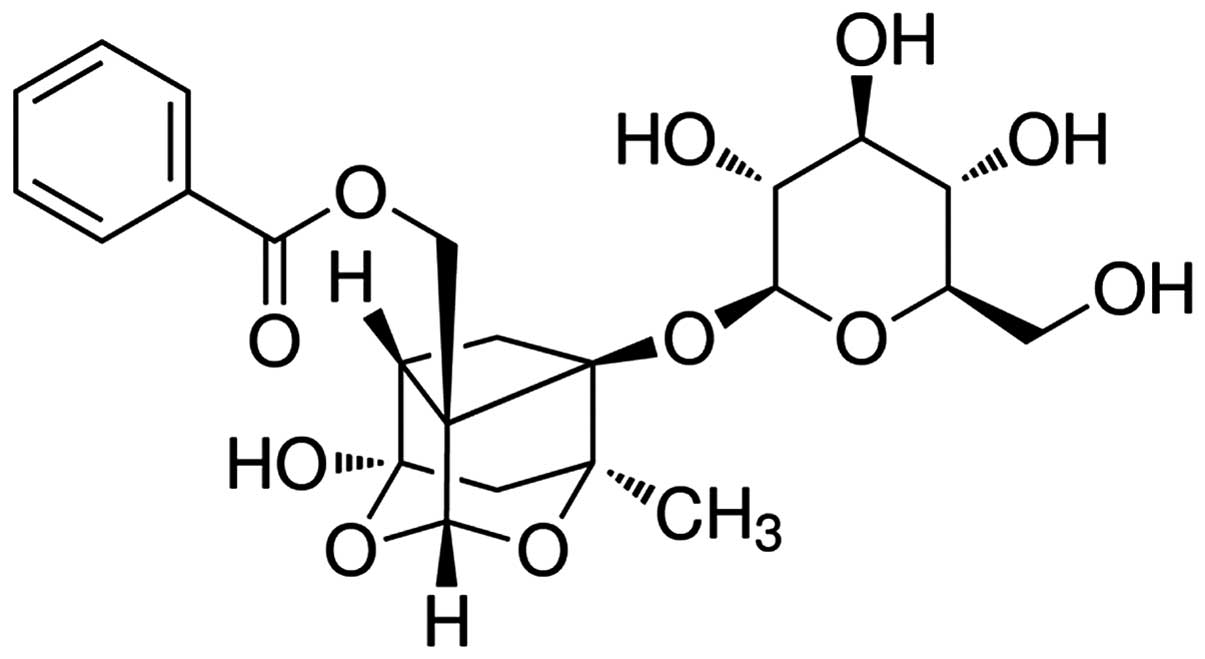
Paeoniflorin (purity, ≥98%) was purchased from
Sigma-Aldrich (St. Louis, MO, USA) and its chemical structure is
presented in Fig. 1. Nuclear
factor-κB (NF-κB) p65 unit (cat. no. H202; Nanjing Jiancheng
Bioengineering Institute, Nanjing, China), tumor necrosis factor-α
(TNF-α; cat. no. E-CL-R0019c; Wuhan Elabscience, Biotechnology Co.,
Ltd., Wuhan China), IL-1β (cat. no. H002; Nanjing Jiancheng
Bioengineering Institute), IL-6 (cat. no. H007; Nanjing Jiancheng
Bioengineering Institute) and caspase-3 (cat. no. C1115; Beyotime
Institute of Biotechnology, Nanjing, China) commercial kits were
purchased. A bicinchoninic acid protein quantification kit was
purchased from Sigma-Aldrich (cat. no. BCA1-1KT).
Transgenic mice
The present study was approved by the Institutional
Animal Care and Use Committee at Dalian University (Dalian, China).
Transgenic mice (n=16; Cyagen Biosciences, Guangzhou, China)
expressing the human mutant PS2 and under the control of
neuron-specific enolase (NSE) were maintained in the genetic
background of C57BL/6 x DBA/2 mice. All mice were maintained in the
laboratory for 2 weeks under a 12-h light/dark cycle (housed with
the mice of the same group at 23±1°C with 50% relative humidity),
fed a standard laboratory diet and had access to water ad
libitum.
Animal grouping
Control non-transgenic mice were divided into two
groups, as follows: i) The control group (Con; n=8), non-transgenic
mice receiving sodium pentobarbital [0.1 ml/100 g administered
intraperitoneally (i.p.)]; and ii) the control-paeoniflorin group
(Con-Pae; n=8), non-transgenic mice receiving 2.0 mg/kg
paeoniflorin for 24 h. Transgenic mice were divided into two
groups, as follows: i) AD group (Alz; n=8), transgenic mice
receiving sodium pentobarbital (0.1 ml/100 g i.p.); and ii) AD
paeoniflorin group (Alz-Pae; n=8), transgenic mice receiving 2.0
mg/kg paeoniflorin for 24 h.
Morris water maze test
Following treatment with paeoniflorin for 24 h,
Morris water maze tests were performed, as described in a previous
study (19). All the mice were
administered the non-visible platform trial twice per day for the
first five days, a probe trial on the sixth day, and a visible
platform trial on the seventh day. All the mice learned to use
visual cues in the room to navigate to an escape platform located
at a fixed position and hidden or submerged 1 cm below the surface
of the water. All the mice were placed in the pool from different
quadrants for training periods of 120 sec. If the mice did not find
the platform within 120 sec, the latency was recorded as 120 sec.
All mice were replaced on the platform for 20 sec, and the next
training period was performed following 120 sec of rest. On each of
the five acquisition days, the platform was removed, and the number
of crossings of the platform location within 120 sec (crossing
number) was recorded.
Evaluation of inflammation and caspase-3
activity
Following treatment with paeoniflorin for 24 h, the
mice were sacrificed by cervical dislocation under anaesthesia
(pentobarbital), and the cerebral cortex samples were rapidly
removed. The cortex samples were snap-frozen on dry ice and stored
at −80°C. The cortex samples were homogenized in physiological
saline (0.1 ml/100 g; Dalian Yuanda Pharmaceutical Co., Ltd.,
Dalian, China) and centrifuged at 12,000 × g for 10 min at 4°C. The
liquid supernatant was collected to analyze the activity of NF-κB
p65, TNF-α, IL-1β, IL-6 and caspase-3 activities according to the
manufacturer's protocols (Westang Biotech, Co., Ltd.).
Western blot analysis of protein
expression levels
Following treatment with paeoniflorin for 24 h,
cerebral cortex samples were homogenized with PRO-PREP™ protein
extraction solution (Invitrogen; Thermo Fisher Scientific, Inc.,
Waltham, MA, USA) and centrifuged at 12,000 × g at 4°C for 10 min.
The protein concentration was determined using a bicinchoninic acid
protein quantification kit. Equal protein quantities (50 µg)
were loaded onto a 10% polyacrylamide gel (Beyotime Institute of
Biotechnology) for 90 min for electrophoresis (100 V) and
subsequently transferred to polyvinylidene fluoride membranes (0.22
mm; EMD Millipore, Billerica, MA, USA). Following blocking of
nonspecific binding with Tris-buffered saline (Beyotime Institute
of Biotechnology) containing skimmed milk, the membranes were
incubated with the following primary antibodies overnight at 4°C:
Bcl-2 (cat. no. sc-578; 1:2,000; Santa Cruz Biotechnology, Inc.,
Dallas, TX, USA), Bax (cat. no. sc-20067; 1:1,500, Santa Cruz
Biotechnology, Inc.), phosphorylated (p)-Akt (cat. no. sc-293125;
1:1,000; Santa Cruz Biotechnology, Inc.), p38 mitogen-activated
protein kinase (p38 MAPK; cat. no. sc-398305; 1:1,000; Santa Cruz
Biotechnology, Inc.), p-p38 MAPK (cat. no. sc-7973; 1:1,000; Santa
Cruz Biotechnology, Inc.) and β-actin (cat. no. sc-8432; 1:500;
Sangon Biotech Co., Ltd., Shanghai, China). The membranes were
washed three times with washing buffer (Beyotime Institute of
Biotechnology) and incubated for 1 h at 37°C with secondary
antibodies (sc-53804; 1:5,000; Santa Cruz Biotechnology, Inc.). The
membrane blots were developed using enhanced chemiluminescence
reagents (cat. no. P0018A; Applygen Technologies, Inc., Beijing,
China). The band intensity was resolved using a gel image analysis
system (Optiquant; Bio-Rad Laboratories, Inc., Hercules, CA,
USA).
Statistical analysis
The data are presented as the mean ± standard error
and assessed using one-way analysis of variance with a 95%
confidence interval. P<0.05 was considered to indicate a
statistically significant difference.
Results
Protective effect of paeoniflorin
improves cognitive function in AD mice
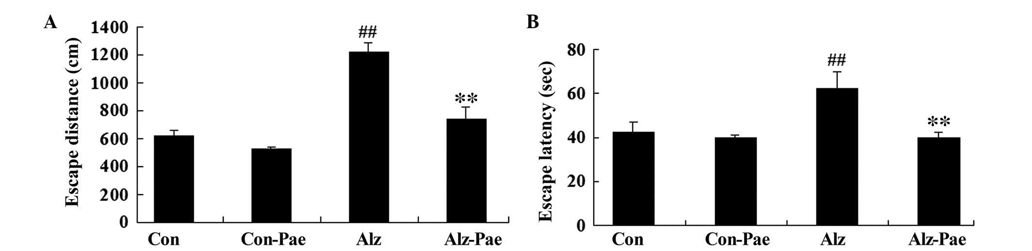
To investigate whether the protective effect of
paeoniflorin improves cognitive function in AD mice, the Morris
water maze test was performed. Patterns of escape distance and
latency were significantly increased in the transgenic mice,
compared with those of the control group (P<0.05; Fig. 2). However, these values were
significantly decreased by treatment with paeoniflorin (Alz-Pae),
compared with the AD group (Alz; P<0.05; Fig. 2). These results suggest that
paeoniflorin may improve cognitive function of transgenic mice.
Protective effect of paeoniflorin
decreases inflammation in AD mice
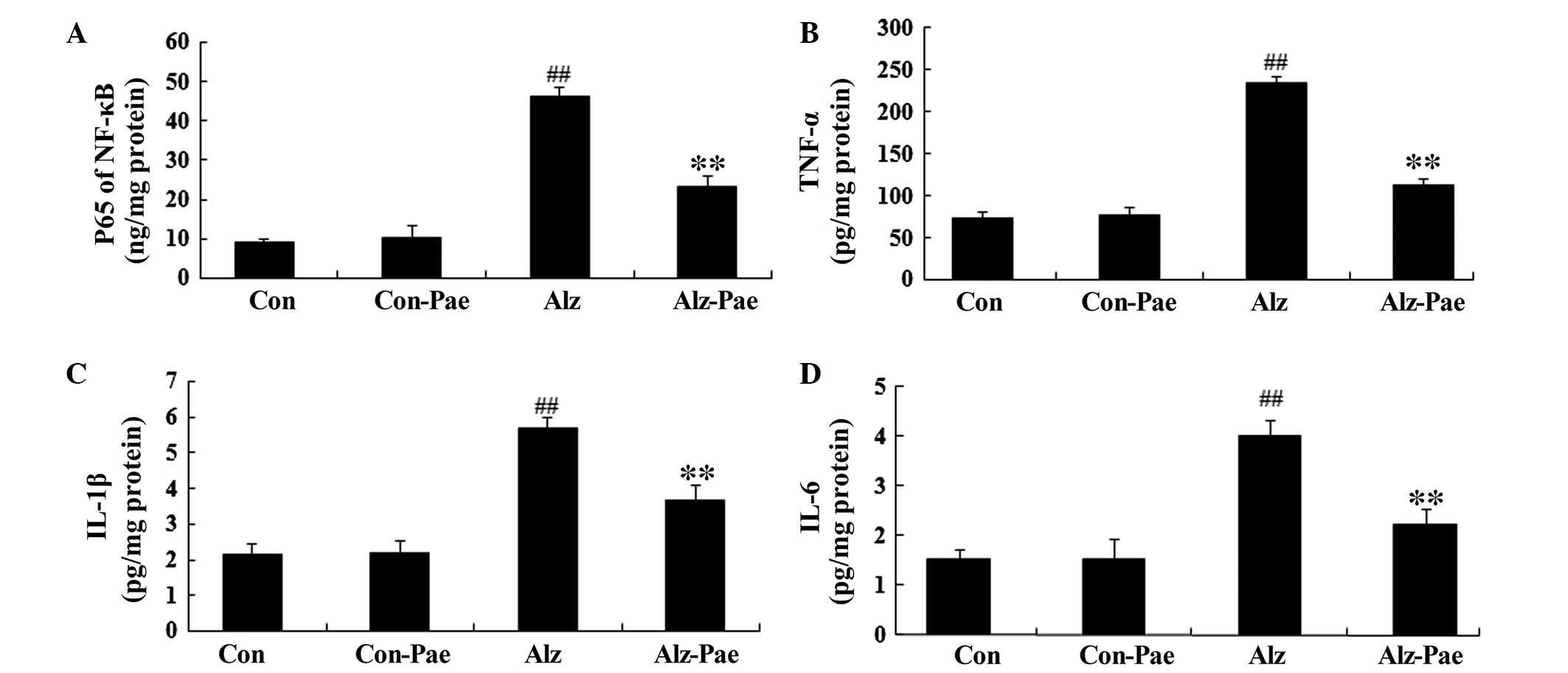
To investigate whether the protective effect of
paeoniflorin decreased inflammation in AD mice, the activity of
NF-κB p65, TNF-α, IL-1β and IL-6 was analyzed. These inflammatory
factors were significantly increased in the Alz, compared with the
Con group (P<0.05; Fig. 3).
Notably, paeoni-florin treatment (Alz-Pae) significantly decreased
the activity of inflammatory factors in the transgenic mice,
compared with the Alz group (P<0.05; Fig. 3).
Protective effect of paeoniflorin
influences caspase-3 activity in AD mice
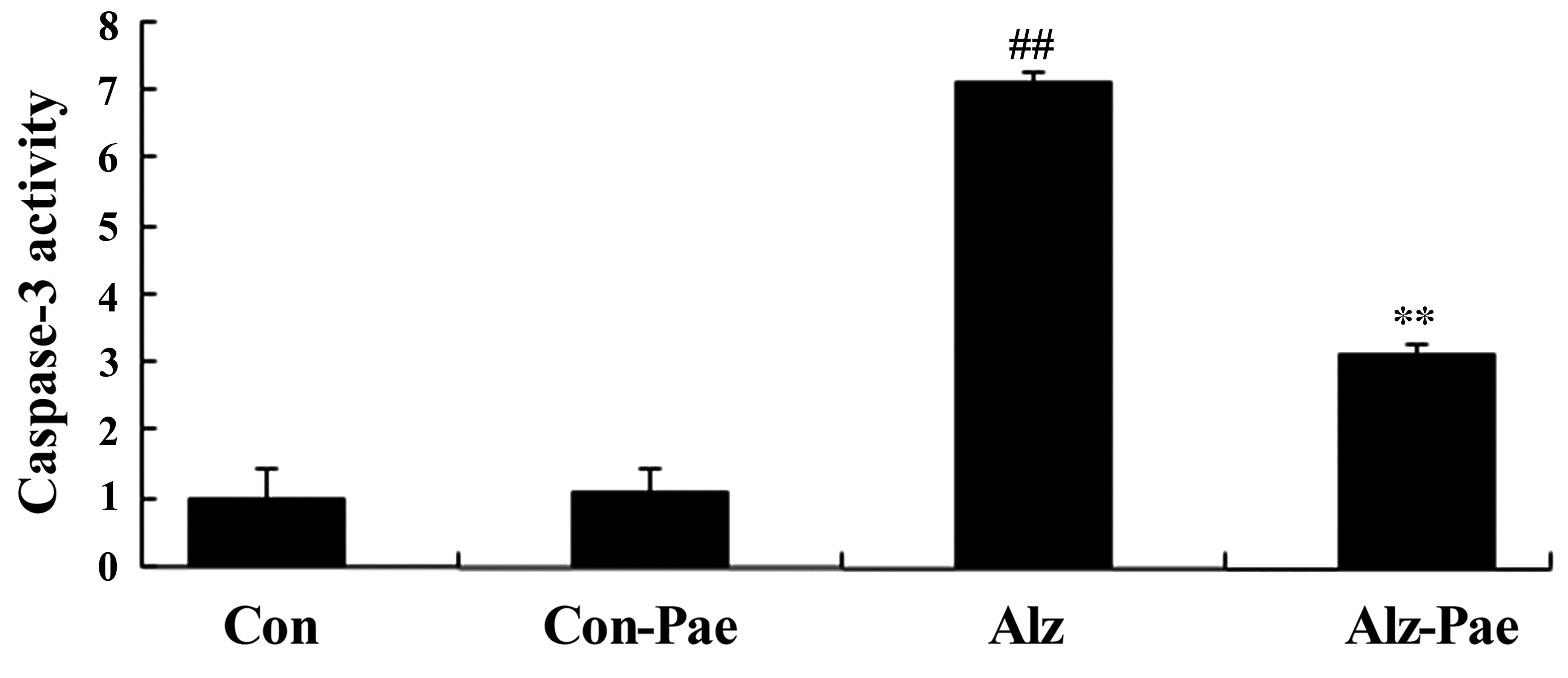
To investigate whether the protective effect of
paeoniflorin influences caspase-3 in AD mice, caspase-3 activity
was analyzed. Caspase-3 activity was significantly increased in the
Alz group, compared with the Con group (P<0.05; Fig. 4). Administration of paeoniflorin
(Alz-Pae) significantly decreased the caspase-3 activity of
transgenic mice, compared with the Alz group (P<0.05; Fig. 4).
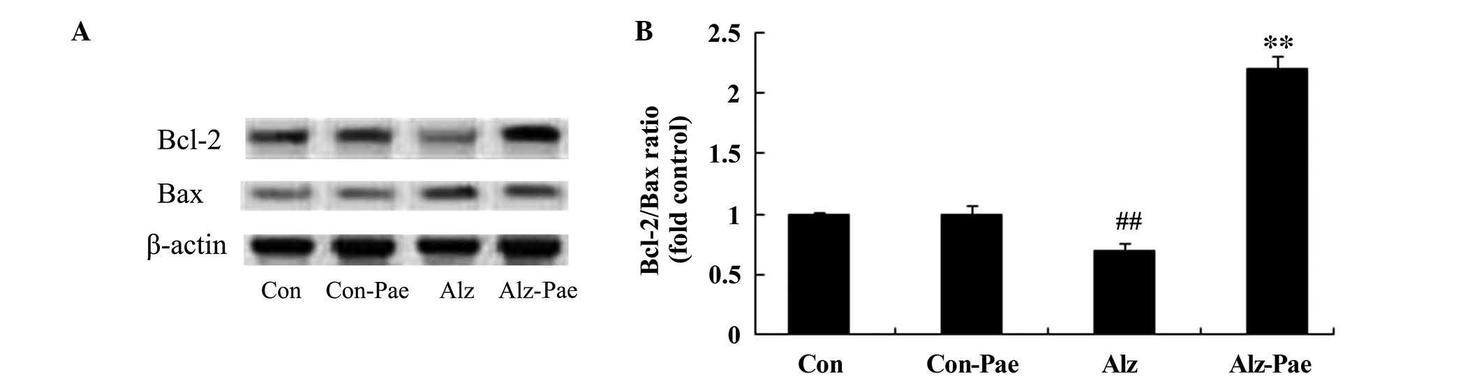
Protective effect of paeoniflorin
influences the Bcl-2/Bax ratio in AD mice
To investigate the protective effect of paeoniflorin
on Bcl-2/Bax ratio in the AD mice, the Bcl-2 and Bax protein
expression levels were detected by western blot analysis. Bcl-2
protein expression was significantly suppressed and Bax protein
expression was increased in the Alz group, compared with the Con
group (P<0.05; Fig. 5A).
Paeoniflorin treatment (Alz-Pae) significantly reversed Bcl-2/Bax
protein expression in transgenic mice, which exhibited increased
Bcl-2 protein expression levels and decreased Bax protein
expression levels, compared with the Alz group (P<0.05; Fig. 5A). The Bcl-2/Bax ratio was
decreased in the Alz group compared with the Con group, and
increased in the Alz-Pae group compared with the Alz group
(Fig. 5B).
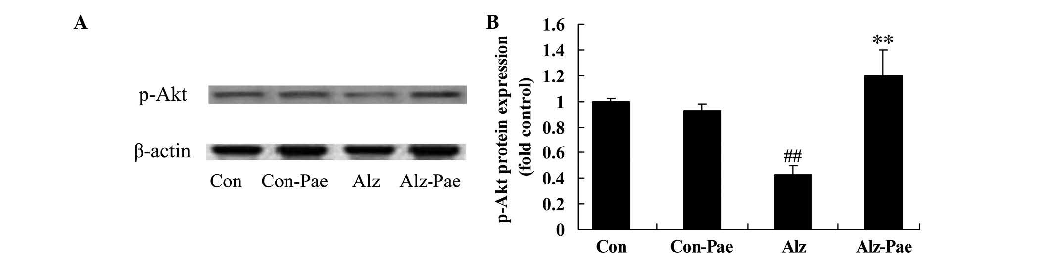
Protective effect of paeoniflorin
influences p-Akt in AD mice
To investigate the protective effect of paeoniflorin
on p-Akt in AD mice, p-Akt protein expression levels were evaluated
by western blot analysis. The western blots indicate that the
expression levels of p-Akt were significantly reduced in the Alz
group compared with the Con group (P<0.05; Fig. 6A). However, treatment with
paeoniflorin significantly increased p-Akt expression in the
Alz-Pae mice compared with the Alz group (P<0.05; Fig. 6B).
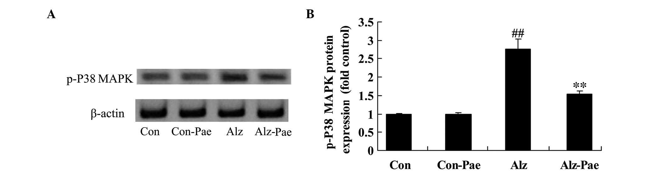
Protective effect of paeoniflorin
influences p-p38 MAPK in AD mice
To further analyze the protective effect of
paeoniflorin on MAPK in AD mice, the p-p38 MAPK protein expression
levels were examined by western blot analysis. The results of the
western blotting indicated that the p-p38 MAPK protein expression
was significantly increased in the Alz group compared with the Con
group (P<0.05; Fig. 7). The
p-p38 MAPK protein expression was significantly decreased by
treatment with paeoniflorin (Alz-Pae), compared with the Alz group
(P<0.05; Fig. 7B).
Discussion
AD has a high incidence that will continue to
increase due to prolonged average life expectancy, an aging
population in China and an increase in the number of elderly
individuals (20). Early diagnosis
is difficult, there is a lack of effective therapeutic agents and
current treatment strategies are not effective, thus AD requires
further research. Recent studies have demonstrated that certain
types of Chinese medicine may have an effect on AD (21). However, due to the subjectivity,
lack of quantitative indicators and clinical difficulty in
administration, their therapeutic applications are limited
(22). The present study observed
that the neuroprotective effect of paeoniflorin significantly
improved cognitive function and reduced patterns of escape distance
and latency in AD mice. Kapoor (23) reported that the neuroprotective
effects of paeoniflorin protect against glutamate-induced
neurotoxicity via the Bcl-2/Bax signaling pathway in PC12 cells.
Guo et al (24)
demonstrated that paeoniflorin may be a potential neuroprotective
agent for stroke and protected against ischemia-induced brain
damage in mice.
In recent years, it has been demonstrated that Aβ
protein-induced inflammation may lead to AD pathogenesis (25). AD nerve inflammation is an immune
reaction involving the microglia and astrocytes of the brain
(26). Activated microglia and
astrocytes express a large quantity of inflammatory cytokines, such
as TNF-α, IL-1β and IL-6, and specific receptors on the cell
surface are involved in inflammation and death of neighboring cells
in the brain (27). Aβ proteins
accumulate in the brain of patients with AD, which increase the
number of receptors on microglial cells. The ligand is more easily
integrated into the cell, resulting in nerve cell damage (28). Data from the current study
demonstrated that the neuroprotective effects of paeoniflorin
significantly decreased the activity of NF-κB p65, TNF-α, IL-1β and
IL-6 in AD mice. Sun et al (29) indicated that paeoniflorin
suppressed inflammation of asthmatic mice, and Jiang et al
(30) reported that the
anti-inflammatory effect of paeoniflorin inhibits systemic
inflammation and activation of NF-κB in experimental sepsis.
The occurrence of AD is associated with apoptosis,
as abnormal expression of Bcl-2, Bax and caspase-3 are directly
involved in apoptosis (31). Bcl-2
inhibits apoptosis to protect cell survival, rather than promoting
cell proliferation, by stabilizing the mitochondrial membrane,
preventing its release of caspases, apoptosis-associated factors
and cytochrome c (32,33).
The Bcl-2 family includes Bcl-2, which inhibits apoptosis, and Bax,
which promotes apoptosis. However, Bax has an inhibitory effect on
Bcl-2 and promotes the release of cytochrome c, thus
activating caspases and accelerating the induction of apoptosis
(31). The regulatory effects of
Bcl-2 and Bax on apoptosis are in opposition, thus, they are
regarded as co-regulators of apoptosis. In the present study,
administration of paeoniflorin effectively attenuated the activity
of caspase-3 and increased Bcl-2/Bax protein expression levels in
the AD mice. Sun et al (34) reported that the effect of
paeoniflorin protects against glutamate-induced neurotoxicity via
Bcl-2/Bax signaling pathways in PC12 cells.
Phosphatidylinositol-3-kinases (PI3K) are important
in signal transduction pathways in cells, Akt (also termed protein
kinase B) is key in the signaling pathway (35). PI3K/Akt signaling pathways are
involved in the regulation of cell apoptosis, proliferation and
differentiation, as well as a series of physiological activities
and metabolism (36). MAPK is a
type of serine and threonine protein kinase in cells, common in a
variety of organisms (including yeast and mammalian cells),
involved in mediating the growth, development, division,
differentiation, death and synchronization of multiple cellular
processes (37). In the present
study, pretreatment with paeoniflorin increased p-Akt and decreased
p-p38 MAPK protein expression levels in AD mice. Xu et al
(38) demonstrated that
paeoniflorin promotes the phosphorylation of Akt and attenuates
lipopolysaccharide-induced permeability of endothelial cells.
Wankun et al (39)
indicated that paeoniflorin protects against oxidative stress and
suppresses H2O2-induced p38 MAPK in human
retinal pigment epithelium cells.
In conclusion, the current study demonstrates that
the neuroprotective effect of paeoniflorin improves AD via
influencing inflammation and Bcl-2/Bax protein expression in the
cerebral cortex of transgenic mice models of AD. In addition, the
results suggest that paeoniflorin ameliorated the cognitive
dysfunction in AD mice.
Acknowledgments
The present study was supported by the National
Natural Science Foundation of China (grant no. 81000575).
References
|
1
|
Saine K, Cullum CM, Martin-Cook K, Hynan
L, Svetlik DA and Weiner MF: Comparison of functional and cognitive
donepezil effects in Alzheimer's disease. Int Psychogeriatr.
14:181–185. 2002. View Article : Google Scholar : PubMed/NCBI
|
|
2
|
Wang DS, Dickson DW and Malter JS: Tissue
transglutaminase, protein cross-linking and Alzheimer's disease:
Review and views. Int J Clin Exp Pathol. 1:5–18. 2008.PubMed/NCBI
|
|
3
|
Cai Z: Monoamine oxidase inhibitors:
Promising therapeutic agents for Alzheimer's disease (Review). Mol
Med Rep. 9:1533–1541. 2014.PubMed/NCBI
|
|
4
|
Mufson EJ, Mahady L, Waters D, Counts SE,
Perez SE, DeKosky ST, Ginsberg SD, Ikonomovic MD, Scheff SW and
Binder LI: Hippocampal plasticity during the progression of
Alzheimer's disease. Neuroscience. 2015.Epub ahead of print.
View Article : Google Scholar : PubMed/NCBI
|
|
5
|
Collins JM, King AE, Woodhouse A,
Kirkcaldie MT and Vickers JC: The effect of focal brain injury on
beta-amyloid plaque deposition, inflammation and synapses in the
APP/PS1 mouse model of Alzheimer's disease. Exp Neurol.
267:219–229. 2015. View Article : Google Scholar : PubMed/NCBI
|
|
6
|
Marx F, Blasko I, Pavelka M and
Grubeck-Loebenstein B: The possible role of the immune system in
Alzheimer's disease. Exp Gerontol. 33:871–881. 1998. View Article : Google Scholar
|
|
7
|
Ho GJ, Drego R, Hakimian E and Masliah E:
Mechanisms of cell signaling and inflammation in Alzheimer's
disease. Curr Drug Targets Inflamm Allergy. 4:247–256. 2005.
View Article : Google Scholar : PubMed/NCBI
|
|
8
|
Armato U, Chakravarthy B, Pacchiana R and
Whitfield JF: Alzheimer's disease: An update of the roles of
receptors, astrocytes and primary cilia (review). Int J Mol Med.
31:3–10. 2013.
|
|
9
|
Kim JH: Brain-derived neurotrophic factor
exerts neuroprotective actions against amyloid β-induced apoptosis
in neuroblastoma cells. Exp Ther Med. 8:1891–1895. 2014.PubMed/NCBI
|
|
10
|
Ferrer I: Altered mitochondria, energy
metabolism, voltage-dependent anion channel, and lipid rafts
converge to exhaust neurons in Alzheimer's disease. J Bioenerg
Biomembr. 41:425–431. 2009. View Article : Google Scholar : PubMed/NCBI
|
|
11
|
Wang Y, Xu S, Cao Y, Xie Z, Lai C, Ji X
and Bi J: Folate deficiency exacerbates apoptosis by inducing
hypomethylation and resultant overexpression of DR4 together with
altering DNMTs in Alzheimer's disease. Int J Clin Exp Med.
7:1945–1957. 2014.PubMed/NCBI
|
|
12
|
Xie X, Wang HT, Li CL, Gao XH, Ding JL,
Zhao HH and Lu YL: Ginsenoside Rb1 protects PC12 cells against
β-amyloid-induced cell injury. Mol Med Rep. 3:635–639. 2010.
|
|
13
|
Ramnath V, Rekha PS, Kuttan G and Kuttan
R: Regulation of Caspase-3 and Bcl-2 Expression in Dalton's
Lymphoma Ascites Cells by Abrin. Evid Based Complement Alternat
Med. 6:233–238. 2009. View Article : Google Scholar :
|
|
14
|
Shu YZ, Hattori M, Akao T, Kobashi K,
Kagei K, Fukuyama K, Tsukihara T and Namba T: Metabolism of
paeoniflorin and related compounds by human intestinal bacteria.
II. Structures of 7S- and 7R-paeonimetabolines I and II formed by
Bacteroides fragilis and Lactobacillus brevis. Chem Pharm Bull
(Tokyo). 35:3726–3733. 1987. View Article : Google Scholar
|
|
15
|
Wang H, Zhou H, Wang CX, Li YS, Xie HY,
Luo JD and Zhou Y: Paeoniflorin inhibits growth of human colorectal
carcinoma HT 29 cells in vitro and in vivo. Food Chem Toxicol.
50:1560–1567. 2012. View Article : Google Scholar : PubMed/NCBI
|
|
16
|
Chen T, Guo ZP, Jiao XY, Jia RZ, Zhang YH,
Li JY, Huang XL and Liu HJ: Peoniflorin suppresses tumor necrosis
factor-α induced chemokine production in human dermal microvascular
endothelial cells by blocking nuclear factor-κB and ERK pathway.
Arch Dermatol Res. 303:351–360. 2011. View Article : Google Scholar
|
|
17
|
Hwang YH, Kim T, Cho WK, Jang D, Ha JH and
Ma JY: Food- and gender-dependent pharmacokinetics of paeoniflorin
after oral administration with Samul-tang in rats. J
Ethnopharmacol. 142:161–167. 2012. View Article : Google Scholar : PubMed/NCBI
|
|
18
|
Hu ZY, Xu L, Yan R, Huang Y, Liu G, Zhou
WX and Zhang YX: Advance in studies on effect of paeoniflorin on
nervous system. Zhongguo Zhong Yao Za Zhi. 38:297–301. 2013.In
Chinese. PubMed/NCBI
|
|
19
|
Cho JY, Hwang DY, Kang TS, Shin DH, Hwang
JH, Lim CH, Lee SH, Lim HJ, Min SH, Seo J, et al: Use of
NSE/PS2m-transgenic mice in the study of the protective effect of
exercise on Alzheimer's disease. J Sports Sci. 21:943–951. 2003.
View Article : Google Scholar : PubMed/NCBI
|
|
20
|
Jiang P, Li C, Xiang Z and Jiao B:
Tanshinone IIA reduces the risk of Alzheimer's disease by
inhibiting iNOS, MMP2 and NF-κBp65 transcription and translation in
the temporal lobes of rat models of Alzheimer's disease. Mol Med
Rep. 10:689–694. 2014.PubMed/NCBI
|
|
21
|
Sulistio YA and Heese K: Proteomics in
Traditional Chinese Medicine with an Emphasis on Alzheimer's
Disease. Evid Based Complement Alternat Med. 2015:3935102015.
View Article : Google Scholar : PubMed/NCBI
|
|
22
|
Huang HJ, Lee CC and Chen CY: Lead
discovery for Alzheimer's disease related target protein RbAp48
from traditional Chinese medicine. BioMed Res Int. 2014:7649462014.
View Article : Google Scholar : PubMed/NCBI
|
|
23
|
Kapoor S: Neuroprotective effects of
paeoniflorin: An emerging concept in neurology. Folia Neuropathol.
51:922013. View Article : Google Scholar : PubMed/NCBI
|
|
24
|
Guo RB, Wang GF, Zhao AP, Gu J, Sun XL and
Hu G: Paeoniflorin protects against ischemia-induced brain damages
in rats via inhibiting MAPKs/NF-κB-mediated inflammatory responses.
PLoS One. 7:e497012012. View Article : Google Scholar
|
|
25
|
Galimberti D and Scarpini E: Genetics and
biology of Alzheimer's disease and frontotemporal lobar
degeneration. Int J Clin Exp Med. 3:129–143. 2010.PubMed/NCBI
|
|
26
|
Nelson L, Gard P and Tabet N: Hypertension
and inflammation in Alzheimer's disease: Close partners in disease
development and progression! J Alzheimers Dis. 41:331–343.
2014.PubMed/NCBI
|
|
27
|
Roussos P, Katsel P, Fam P, Tan W, Purohit
DP and Haroutunian V: The triggering receptor expressed on myeloid
cells 2 (TREM2) is associated with enhanced inflammation,
neuropathological lesions and increased risk for Alzheimer's
dementia. Alzheimers Dement. 11:1163–1170. 2014. View Article : Google Scholar : PubMed/NCBI
|
|
28
|
Calderón-Garcidueñas L, Maronpot RR,
Torres-Jardon R, Henríquez-Roldán C, Schoonhoven R, Acuña-Ayala H,
Villarreal-Calderón A, Nakamura J, Fernando R, Reed W, et al: DNA
damage in nasal and brain tissues of canines exposed to air
pollutants is associated with evidence of chronic brain
inflammation and neurodegeneration. Toxicol Pathol. 31:524–538.
2003. View Article : Google Scholar : PubMed/NCBI
|
|
29
|
Sun J, Wu J, Xu C, Luo Q, Li B and Dong J:
Paeoniflorin attenuates allergic inflammation in asthmatic mice.
Int Immunopharmacol. 24:88–94. 2015. View Article : Google Scholar
|
|
30
|
Jiang WL, Chen XG, Zhu HB, Gao YB, Tian JW
and Fu FH: Paeoniflorin inhibits systemic inflammation and improves
survival in experimental sepsis. Basic Clin Pharmacol Toxicol.
105:64–71. 2009. View Article : Google Scholar : PubMed/NCBI
|
|
31
|
Kong J, Ren G, Jia N, Wang Y, Zhang H,
Zhang W, Chen B and Cao Y: Effects of nicorandil in neuroprotective
activation of PI3K/AKT pathways in a cellular model of Alzheimer's
disease. Eur Neurol. 70:233–241. 2013. View Article : Google Scholar : PubMed/NCBI
|
|
32
|
Um HD: Bcl-2 family proteins as regulators
of cancer cell invasion and metastasis: A review focusing on
mitochondrial respiration and reactive oxygen species. Oncotarget.
2015.
|
|
33
|
Kang MH, Kim IH and Nam TJ: Phloroglucinol
induces apoptosis via apoptotic signaling pathways in HT-29 colon
cancer cells. Oncol Rep. 32:1341–1346. 2014.PubMed/NCBI
|
|
34
|
Sun R, Wang K, Wu D, Li X and Ou Y:
Protective effect of paeoniflorin against glutamate-induced
neurotoxicity in PC12 cells via Bcl-2/Bax signal pathway. Folia
Neuropathol. 50:270–276. 2012. View Article : Google Scholar : PubMed/NCBI
|
|
35
|
Dong M, Yang G, Liu H, Lin S, Sun D and
Wang Y: Aged black garlic extract inhibits HT29 colon cancer cell
growth via the PI3K/Akt signaling pathway. Biomed Rep. 2:250–254.
2014.PubMed/NCBI
|
|
36
|
Gu Y, Liu SL, Ju WZ, Li CY and Cao P:
Analgesic-antitumor peptide induces apoptosis and inhibits the
proliferation of SW480 human colon cancer cells. Oncol Lett.
5:483–488. 2013.PubMed/NCBI
|
|
37
|
Zhang Y, Uguccioni G, Ljubicic V, et al:
Multiple signaling pathways regulate contractile activity-mediated
PGC-1α gene expression and activity in skeletal muscle cells.
Physiol Rep. 2:e120082014. View Article : Google Scholar
|
|
38
|
Xu H, Song J, Gao X, Xu Z, Xu X, Xia Y and
Dai Y: Paeoniflorin attenuates lipopolysaccharide-induced
permeability of endothelial cells: Involvements of F-actin
expression and phosphorylations of PI3K/Akt and PKC. Inflammation.
36:216–225. 2013. View Article : Google Scholar
|
|
39
|
Wankun X, Wenzhen Y, Min Z, Weiyan Z, Huan
C, Wei D, Lvzhen H, Xu Y and Xiaoxin L: Protective effect of
paeoniflorin against oxidative stress in human retinal pigment
epithelium in vitro. Mol Vis. 17:3512–3522. 2011.
|