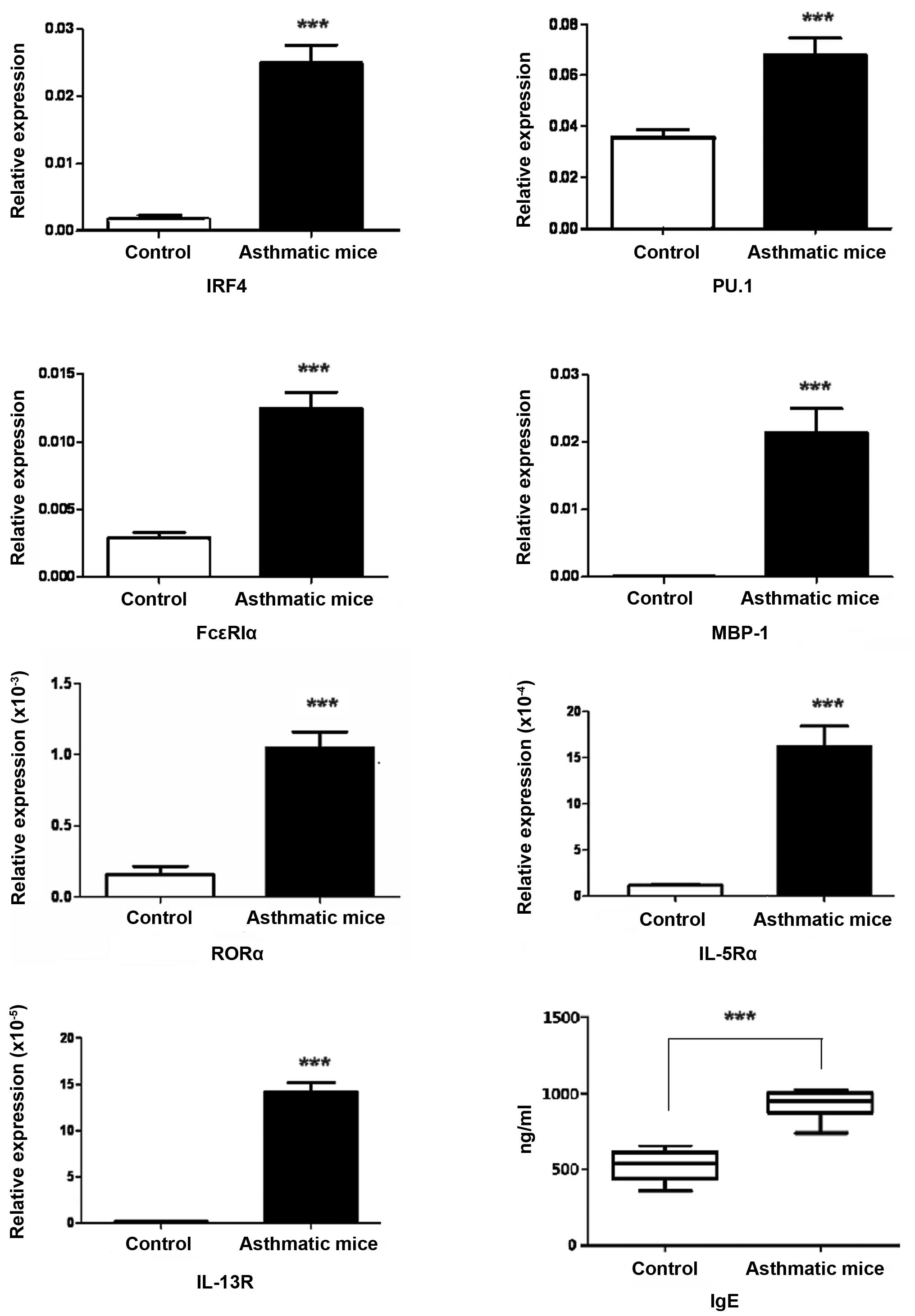
|
1
|
Guo HW, Yun CX, Hou GH, Du J, Huang X, Lu
Y, Keller ET, Zhang J and Deng JG: Mangiferin attenuates Th1/Th2
cytokine imbalance in an ovalbumin-induced asthmatic mouse model.
PLOS One. 9:e1003942014. View Article : Google Scholar : PubMed/NCBI
|
|
2
|
Chen W, Sivaprasad U, Gibson AM,
Cunningham CM, Bass SA, Kinker KG, Finkelman FD, Wills-Karp M and
Khurana Hershey GK: IL-13 receptor α2 contributes to development of
experimental allergic asthma. J Allergy Clin Immunol. 132:951–958.
2013. View Article : Google Scholar
|
|
3
|
Farahani R, Sherkat R, Hakemi MG,
Eskandari N and Yazdani R: Cytokines (interleukin-9, IL-17, IL-22,
IL-25 and IL-33) and asthma. Adv Biomed Res. 3:1272014. View Article : Google Scholar : PubMed/NCBI
|
|
4
|
Horka H, Staudt V, Klein M, Taube C,
Reuter S, Dehzad N, Andersen JF, Kopecky J, Schild H, Kotsyfakis M,
et al: The tick salivary protein sialostatin L inhibits the
Th9-derived production of the asthma-promoting cytokine IL-9 and is
effective in the prevention of experimental asthma. J Immunol.
188:2669–2676. 2012. View Article : Google Scholar : PubMed/NCBI
|
|
5
|
Besnard AG, Sabat R, Dumoutier L, Renauld
JC, Willart M, Lambrecht B, Teixeira MM, Charron S, Fick L, Erard
F, et al: Dual Role of IL-22 in allergic airway inflammation and
its cross-talk with IL-17A. Am J Respir Crit Care Med.
183:1153–1163. 2011. View Article : Google Scholar : PubMed/NCBI
|
|
6
|
Ma L, Xue HB, Guan XH, Shu CM, Zhang JH
and Yu J: Possible pathogenic role of T helper type 9 cells and
interleukin (IL)-9 in atopic dermatitis. Clin Exp Immunol.
175:25–31. 2014. View Article : Google Scholar :
|
|
7
|
Pilette C: Pathophysiology of asthma: Data
concerning regulation of IGE and Th2 responses in the lung. Bull
Mem Acad R Med Belg. 166:280–287. 2011.In French.
|
|
8
|
Chen Y, Thai P, Zhao YH, Ho YS, DeSouza MM
and Wu R: Stimulation of airway mucin gene expression by
interleukin (IL)-17 through IL-6 paracrine/autocrine loop. J Biol
Chem. 278:17036–17043. 2003. View Article : Google Scholar : PubMed/NCBI
|
|
9
|
Gasch M, Goroll T, Bauer M, Hinz D,
Schütze N, Polte T, Kesper D, Simon JC, Hackermüller J, Lehmann I
and Herberth G: Generation of IL-8 and IL-9 producing
CD4+ T cells is affected by Th17 polarizing conditions
and AHR ligands. Mediators Inflamm. 1825492014.
|
|
10
|
Kung TT, Luo B, Crawley Y, Garlisi CG,
Devito K, Minnicozzi M, Egan RW, Kreutner W and Chapman RW: Effect
of anti-mIL-9 antibody on the development of pulmonary inflammation
and airway hyperresponsiveness in allergic mice. Am J Respir Cell
Mol Biol. 25:600–605. 2001. View Article : Google Scholar : PubMed/NCBI
|
|
11
|
Spits H, Artis D, Colonna M, Diefenbach A,
Di Santo JP, Eberl G, Koyasu S, Locksley RM, McKenzie AN, Mebius
RE, et al: Innate lymphoid cells-a proposal for uniform
nomenclature. Nat Rev Immunol. 13:145–149. 2013. View Article : Google Scholar : PubMed/NCBI
|
|
12
|
Lund S, Walford HH and Doherty TA: Type 2
innate lymphoid cells in allergic disease. Curr Immunol Rev.
9:214–221. 2013. View Article : Google Scholar
|
|
13
|
Fukuoka A, Futatsugi-Yumikura S, Takahashi
S, Kazama H, Iyoda T, Yoshimoto T, Inaba K, Nakanishi K and
Yonehara S: Identification of a novel type 2 innate immunocyte with
the ability to enhance IgE production. Int Immunol. 25:373–382.
2013. View Article : Google Scholar : PubMed/NCBI
|
|
14
|
Gold MJ, Antignano F, Halim TY, Hirota JA,
Blanchet MR, Zaph C, Takei F and McNagny KM: Group 2 innate
lymphoid cells facilitate sensitization to local, but not systemic,
TH2-inducing allergen exposures. J Allergy Clin Immunol.
133:1142–1148. 2014. View Article : Google Scholar : PubMed/NCBI
|
|
15
|
Wang SY, Yang M, Xu XP, Qiu GF, Ma J, Wang
SJ, Huang XX and Xu HX: Intranasal delivery of T-bet modulates the
profile of helper T cell immune responses in experimental asthma. J
Investig Allergol Clin Immunol. 18:357–365. 2008.PubMed/NCBI
|
|
16
|
He Z, Shotorban SS, Jiao Z, Su Z, Tong J,
Liu Y, Shen P, Ma J, Gao J, Wang T, et al: HMGB1 promotes the
differentiation of Th17 via up-regulating TLR2 and IL-23 of CD14+
monocytes from patients with rheumatoid arthritis. Scand J Immunol.
76:483–490. 2012. View Article : Google Scholar : PubMed/NCBI
|
|
17
|
Wu Y, Yan Y, Su Z, Bie Q, Wu J, Wang S, Yu
Y, Ding H, Lu P and Xu H: Enhanced circulating ILC2s accompany by
upregulated MDSCs in patients with asthma. Int J Clin Exp Pathol.
8:3568–3579. 2015.PubMed/NCBI
|
|
18
|
Erpenbeck VJ, Hohlfeld JM, Discher M,
Krentel H, Hagenberg A, Braun A and Krug N: Increased expression of
interleukin-9 messenger RNA after segmental allergen challenge in
allergic asthmatics. Chest. 123:370S2003.PubMed/NCBI
|
|
19
|
Shimbara A, Christodoulopoulos P,
Soussi-Gounni A, Olivenstein R, Nakamura Y, Levitt RC, Nicolaides
NC, Holroyd KJ, Tsicopoulos A, Lafitte JJ, et al: IL-9 and its
receptor in allergic and nonallergic lung disease: Increased
expression in asthma. J Allergy Clin Immunol. 105:108–115. 2000.
View Article : Google Scholar : PubMed/NCBI
|
|
20
|
Dardalhon V, Awasthi A, Kwon H, Galileos
G, Gao W, Sobel RA, Mitsdoerffer M, Strom TB, Elyaman W, Ho IC, et
al: IL-4 inhibits TGF-beta-induced Foxp3+ T cells and, together
with TGF-beta, generates IL-9+ IL-10+ Foxp3(−) effector T cells.
Nat Immunol. 9:1347–1355. 2008. View
Article : Google Scholar : PubMed/NCBI
|
|
21
|
Veldhoen M, Uyttenhove C, van Snick J,
Helmby H, Westendorf A, Buer J, Martin B, Wilhelm C and Stockinger
B: Transforming growth factor-beta 'reprograms' the differentiation
of T helper 2 cells and promotes an interleukin 9-producing subset.
Nat Immunol. 9:1341–1346. 2008. View
Article : Google Scholar : PubMed/NCBI
|
|
22
|
Chang HC, Sehra S, Goswami R, Yao W, Yu Q,
Stritesky GL, Jabeen R, McKinley C, Ahyi AN, Han L, et al: The
transcription factor PU.1 is required for the development of
IL-9-producing T cells and allergic inflammation. Nat Immunol.
11:527–534. 2010. View
Article : Google Scholar : PubMed/NCBI
|
|
23
|
Kaplan MH: Th9 cells: differentiation and
disease. Immunol Rev. 252:104–115. 2013. View Article : Google Scholar : PubMed/NCBI
|
|
24
|
Eiwegger T and Akdis CA: IL-33 links
tissue cells, dendritic cells and Th2 cell development in a mouse
model of asthma. Eur J Immunol. 41:1535–1538. 2011. View Article : Google Scholar : PubMed/NCBI
|
|
25
|
Bie Q, Zhang P, Su Z, Zheng D, Ying X, Wu
Y, Yang H, Chen D, Wang S and Xu H: Polarization of ILC2s in
peripheral blood might contribute to immunosuppressive
microenvironment in patients with gastric cancer. J Immunol Res.
9231352014.PubMed/NCBI
|
|
26
|
Fahy JV and Locksley RM: The airway
epithelium as a regulator of Th2 responses in asthma. Am J Respir
Crit Care Med. 184:390–392. 2011. View Article : Google Scholar : PubMed/NCBI
|
|
27
|
Juncadella IJ, Kadl A, Sharma AK, Shim YM,
Hochreiter-Hufford A, Borish L and Ravichandran KS: Apoptotic cell
clearance by bronchial epithelial cells critically influences
airway inflammation. Nature. 493:547–551. 2013. View Article : Google Scholar :
|
|
28
|
Mikami N, Miyagi Y, Sueda K, Takatsuji M,
Fukada S, Yamamoto H and Tsujikawa K: Calcitonin gene-related
peptide and cyclic adenosine 5′-monophosphate/protein kinase A
pathway promote IL-9 production in Th9 differentiation process. J
Immunol. 190:4046–4055. 2013. View Article : Google Scholar : PubMed/NCBI
|
|
29
|
Temann UA, Ray P and Flavell RA: Pulmonary
overexpression of IL-9 induces Th2 cytokine expression, leading to
immunepathology. J Clin Invest. 109:29–39. 2002. View Article : Google Scholar : PubMed/NCBI
|
|
30
|
Corren J: Cytokine inhibition in severe
asthma: Current knowledge and future directions. Curr Opin Pulm
Med. 17:29–33. 2011. View Article : Google Scholar : PubMed/NCBI
|
|
31
|
O'Flaherty BM, Soni T, Wakeman BS and
Speck SH: The murine gammaherpesvirus immediate-early Rta
synergizes with IRF4, targeting expression of the viral M1
superantigen to plasma cells. PLoS Pathog. 10:e10043022014.
View Article : Google Scholar : PubMed/NCBI
|
|
32
|
Holgate ST: New strategies with anti-IgE
in allergic diseases. World Allergy Organ J. 7:172014. View Article : Google Scholar : PubMed/NCBI
|
|
33
|
Turner JE, Morrison PJ, Wilhelm C, Wilson
M, Ahlfors H, Renauld JC, Panzer U, Helmby H and Stockinger B:
IL-9-mediated survival of type 2 innate lymphoid cells promotes
damage control in helminth-induced lung inflammation. J Exp Med.
210:2951–2965. 2013. View Article : Google Scholar : PubMed/NCBI
|