|
1
|
Beyer Nardi N and da Silva Meirelles L:
Mesenchymal stem cells: Isolation, in vitro expansion and
characterization. Handb Exp Pharmacol:. 249–282. 2006. View Article : Google Scholar
|
|
2
|
Chen WC, Péault B and Huard J:
Regenerative translation of human blood-vessel-derived MSC
precursors. Stem Cells Int. 2015:3751872015. View Article : Google Scholar : PubMed/NCBI
|
|
3
|
Deng S, Huang R, Wang J, Zhang S, Chen Z,
Wu S, Jiang Y, Peng Q, Cai X and Lin Y: Miscellaneous animal models
accelerate the application of mesenchymal stem cells for cartilage
regeneration. Curr Stem Cell Res Ther. 9:223–233. 2014. View Article : Google Scholar : PubMed/NCBI
|
|
4
|
Landa-Solís C, Granados-Montiel J,
Olivos-Meza A, Ortega-Sánchez C, Cruz-Lemini M, Hernández-Flores C,
Chang-González ME, García RG, Olivos-Díaz B, Velasquillo-Martínez
MC, et al: Cryopreserved CD90+ cells obtained from mobilized
peripheral blood in sheep: a new source of mesenchymal stem cells
for preclinical applications. Cell Tissue Bank. July 29–2015.Epub
ahead of print.
|
|
5
|
Caplan AI: Adult mesenchymal stem cells
for tissue engineering versus regenerative medicine. J Cell
Physiol. 213:341–347. 2007. View Article : Google Scholar : PubMed/NCBI
|
|
6
|
Lee SI, Yeo SI, Kim BB, Ko Y and Park JB:
Formation of size-controllable spheroids using gingiva-derived stem
cells and concave microwells: Morphology and viability tests.
Biomed Rep. 4:97–101. 2016.PubMed/NCBI
|
|
7
|
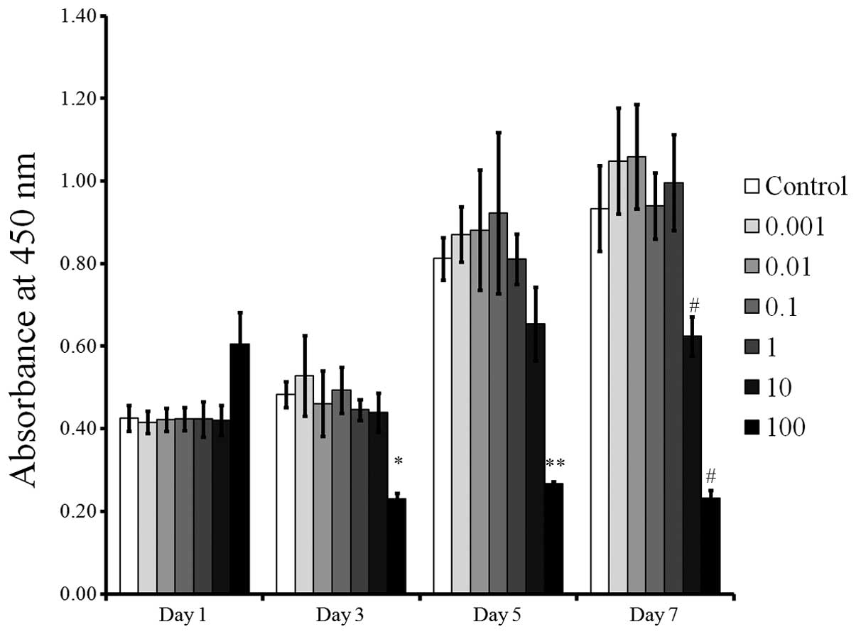
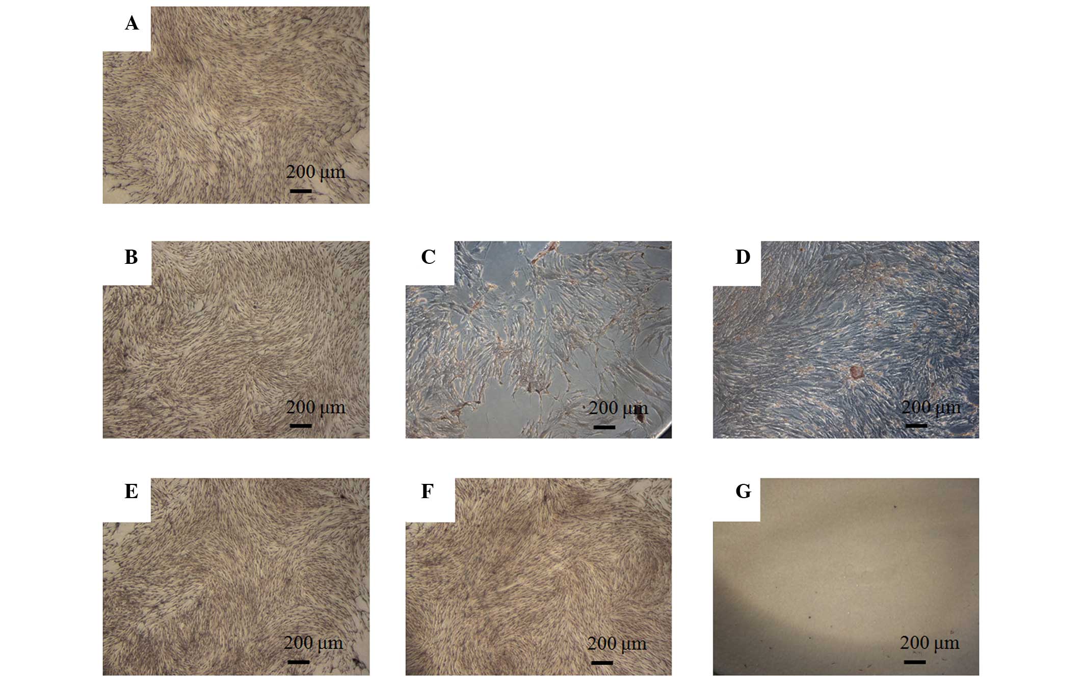
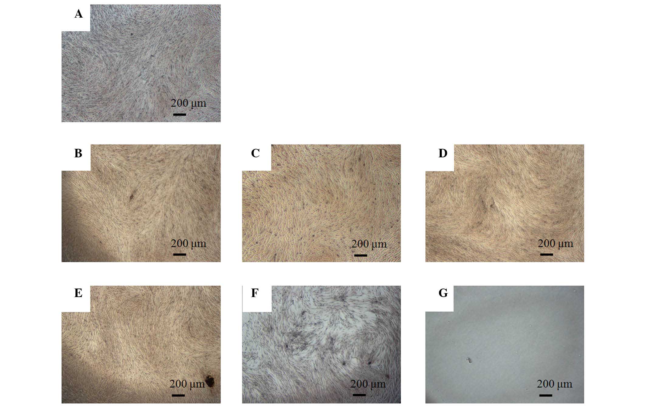
Jeong SH, Lee JE, Kim BB, Ko Y and Park
JB: Evaluation of the effects of Cimicifugae Rhizoma on the
morphology and viability of mesenchymal stem cells. Exp Ther Med.
10:629–634. 2015.PubMed/NCBI
|
|
8
|
Tamura S, Ohike A, Ibuki R, Amidon GL and
Yamashita S: Tacrolimus is a class II low-solubility
high-permeability drug: The effect of P-glycoprotein efflux on
regional permeability of tacrolimus in rats. J Pharm Sci.
91:719–729. 2002. View Article : Google Scholar : PubMed/NCBI
|
|
9
|
Scholten EM, Cremers SC, Schoemaker RC,
Rowshani AT, van Kan EJ, den Hartigh J, Paul LC and de Fijter JW:
AUC-guided dosing of tacrolimus prevents progressive systemic
overexposure in renal transplant recipients. Kidney Int.
67:2440–2447. 2005. View Article : Google Scholar : PubMed/NCBI
|
|
10
|
Felipe CR, Garcia C, Moreira S, Olsen N,
Silva HT and Pestana OM: Choosing the right dose of new
immunossuppressive drugs for new populations: Importance of
pharmacokinetic studies. Transplant Proc. 33:1095–1096. 2001.
View Article : Google Scholar : PubMed/NCBI
|
|
11
|
van Hooff JP, Boots JM, van Duijnhoven EM
and Christiaans MH: Dosing and management guidelines for tacrolimus
in renal transplant patients. Transplant Proc. 31:54S–57S. 1999.
View Article : Google Scholar : PubMed/NCBI
|
|
12
|
Taylor DO, Barr ML, Radovancevic B,
Renlund DG, Mentzer RM Jr, Smart FW, Tolman DE, Frazier OH, Young
JB and VanVeldhuisen P: A randomized, multicenter comparison of
tacrolimus and cyclosporine immunosuppressive regimens in cardiac
transplantation: Decreased hyperlipidemia and hypertension with
tacrolimus. J Heart Lung Transplant. 18:336–345. 1999. View Article : Google Scholar : PubMed/NCBI
|
|
13
|
Busuttil RW, Klintmalm GB, Lake JR, Miller
CM and Porayko M: General guidelines for the use of tacrolimus in
adult liver transplant patients. Transplantation. 61:845–847. 1996.
View Article : Google Scholar : PubMed/NCBI
|
|
14
|
Staatz CE and Tett SE: Clinical
pharmacokinetics and pharmacodynamics of tacrolimus in solid organ
transplantation. Clin Pharmacokinet. 43:623–653. 2004. View Article : Google Scholar : PubMed/NCBI
|
|
15
|
Laskow DA, Neylan JF III, Shapiro RS,
Pirsch JD, Vergne-Marini PJ and Tomlanovich SJ: The role of
tacrolimus in adult kidney transplantation: A review. Clin
Transplant. 12:489–503. 1998.PubMed/NCBI
|
|
16
|
Kugimiya F, Yano F, Ohba S, Igawa K,
Nakamura K, Kawaguchi H and Chung UI: Mechanism of osteogenic
induction by FK506 via BMP/Smad pathways. Biochem Biophys Res
Commun. 338:872–879. 2005. View Article : Google Scholar : PubMed/NCBI
|
|
17
|
Park KM, Hay JE, Lee SG, Lee YJ, Wiesner
RH, Porayko MK and Krom RA: Bone loss after orthotopic liver
transplantation: FK 506 versus cyclosporine. Transplant Proc.
28:1738–1740. 1996.PubMed/NCBI
|
|
18
|
Stempfle HU, Werner C, Echtler S, Assum T,
Meiser B, Angermann CE, Theisen K and Gärtner R: Rapid trabecular
bone loss after cardiac transplantation using FK506
(tacrolimus)-based immunosuppression. Transplant Proc.
30:1132–1133. 1998. View Article : Google Scholar : PubMed/NCBI
|
|
19
|
Tang L, Ebara S, Kawasaki S, Wakabayashi
S, Nikaido T and Takaoka K: FK506 enhanced osteoblastic
differentiation in mesenchymal cells. Cell Biol Int. 26:75–84.
2002. View Article : Google Scholar : PubMed/NCBI
|
|
20
|
Byun YK, Kim KH, Kim SH, Kim YS, Koo KT,
Kim TI, Seol YJ, Ku Y, Rhyu IC and Lee YM: Effects of
immunosuppressants, FK506 and cyclosporin A, on the osteogenic
differentiation of rat mesenchymal stem cells. J Periodontal
Implant Sci. 42:73–80. 2012. View Article : Google Scholar : PubMed/NCBI
|
|
21
|
Hoogduijn MJ, Crop MJ, Korevaar SS,
Peeters AM, Eijken M, Maat LP, Balk AH, Weimar W and Baan CC:
Susceptibility of human mesenchymal stem cells to tacrolimus,
mycophenolic acid and rapamycin. Transplantation. 86:1283–1291.
2008. View Article : Google Scholar : PubMed/NCBI
|
|
22
|
Jin SH, Lee JE, Yun JH, Kim I, Ko Y and
Park JB: Isolation and characterization of human mesenchymal stem
cells from gingival connective tissue. J Periodontal Res.
50:461–467. 2015. View Article : Google Scholar
|
|
23
|
Lechler RI, Sykes M, Thomson AW and Turka
LA: Organ transplantation - how much of the promise has been
realized? Nat Med. 11:605–613. 2005. View
Article : Google Scholar : PubMed/NCBI
|
|
24
|
Kaihara S, Bessho K, Okubo Y, Sonobe J,
Kusumoto K, Ogawa Y and Iizuka T: Effect of FK506 on osteoinduction
by recombinant human bone morphogenetic protein-2. Life Sci.
72:247–256. 2002. View Article : Google Scholar : PubMed/NCBI
|
|
25
|
Utheim TP, Raeder S, Utheim ØA, Cai Y,
Roald B, Drolsum L, Lyberg T and Nicolaissen B: A novel method for
preserving cultured limbal epithelial cells. Br J Ophthalmol.
91:797–800. 2007. View Article : Google Scholar
|
|
26
|
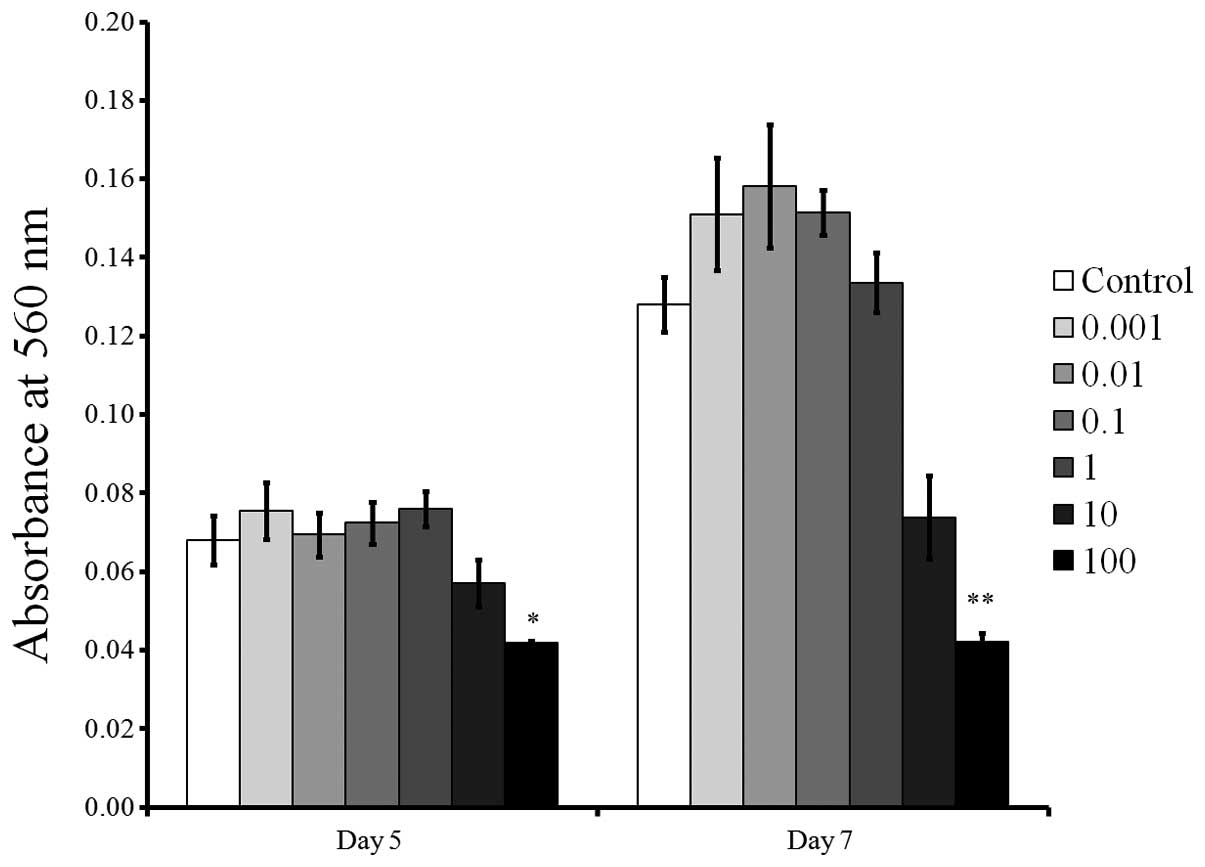
Bang SM, Moon HJ, Kwon YD, Yoo JY, Pae A
and Kwon IK: Osteoblastic and osteoclastic differentiation on SLA
and hydrophilic modified SLA titanium surfaces. Clin Oral Implants
Res. 25:831–837. 2014. View Article : Google Scholar
|
|
27
|
Wang B, Bi M, Zhu Z, Wu L and Wang J:
Effects of the antihypertensive drug benidipine on osteoblast
function in vitro. Exp Ther Med. 7:649–653. 2014.PubMed/NCBI
|
|
28
|
Nakamura T, Shinohara Y, Momozaki S,
Yoshimoto T and Noguchi K: Co-stimulation with bone morphogenetic
protein-9 and FK506 induces remarkable osteoblastic differentiation
in rat dedifferentiated fat cells. Biochem Biophys Res Commun.
440:289–294. 2013. View Article : Google Scholar : PubMed/NCBI
|
|
29
|
Quarles LD, Yohay DA, Lever LW, Caton R
and Wenstrup RJ: Distinct proliferative and differentiated stages
of murine MC3T3-E1 cells in culture: An in vitro model of
osteoblast development. J Bone Miner Res. 7:683–692. 1992.
View Article : Google Scholar : PubMed/NCBI
|
|
30
|
Park JB, Zhang H, Lin CY, Chung CP, Byun
Y, Park YS and Yang VC: Simvastatin maintains osteoblastic
viability while promoting differentiation by partially regulating
the expressions of estrogen receptors α. J Surg Res. 174:278–283.
2012. View Article : Google Scholar
|