Introduction
The incidence of primary malignant brain tumors has
increased by 1.2% each year for the past 30 years, particularly in
the elderly (1). Malignant glioma
accounts for ~70% of primary brain tumor cases, with an annual
incidence of ~5/100,000 and >14,000 new cases each year
(2). Despite recent advances in
therapy, significant progress in prognosis prediction and improved
survival have not been achieved thus far (3), due to its resistance to radio- and
chemotherapy, and a high rate of relapse. Therefore, novel
strategies are required to treat human glioma.
Dietary agents are currently under consideration as
effective and alternative means to control cancer. Previous studies
have demonstrated that capsaicin (Cap), tea polyphenols,
procyanidins and allicin are protective against various types of
human tumor, including prostate cancer, breast cancer, liver cancer
and gastric carcinoma (4–7). Cap and dihydrocapsaicin (DHC) are
homovanillic acid derivatives with similar structures, and are the
principle spicy components of hot chili peppers that are consumed
worldwide, particularly in Southeast Asian and Latin American
countries (8). Cap has been
previously demonstrated to inhibit the proliferation of, or induce
apoptosis in, numerous varieties of human cancer, including lung,
prostate, pancreatic and breast cancer, in vitro and in
vivo (9–12). Cap causes tumor cell cycle arrest
in S or G1/G0 phase in NPC-TW 039 human
nasopharyngeal carcinoma cells, MCF-7 human breast cells, BT-474
cells, SKBR-3 cells, MDA-MB231 cells and SCC-4 human tongue cells,
in model systems in vitro and in vivo (13–15).
Furthermore, Cap triggers apoptosis in >40 distinct tumor cell
lines, primarily through the mitochondrial pathway or death
receptor pathway (16). Cap
induced apoptosis in AsPC-1 and BxPC-3 human pancreatic cancer
cells through the mitochondrial death pathway, which was initiated
by the generation of reactive oxygen species (ROS) and c-Jun
N-terminal kinase (JNK) activation (11). In addition, intragastric
administration of Cap significantly inhibits the growth of AsPC-1
pancreatic xenograft cells (11),
and induces TRPV1-mediated apoptosis in RT4 urothelial cancer cells
through the death receptor pathway by activating Fas cell surface
death receptor (17). Gil and Kang
(18) demonstrated that Cap
inhibits the growth of A172 human glioblastoma cells and induces
apoptosis by downregulation of B cell lymphoma 2 apoptosis
regulator (Bcl-2) and activation of caspase-3. Maity et al
(19) reported that Cap induces
apoptosis in mouse neuro 2a cells via ubiquitin-proteasome system
dysfunction. Notably, normal or noncancerous cells are less
sensitive to the anti-proliferative or apoptotic effects of Cap
compared with cancerous cells (16). DHC, an analog of Cap, inhibits the
proliferation of HCT116, MCF-7 and WI38 cells more potently than
Cap, and induces autophagy in HCT 116 cells (20). Furthermore, DHC induces autophagy
in A549 cells by downregulation of catalase, which leads to ROS
accumulation and attenuation of microtubule-associated protein
light chain 3 conversion (21).
However, the molecular mechanisms of Cap and DHC
induction of apoptosis in U251 human glioma cells are not
sufficiently understood. The present study aimed to investigate the
effect of Cap and DHC on U251 human glioma cells and the mechanisms
of this effect.
Materials and methods
Chemicals and antibodies
Cap, DHC (purity>99%) and trypsin were purchased
from Sigma-Aldrich; Merck Millipore (Darmstadt, Germany). Cell
Counting Kit-8 (CCK-8), Fluo-3AM, GENMED mitochondrial permeability
transition pore (MPTP) living cell fluorescence detection kit and
dimethyl sulfoxide (DMSO) were purchased from Dojindo Molecular
Technologies, Inc. (Kumamoto, Japan). U251 cells were obtained from
the National Platform of Experimental Cell Resources for Sci-Tech
(Beijing, China). L929 cells were obtained from the Institute of
Biochemistry and Cell Biology (Shanghai, China). Annexin
V-fluorescein isothiocyanate (FITC) Apoptosis Detection kit and
Cell Cycle Detection kit were purchased from Nanjing KeyGen Biotech
Co., Ltd. (Nanjing, China). Caspase-3 activity assay kit, caspase-9
activity assay kit, Rhodamine 123 (Rh123), ROS assay kit and
cytochrome C (cyto c) antibody (catalog no. AC909) were purchased
from Beyotime Institute of Biotechnology (Haimen, China).
UltraSensitive™ surface protein array (mouse/rabbit)
immunohistochemistry (IHC) kit and 3,3′-diaminobenzidine (DAB) kit
were obtained from Fuzhou Maixin Biotech Co., Ltd. (Fuzhou, China).
Inverted fluorescence microscope and confocal laser scanning
microscope were obtained from Nikon Corporation (Tokyo, Japan).
Flow cytometry equipment was obtained from BD Biosciences (Franklin
Lakes, NJ, USA).
Cell culture
U251 human glioma cells were maintained in
Dulbecco's modified Eagle's medium (DMEM; Gibco; Thermo Fisher
Scientific, Inc., Waltham, MA, USA) supplemented with 10% fetal
bovine serum (FBS; Thermo Fisher Scientific, Inc.), 10 mM
4-(2-hydroxyethyl)-1-piperazineethanesulfonic acid (HEPES), 2 mM
L-glutamine and 1% penicillin-streptomycin solution. L929 murine
fibroblast cells were maintained in RPMI-1640 (Gibco; Thermo Fisher
Scientific, Inc.) supplemented with 10% FBS, 10 mM HEPES, 2 mM
L-glutamine and 1% penicillin-streptomycin solution. All cultures
were maintained at 37°C in a humidified chamber of 95% air and 5%
CO2. Cap and DHC were dissolved in 0.5% DMSO
solution.
Cell inhibition rate and cell
survival
Cell inhibition rate and cell survival were assessed
by tetrazolium salt-based colorimetric detection in the CCK-8
assay. Cells were seeded in 96-well plates at an initial density of
5×10³ cells/well. Following exposure to 50, 100, 150 and
200 µM Cap or DHC for 12 (U251 only), 24, 48 and 72 h, 10 µl CCK-8
solution was added to each well and the plate was incubated for 1
h. Cell viability was determined with a microplate reader by
reading the absorbance at 450 nm (A450). The percentage
of viable cells was calculated as follows: Cell viability
(%)=[A450(treated)-A450
(blank)]/[A450(control)-A450(blank)]x100%.
Determination of apoptosis
U251 cells (2×10⁵) were plated in 6-well
plates and allowed to attach for 24 h prior to exposure to 200 µM
Cap and DHC for 12 h at 37°C. Apoptosis induced by Cap or DHC was
evaluated using an Annexin V-FITC Apoptosis Detection kit according
to the manufacturer's protocol. Briefly, U251 cells were detached,
collected and suspended in 500 µl binding buffer, then 5 µl Annexin
V-FITC and 5 µl propidium iodide (PI) was added and cells were
incubated for 10 min at room temperature in the dark. Apoptotic
cells were acquired using a FACS-Calibur flow cytometer (BD
Biosciences) and the data was analyzed using CellQuest™ Pro
software (BD Biosciences) and ModFit LT™ software (Verity Software
House, Inc., Topsham, ME, USE).
Transmission electron microscopy (TEM)
analysis
U251 cells (1×10⁵/ml) were exposed to 200
µM Cap or DHC for 12 h at 37°C, detached and collected, postfixed
in 1% buffered OsO4 for 2 h at 37°C, dehydrated in
graded alcohol and embedded in Epon resin. Sections (60-nm thick)
were cut and observed using TEM. A total of 5 slides were
visualized, with 10 fields analyzed per slide.
Cell cycle analysis
U251 cells (2×10⁵ cells/well) were plated
in 6-well plates and allowed to attach for 24 h prior to exposure
to 200 µM Cap or DHC for 12 h at 37°C. Control cells were treated
with DMSO only. The cell cycle was analyzed using the Cell Cycle
Detection kit according to the manufacturer's protocol. The cells
were harvested, washed with phosphate-buffered saline (PBS),
resuspended in Reagent A (100 µl), and incubated for 10 min at room
temperature. Reagent B (100 µl) was then added, and plates
incubated for a further 10 min at room temperature. Cells were
stained with 150 µl PI for 15 min at room temperature in the dark,
then fluorescence measured with a Becton-Dickinson FACS-Calibur
flow cytometer and the data was analyzed using CellQuest Pro
software and ModFit LT software.
ROS generation
Changes in intracellular ROS levels were detected by
recording the oxidative conversion of cell permeable
2′,7′,-dichlorofluorescin diacetate (DCFH-DA) to fluorescent
dichlorofluorescein (DCF) by flow cytometry. Following treatment
with 200 µM Cap or DHC for 12 h, 2×10⁵ cells were washed
twice with PBS, then incubated with DCFH-DA at 37°C for 20 min.
Cells were subsequently washed three times with serum-free DMEM,
then collected and resuspended in PBS. The fluorescent signal
intensity of DCF was detected using a FACS-Calibur flow cytometer
and the data was analyzed using CellQuest Pro software and ModFit
LT software.
Measurement of intracellular
Ca2+concentration ([Ca2+]i)
Following 12 h treatment with 200 µM Cap or DHC,
2×10⁵ U251 cells were washed 3 times with HEPES-buffered
salt solution (HBSS), then incubated with Fluo 3-AM (final
concentration, 5 µM) at 37°C for 30 min in the dark. Cells were
then washed 3 times with HBSS and incubated in fresh HBSS for a
further 30 min to remove the remaining Fluo 3-AM.
[Ca2+]i was then detected using a laser scanning
confocal microscope, with 6 fields assessed per group.
Measurement of MPTP
MPTP were detected using GENMED MPTP living cell
fluorescence detection kits. U251 cells (2×10⁵
cells/well) were plated in 6-well plates and allowed to attach for
24 h, then exposed to 200 µM Cap or DHC for 12 h at 37°C. Following
washing with Reagent A, the cells were incubated with 500 µl
Reagent B and Reagent C at 37°C for 20 min in the dark.
Subsequently, cells were washed twice with Reagent A, and the
fluorescence detected with an inverted fluorescence microscope
using 488 nm excitation (Ex) and 505 nm emission (Em) filter
settings, with 6 fields assessed per group.
Measurement of mitochondrial membrane
potential (MMP)
Rh123 was used to estimate MMP. Treated U251 cells
(1×10⁵ cells treated with 200 µM Cap or DHC for 12 h at
37°C) were harvested and washed with PBS, then incubated with Rh123
(10 µl/ml) in PBS for 30 min in the dark at 37°C. Following 2
washes with PBS, the fluorescence was measured with the laser
scanning confocal microscope using 507 nm Ex and 529 nm Em filter
settings.
Caspase-9 and caspase-3 activity
measurement
Caspase-9 and caspase-3 activity was detected with
caspase-9 and caspase-3 activity kits according to the
manufacturer's protocols, based on the ability of the enzymes to
hydrolyze acetyl-Asp-Glu-Val-Asp phospho (p)-nitroaniline
(caspase-3) or acetyl-Leu-Glu-His-Asp-p-nitroanilide (caspase-9)
into the yellow formazan product, p-nitroanilide. U251 cells
(2×10⁵ cells/well) were plated in 6-well plates and
allowed to attach for 24 h, followed by exposure to 200 µM Cap or
DHC for 12 h at 37°C. Treated cells were washed with cold PBS, then
lysed with lysis buffer (100 µl per 2×10⁶ cells) for 15
min on ice, and centrifuged for 15 min at 4°C at 16,000 × g.
Supernatant (10 µl) was then mixed with 80 µl reaction buffer and
10 µl 2 mM caspase-9 and caspase-3 substrate in 96-well plates, and
incubated at 37°C for 2 h. The caspase-9 and caspase-3 relative
activity was evaluated with a microplate reader at 405 nm.
Xenograft tumor model
A total of 40 male BALB/c nude mice (weight, 18–22
g) were purchased from Vital River Laboratory Animal Technology Co.
Ltd. (Beijing, China). The use of nude mice and their treatment was
approved by the Institutional Animal Care and Use Committee,
Tsinghua University (Beijing, China), and all experiments were
carried out in strict compliance with their regulations. Nude mouse
xenograft models of human glioma were established by injecting
1.0×10⁷ U251 cells subcutaneously in the right hind leg
of BALB/c nude mice, with treatment beginning after one week. Mice
were divided randomly into 6 groups with 6–7 mice in each group.
Cap/DHC was dissolved in 5% ethanol and 5% Tween-80. ‘Control
group’ mice received PBS (0.1 ml/20 g) by oral gavage. ‘Vehicle
group’ mice received the same volume (0.1 ml/20 g) of vehicle (5%
ethanol+5% Tween-80). ‘Cap (5 mg/kg) group’ mice received 5 mg
Cap/kg by oral gavage every three days. ‘Cap (10 mg/kg) group’ mice
received 10 mg Cap/kg by oral gavage every three days. ‘DHC (5
mg/kg) group’ mice received 5 mg DHC/kg by oral gavage every three
days. ‘DHC (10 mg/kg) group’ mice received 10 mg DHC/kg by oral
gavage every three days. Tumors were measured by vernier calipers
and tumor size was calculated using the following formula: Volume
(mm3)=π(axb2)/6, where a is the long diameter
and b is the short diameter. Each mouse was weighed every three
days. The mice were sacrificed with CO2 3 weeks
following treatment. The tumor, stomach, heart, spleen, lung,
liver, and kidney were weighed and fixed in 10% formalin solution
for histological analysis by hematoxylin and eosin (H&E)
staining. Sections (5-µm thick) were stained as previously
described (22). Sections were
observed under a TH4-200 light microscope (Olympus Corporation,
Tokyo, Japan); 10 fields were examined per mouse.
Cyto c expression in tumor
xenografts
For IHC analysis of cyto c, sections (5 µm thick)
were cut, dewaxed, rehydrated and immersed in a 10 nM sodium
citrate buffer (pH 6.0) and boiled for 30 min in a pressure cooker
while maintaining the pressure of 181 kpa, then cooled and washed
in PBS. Endogenous peroxidase activity was blocked with Immunol
Staining Blocking buffer (Beyotime Institute of Biotechnology) for
1 h at room temperature to reduce nonspecific background staining.
Subsequently, cyto C primary antibody diluted 1:1,000 in Immunol
Staining Primary Antibody Dilution buffer (Beyotime Institute of
Biotechnology) was added to the slides, incubated for 1 h at room
temperature, then washed in PBS. Biotinylated anti-mouse
immunoglobulin diluted 1:1,000 in Immunol Staining Secondary
Antibody Dilution buffer (Beyotime Institute of Biotechnology) and
streptavidin conjugated to horseradish peroxidase, from the cyto C
antibody kit, were subsequently applied for 30 min at room
temperature. Finally, DAB was used for color development according
to the manufacturer's protocol and sections were counterstained
with hematoxylin. Negative control slides in the absence of primary
antibody were included for each staining. Sections were observed
under a TH4-200 light microscope; 6 fields were examined per
mouse.
Statistical analysis
Data are presented as the mean ± standard error for
the indicated number of independently performed experiments. Data
analyses were performed using SPSS v.20.0 (IBM SPSS, Armonk, NY,
USA). Statistical significance was evaluated by one-way analysis of
variation, followed by the least significant difference
post-hoc test. P<0.05 was considered to indicate a
statistically significant difference.
Results
Effects of Cap and DHC on cell
viability and induction of apoptosis in U251 human glioma cells and
L929 normal murine fibroblast cells
The effects of Cap and DHC on the viability of U251
cells were examined by CCK-8 assay. Cap and DHC both significantly
inhibited the proliferation of U251 cell lines in a dose- and
time-dependent manner (Fig. 1A).
Treatment of U251 cells for 72 h with 200 µM Cap and 200 µM DHC,
resulted in 3.08±1.60 and 3.49±1.50% survival, respectively
(P<0.001 and P<0.001, respectively; Fig. 1A), indicating that the majority of
cells were killed by exposure to high concentrations of Cap and
DHC. Cell viability of vehicle-treated cells (0.5% DMSO) remained
>94.17%, indicating that 0.5% DMSO is not toxic to U251 cells
(Fig. 1A). However, the viability
of L929 normal mouse fibroblasts was not significantly affected by
exposure to the equivalent concentrations of Cap and DHC that were
highly cytotoxic to cancer cells (Fig.
1B).
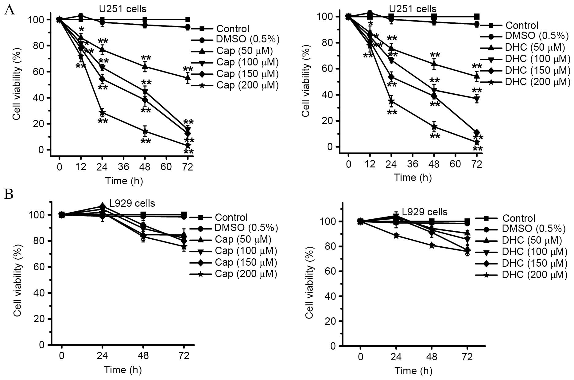 | Figure 1.Effect of Cap and DHC on cell
viability of U251 cells and L929 cells. (A) Viability of human
glioma U251 cells was analyzed by CCK-8 assay following culture in
the presence of various concentrations of Cap and DHC (50, 100, 150
and 200 µM) for 12, 24, 48 and 72 h. (B) Viability of L929 normal
murine fibroblast cells was analyzed by CCK-8 assay following
culture in the presence of various concentration of Cap and DHC
(50, 100, 150 and 200 µM) for 24, 48 and 72 h. Data are presented
as the mean ± standard error of 3 independent experiments.
*P<0.05, **P<0.01 vs. control. CCK-8, Cell Counting Kit-8;
DMSO, dimethyl sulfoxide; Cap, capsaicin; DHC,
dihydrocapsaicin. |
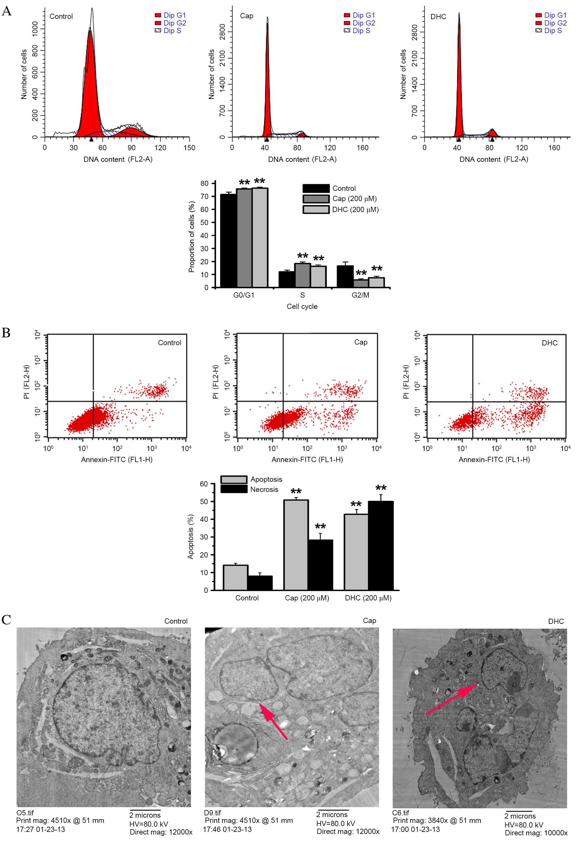
Effects of Cap and DHC on cell cycle
and apoptosis of human glioma U251 cells
Following exposure to Cap or DHC (200 µM) for 12 h,
the proportion of U251 cells in the G2/M phase
significantly decreased from 16.50% in the control group to 5.78%
with Cap (P=0.001; Fig. 2A) and
7.43% with DHC (P=0.001; Fig. 2A).
However, the proportion of cells in the G0/G1
phase significantly increased from 71.47% in the control group to
75.78% with Cap (P=0.006; Fig. 2A)
and 76.32% with DHC (P=0.003; Fig.
2A), while the proportion of cells in S phase also
significantly increased from 12.02% in the control group to 18.43%
with Cap (P=0.001; Fig. 2A) and
16.25% with DHC (P=0.006; Fig.
2A). The anti-tumor effect of Cap and DHC was, therefore,
associated with G0/G1 and S phase cell-cycle
arrest.
Annexin V-FITC/PI double staining assays were also
performed to analyze cell apoptosis following 12 h treatment with
200 µM Cap or DHC. The proportion of apoptosed cells significantly
increased from 14.16% in the DMSO-treated control group to 50.81%
with Cap (P<0.001; Fig. 2B) and
42.79% with DHC (P<0.001; Fig.
2B). Necrosis was also significantly increased from 3.45% in
the control group to 7.17% with Cap (P=0.001; Fig. 2B) and 11.15% with DHC (P<0.001;
Fig. 2B).
TEM also revealed that treatment with either 200 µM
Cap or 200 µM DHC for 12 h resulted in condensed cytoplasm, nuclei
displaying peripheral chromatin condensation, which had broken up
into rounded bodies, and cells developing into apoptotic bodies,
compared with the integrated cellular morphology of the untreated
cells (Fig. 2C).
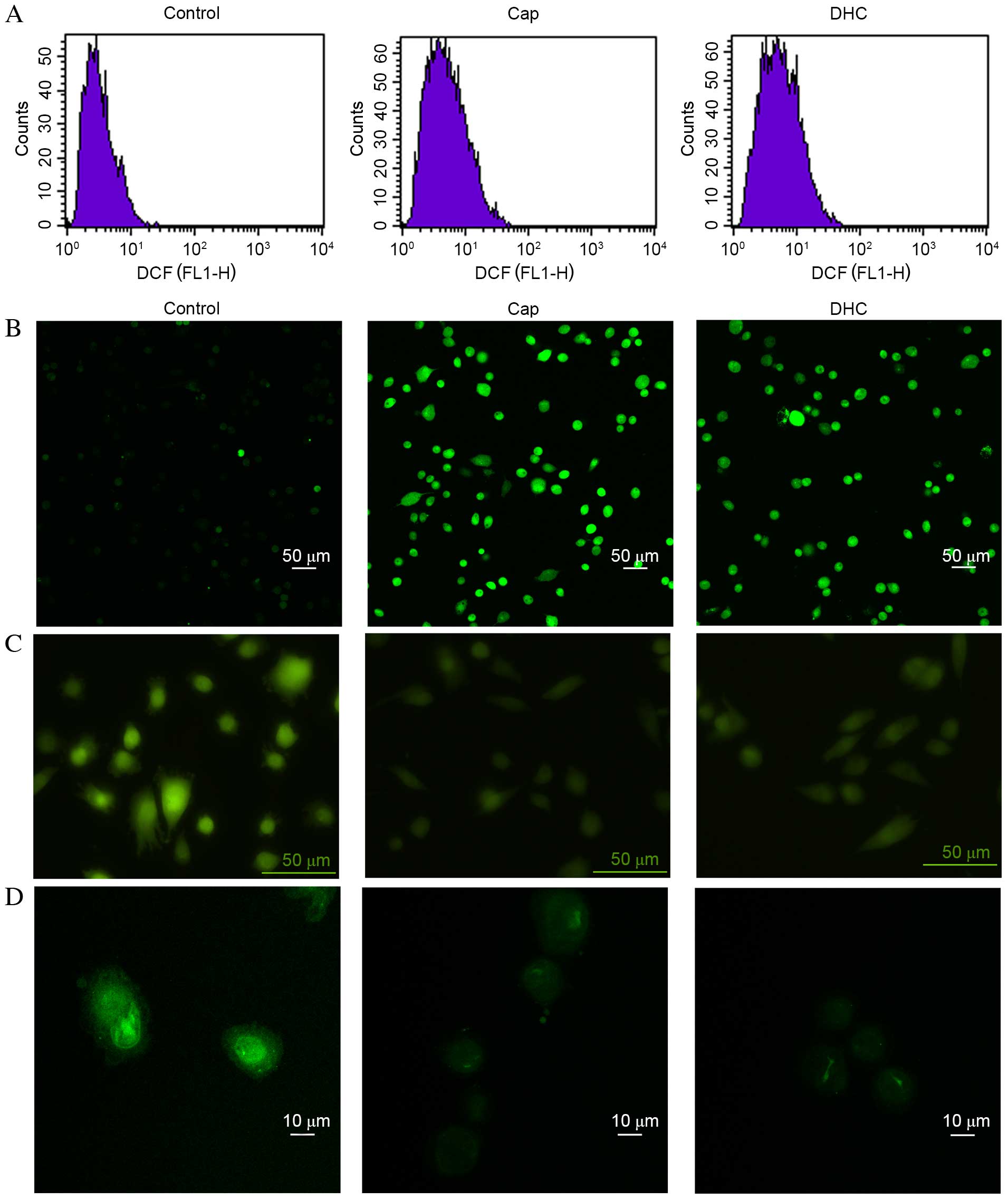
Cap and DHC treatment increases ROS
generation and [Ca2+]i in U251 cells
ROS can initiate the loss of MMP, mitochondrial
translocation of pro-apoptotic proteins, including Bcl-2 associated
protein X apoptosis regulator (Bax) and Bcl-2 associated agonist of
cell death (Bad), and release of cyto c (23). Intracellular ROS levels in U251
cells were, therefore, evaluated by DCFH-DA assays and flow
cytometry detection. DCFH-DA is intracellularly hydrolyzed to DCFH,
which is oxidized by ROS to generate fluorescent DCF. Following 12
h exposure to Cap or DHC, mean DCF fluorescence intensity in U251
cells increased from 3.45 in the control group to 5.84 with Cap and
6.41 with DHC (Fig. 3A).
High levels of Ca2+ can open
mitochondrial permeability transition pores, depolarize
mitochondrial membrane potential, activate caspase-9 and caspase-3,
initiate the mitochondrial apoptosis pathway, to induce cell
apoptosis (24). Fluo 3-AM was,
therefore, used to observe the intracellular concentration of
Ca2+. The mean fluorescence intensity in U251 cells
increased from 333.79 in the control group to 1,116.46 (P<0.001)
with Cap and 1,240.66 (P<0.001) with DHC, respectively (Fig. 3B), indicating an increase in the
concentration of intracellular Ca2+.
Cap and DHC treatment increases MPTP
formation and reduces MMP
MPTP formation is one of the principle regulators of
the mitochondrion during cell apoptosis. The opening of MPTPs can
lead to matrix swelling, rupture of the outer membrane of
mitochondrion and a release of intermembrane space proteins into
the cytoplasm, ultimately resulting in a loss of MMP. The presence
of MPTPs was, therefore, detected using a GENMED MPTP living cell
fluorescence detection kit. Following exposure to Cap or DHC for 12
h, the fluorescence intensity in U251 cells was decreased compared
with the control group (Fig. 3C),
indicating the formation of MPTPs.
Depolarization of the MMP induces release of cyto c
and apoptosis-inducing factor (AIF) from the intermembrane space
into the cytoplasm, resulting in fragmentation of nucleus and cell
apoptosis (25). The mean
fluorescence intensity in U251 cells decreased from 980.90 in the
control group to 216.13 following 12 h exposure to 200 µM Cap
(P<0.001) and 222.48 with 200 µM DHC (P<0.001), respectively
(Fig. 3D), indicating the loss of
MMP.
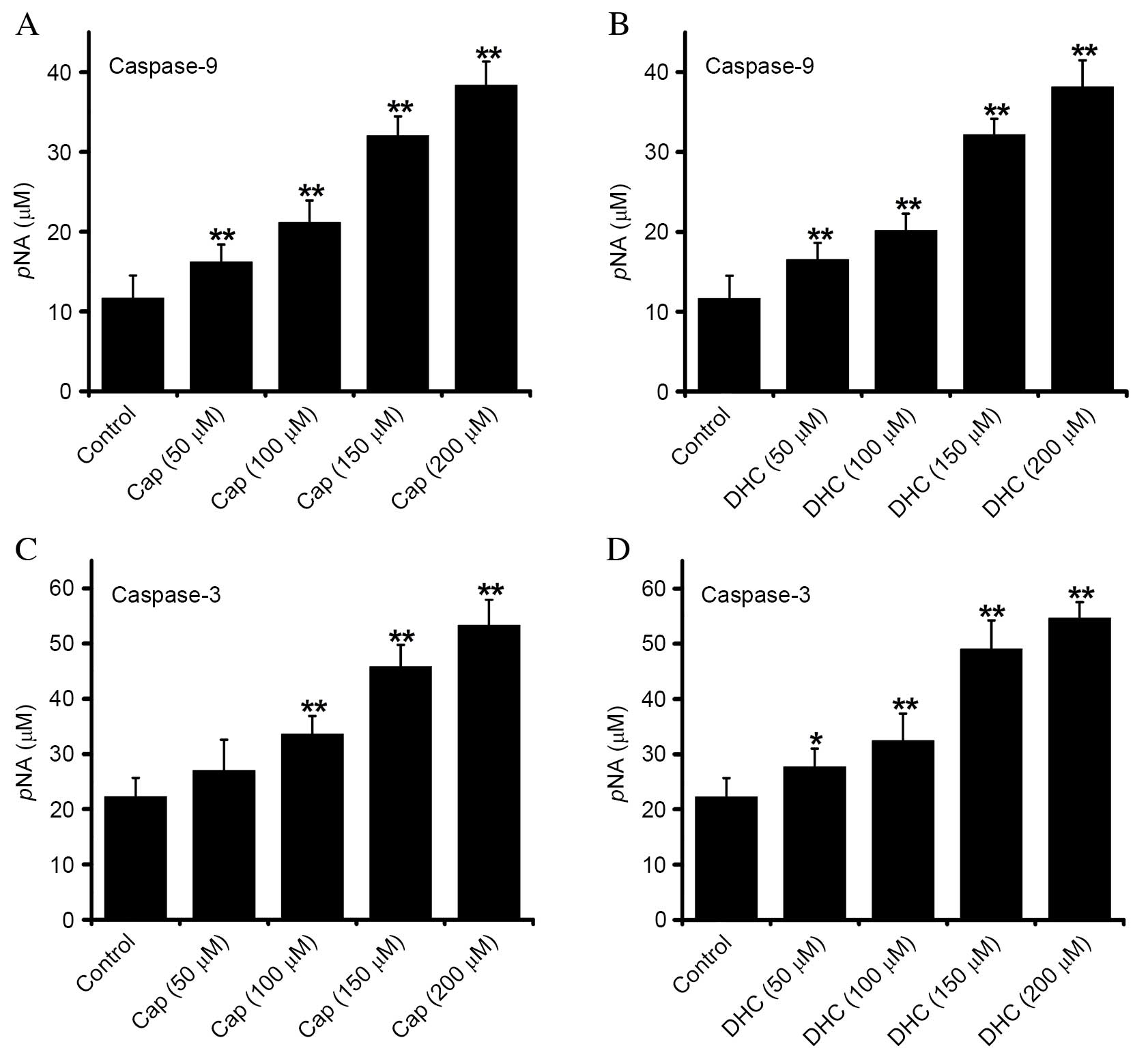
Cap and DHC treatment activates
caspase-9 and −3
Apoptotic cell death is inevitable following
caspase-3 activation, and is a vital factor in mitochondrial
pathway-induced apoptosis. Caspase-9 (Fig. 4A and B) and −3 (Fig. 4C and D) activity were
dose-dependently increased in U251 cells exposed to Cap or DHC for
12 h, compared with control group, indicating that Cap- and
DHC-induced apoptosis is associated with the activation of
caspase-9 and −3.
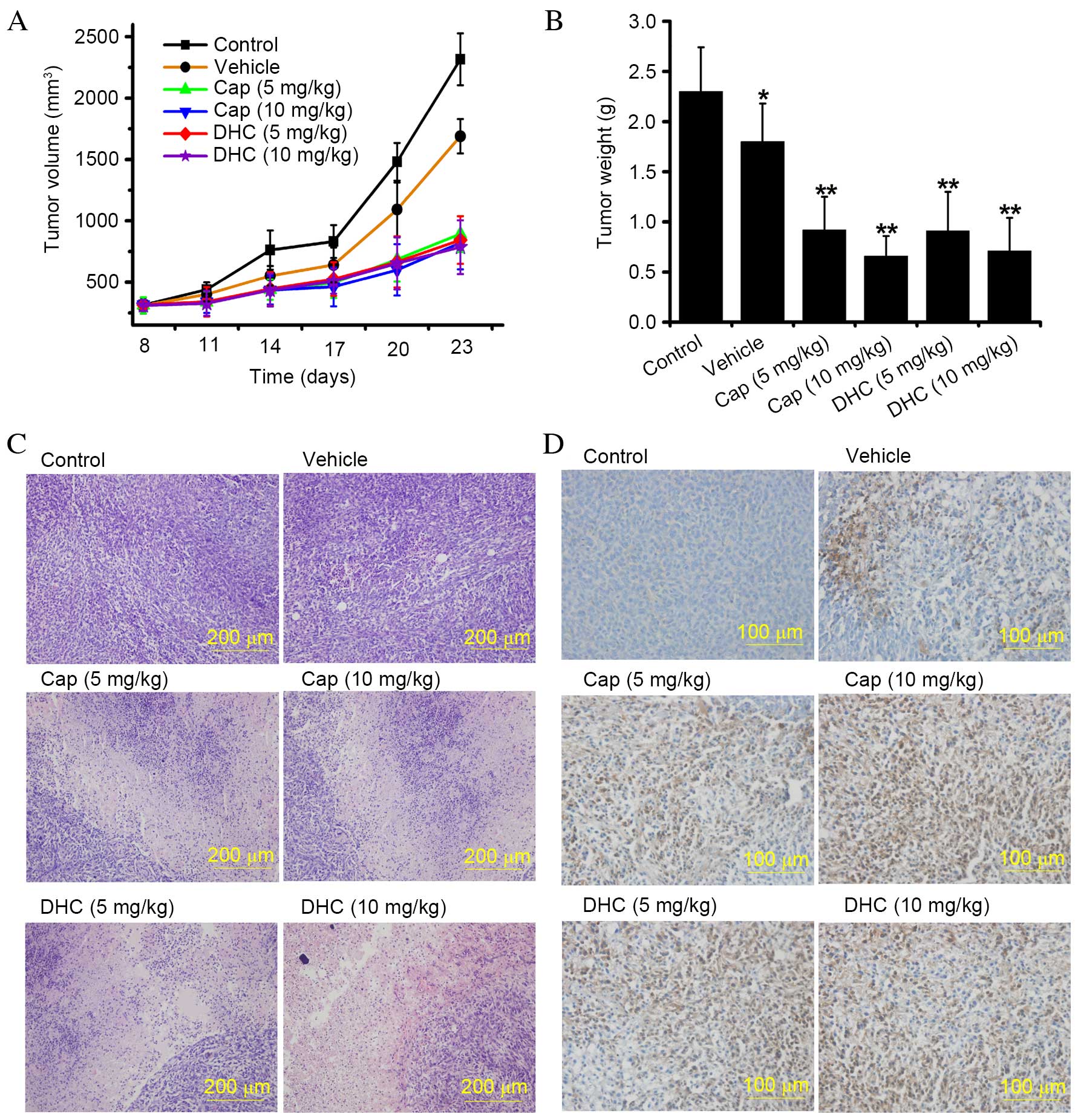
Cap and DHC inhibits the growth of
U251 human glioma tumor xenografts
In vitro experiments are valuable for quick,
convenient, and large-scale screening of latent anti-tumor agents,
however the effects observed in cell-based assays require further
investigation in suitable pre-clinical in vivo models prior
to clinical trials. Tumor xenografts in nude mice is an accepted
animal model for pre-clinical evaluation of potential anti-tumor
agents. The effect of Cap and DHC on the development of U251 glioma
tumor xenografts was, therefore, evaluated in nude mice. The rate
of U251 glioma tumor xenograft development was attenuated in mice
administered with Cap or DHC in both the low dose group (5 mg/kg,
every 3 days) and high dose group (10 mg/kg, every 3 days; Fig. 5A). The mean tumor volume at 23 days
in control mice (2316.48±211.28 mm3) was ~2.8-fold
larger than tumors from the Cap (10 mg/kg)-treated group
(821.15±215.63 mm3; P<0.001) and ~2.95-fold larger
than tumors from the DHC (10 mg/kg)-treated group (785.83±218.31
mm3; P<0.001; Fig.
5A). Similar results were obtained with mice treated with 5
mg/kg Cap (P<0.001) and DHC (P<0.001) treated mice compared
with control mice (Fig. 5A). The
mean wet weight of tumors from control mice was 2.30±0.44 g,
compared with 0.92±0.33 g in Cap (10 mg/kg)-treated mice
(P<0.001; Fig. 5B) and
0.71±0.34 g in DHC (10 mg/kg)-treated mice (P<0.001; Fig. 5B). A similarly significant
anti-tumor effect was demonstrated in mice treated with 5 mg/kg Cap
or DHC compared with control mice (P<0.001 and P<0.001,
respectively; Fig. 5B). However,
the differences between high-dose and low-dose treatments with the
same drug were not statistically significant (Fig. 5B). As previous reported, alcohol
may certain antitumor effects (26). In addition, no difference in
average body weight was observed between the control group, Cap-
and DHC-treated mice throughout the experiment (data not shown).
The liver index (liver weight/body weightx100) and spleen index
(spleen weight/body weightx100) in Cap- and DHC-treated mice were
also not significantly different compared with the control group
(data not shown), indicating that Cap and DHC are not toxic to
mice. Histological analysis of the tumors by H&E staining
revealed liquefaction and necrosis in tumors from Cap- and
DHC-treated mice, compared with vigorous growth in control and
vehicle-treated mice (Fig. 5C). No
ulcers or erosion, were observed in the stomachs of any mice, nor
were any apparent side effects observed during the histological
analysis of the hearts, spleens, lungs, livers and kidneys of the
mice (data not shown). IHC analysis of cyto c expression revealed
numerous cyto c-positive cells in the tumors of Cap- and
DHC-treated mice, but few in the control or vehicle-treated groups
(Fig. 5D), indicating that cyto c
was released from mitochondria to the cytoplasmic matrix, inducing
U251 cell apoptosis. Cap and DHC have, therefore, been demonstrated
to significantly reduce U251 human glioma tumor development with no
apparent negative side effects on the organs.
Discussion
Human glioma is the most common type of primary
brain tumor, and one of the most invasive and aggressive tumors,
which, even with treatments, including surgery, radiotherapy and
chemotherapy, often relapses and exhibits resistance to
conventional treatment methods. Apoptosis and angiogenesis are two
potential targets for novel strategies to treat this disease
(27). Resistance to radiotherapy
and chemotherapy are common consequences of the dysregulation of
apoptosis, therefore, inducing apoptosis is an important feature of
various chemotherapy drugs, including temozolomide and cisplatin
(28,29). Cap, the most abundant pungent chili
pepper component, has been widely examined as an anti-cancer agent,
and induces cell apoptosis in numerous cancer cell lines, in
vitro and when explanted into rodents (11,30,31).
The present study demonstrated that Cap and DHC, the second most
abundant capsaicinoids in chili pepper, are anti-cancer agents
against U251 human glioma cells and are not toxic to L929 normal
murine fibroblast cells. The apoptotic effects of Cap and DHC in
U251 cells were associated with the generation of ROS, increased
intracellular Ca2+, disruption of MMP and the release of
cyto c to the cytosol, which ultimately resulted in the activation
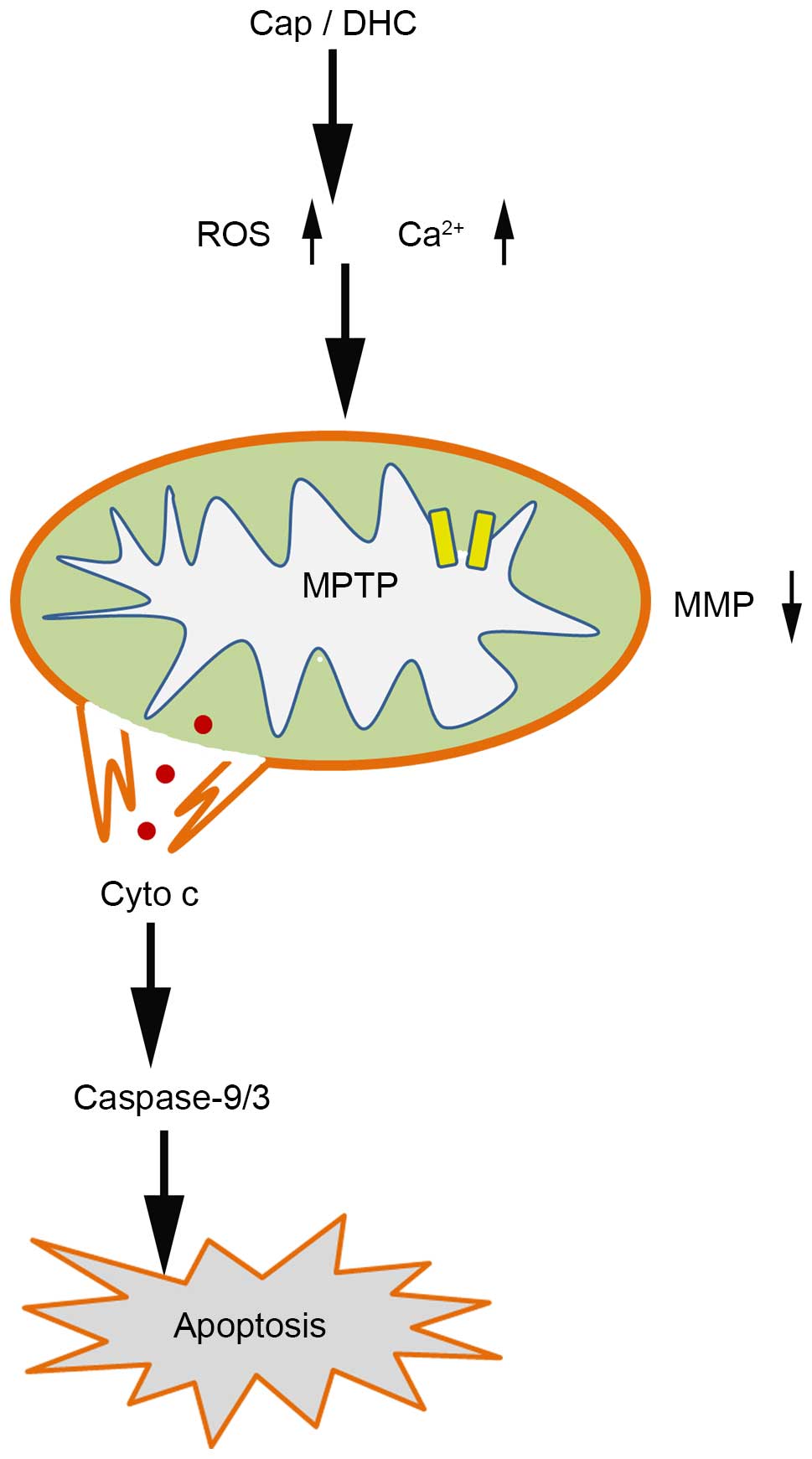
of caspase-9 and caspase-3 cascade (Fig. 6).
Cell cycle arrest is a prospective therapeutic
target in cancer as it is a downstream focal point for oncogenic
signaling pathways (32). Previous
reports have demonstrated that Cap inhibits cell proliferation by
cell-cycle arrest: MCF-7 human breast cancer cells and BT-20 cells
were arrested in the S phase following 72 h treatment with 200 µM
Cap (12). Thoennissen et
al (14) demonstrated that Cap
arrests MCF-7, T47D, BT-474, SKBR-3 and MDA-MB231 (ER-positive and
-negative) human breast cancer cells in the
G0/G1 phase at concentrations of 50–200 µM.
Similarly, SCC4 human tongue cancer cells, NPC-TW 039
nasopharyngeal carcinoma cells, RT4 urothelial cancer cells, CE 81
T/VGH esophagus epidermoid carcinoma cells, HL-60 leukemia and NB4
myeloid leukemia cells were also arrested in the
G0/G1 phase following treatment with Cap
(13,15,17,33–35).
In the current study, the anti-proliferation effect of Cap and DHC
was associated with G0/G1 and S phase cell
cycle arrest following 12 h treatment at a concentration of 200 µM
(Fig. 2A).
The most common apoptotic pathway induced by Cap is
reported to be the mitochondrial pathway, which is particularly
susceptible to ROS and Ca2+. A sustained increase in
intracellular Ca2+ increases the production of ROS and
eventually triggers cell apoptosis. Excess
H2O2 inhibits the activity of
Na+/H+ antiporters, which leads to
intracellular acidification and creates a microenvironment for
apoptosis (36). Furthermore, ROS
directly damage the mitochondrial electron transport chain,
activate caspases and induce cell apoptosis (29). Cap boosts the generation of ROS in
human pancreatic cancer cells by inhibiting mitochondrial complex I
and III and destroying mitochondrial functions (37). Initiation of the mitochondrial
pathway by excess ROS and sustained an increase in intracellular
Ca2+ results in the formation of MPTPs, which leads to
the dissipation of the MMP. The opening of MPTPs elevates the
permeability of inner mitochondrial membranes, allowing molecules
<1.5 kDa to enter the mitochondria, resulting in swelling of the
mitochondrial matrix (24,38). The outer mitochondrial membranes
subsequently rupture, releasing numerous pro-apoptotic proteins
from the intermembrane space into the cytosol (24,38).
These pro-apoptotic proteins are divided into two groups: One group
includes AIF and endonuclease G, which act as through a
caspase-independent pathway to induce cell apoptosis; the other
group, containing cyto c and second mitochondrial-derived activator
of caspases (Smac/Diablo), activate caspases. Cyto c induces the
oligomerization of apoptotic protease activating factor 1 (Apaf-1),
which subsequently activates caspase-9. The activated cyto c,
Apaf-1 and caspase-9 then form the apoptosome and, ultimately,
activate caspase-3, resulting in nuclear fragmentation (39). Once caspase-3 is activated, cell
apoptosis is irreversible (29,40).
The present study indicated that Cap and DHC induced apoptosis in
human glioma cells via a ROS- and Ca2+-mediated
mitochondrial pathway, which was associated with MPTP formation,
MMP dissipation and the release of cyto c to the cytosol to
activate caspase-9 and −3 (Figs. 3
and 4).
The anti-neoplastic activity of Cap and DHC was
investigated in vivo by administering Cap or DHC by oral
gavage, producing results that were consistent with previous
studies. Cap (5 mg/kg) has previously been revealed to reduce the
weight and volume of pancreatic xenograft tumors by 30% (11). Thoennissen et al (14) demonstrated that the weight of
invasive, epidermal growth factor receptor-positive, p53 mutant
orthotopic MDA-MB231 breast cancer tumors in female nude mice were
reduced by 70% compared with controls, following oral gavage with
Cap (5 mg/kg per day) 3 times per week for 4 weeks, without
observable side effects. Another study demonstrated that Cap (50
mg/kg), administered daily for 6 days, reduced the leukemia tumor
weight in NOD/SCID mice and increased apoptosis without resulting
in organ damage (35). The daily
consumption of capsicum spices varies worldwide; 2.5 g/person is
consumed per day in India, 5 g/person in Thailand, 15 g/person in
Saudi Arabia and 20 g/person in Mexico (41). The Cap content of different
capsicums also varies dramatically, with a range from 1–42.5%
(42). Furthermore, due to the
unclear pharmacokinetics of Cap, it is difficult to determine how
much Cap should be administered in an animal model. Accordingly,
considering that previous investigations have used 5 mg/kg Cap to
estimate the anti-tumor effect in vivo (43), 5 mg/kg and 10 mg/kg were
administered as preliminary doses in the present study, with the
optimal dose requiring further research.
In conclusion, the present study demonstrated that
Cap and DHC induce apoptosis in human glioma cancer cells through
ROS and Ca2+-mediated induction of the mitochondrial
apoptosis pathway, which caused MPTP formation, dissipation of the
MMP, and release of cyto c to the cytosol, resulting in activation
of caspase-9 and caspase-3 (Fig.
6). In addition, intragastric administration of Cap and DHC
in vivo was demonstrated to suppress the development of U251
human glioma xenograft tumors, with no apparent side effects.
Consequently, these findings provide a basis for developing Cap and
DHC as potential therapeutic approaches against glioma.
Acknowledgements
The present study was supported by the International
Science & Technology Cooperation Program of China (grant no.
2011DFA30620).
References
|
1
|
Brem SS, Bierman PJ, Brem H, Butowski N,
Chamberlain MC, Chiocca EA, DeAngelis LM, Fenstermaker RA, Friedman
A, Gilbert MR, et al: Central nervous system cancers. J Natl Compr
Canc Netw. 9:352–400. 2011.PubMed/NCBI
|
|
2
|
Wen PY and Kesari S: Malignant gliomas in
adults. N Engl J Med. 359:492–507. 2008. View Article : Google Scholar : PubMed/NCBI
|
|
3
|
Maher EA, Furnari FB, Bachoo RM, Rowitch
DH, Louis DN, Cavenee WK and DePinho RA: Malignant glioma: Genetics
and biology of a grave matter. Genes Dev. 15:1311–1333. 2001.
View Article : Google Scholar : PubMed/NCBI
|
|
4
|
Malagarie-Cazenave S, Olea-Herrero N, Vara
D, Morell C and Diaz-Laviada I: The vanilloid capsaicin induces
IL-6 secretion in prostate PC-3 cancer cells. Cytokine. 54:330–337.
2011. View Article : Google Scholar : PubMed/NCBI
|
|
5
|
Deb G, Thakur VS, Limaye AM and Gupta S:
Epigenetic induction of tissue inhibitor of matrix
metalloproteinase-3 by green tea polyphenols in breast cancer
cells. Mol Carcinog. 54:485–499. 2015. View
Article : Google Scholar : PubMed/NCBI
|
|
6
|
Feng LL, Liu BX, Zhong JY, Sun LB and Yu
HS: Effect of grape procyanidins on tumor angiogenesis in liver
cancer xenograft models. Asian Pac J Cancer Prev. 15:737–741. 2014.
View Article : Google Scholar : PubMed/NCBI
|
|
7
|
Zhang X, Zhu Y, Duan W, Feng C and He X:
Allicin induces apoptosis of the MGC-803 human gastric carcinoma
cell line through the p38 mitogen-activated protein
kinase/caspase-3 signaling pathway. Mol Med Rep. 11:2755–2760.
2015.PubMed/NCBI
|
|
8
|
Luo XJ, Peng J and Li YJ: Recent advances
in the study on capsaicinoids and capsinoids. Eur J Pharmacol.
650:1–7. 2011. View Article : Google Scholar : PubMed/NCBI
|
|
9
|
Brown KC, Witte TR, Hardman WE, Luo H,
Chen YC, Carpenter AB, Lau JK and Dasgupta P: Capsaicin displays
anti-proliferative activity against human small cell lung cancer in
cell culture and nude mice models via the E2F pathway. PLoS One.
5:e102432010. View Article : Google Scholar : PubMed/NCBI
|
|
10
|
Sánchez AM, Martínez-Botas J,
Malagarie-Cazenave S, Olea N, Vara D, Lasunción MA and Díaz-Laviada
I: Induction of the endoplasmic reticulum stress protein
GADD153/CHOP by capsaicin in prostate PC-3 cells: A microarray
study. Biochem Biophys Res Commun. 372:785–791. 2008. View Article : Google Scholar : PubMed/NCBI
|
|
11
|
Zhang R, Humphreys I, Sahu RP, Shi Y and
Srivastava SK: In vitro and in vivo induction of apoptosis by
capsaicin in pancreatic cancer cells is mediated through ROS
generation and mitochondrial death pathway. Apoptosis.
13:1465–1478. 2008. View Article : Google Scholar : PubMed/NCBI
|
|
12
|
Chang HC, Chen ST, Chien SY, Kuo SJ, Tsai
HT and Chen DR: Capsaicin may induce breast cancer cell death
through apoptosis-inducing factor involving mitochondrial
dysfunction. Hum Exp Toxicol. 30:1657–1665. 2011. View Article : Google Scholar : PubMed/NCBI
|
|
13
|
Ip SW, Lan SH, Lu HF, Huang AC, Yang JS,
Lin JP, Huang HY, Lien JC, Ho CC, Chiu CF, et al: Capsaicin
mediates apoptosis in human nasopharyngeal carcinoma NPC-TW 039
cells through mitochondrial depolarization and endoplasmic
reticulum stress. Hum Exp Toxicol. 31:539–549. 2012. View Article : Google Scholar : PubMed/NCBI
|
|
14
|
Thoennissen NH, O'Kelly J, Lu D, Iwanski
GB, La DT, Abbassi S, Leiter A, Karlan B, Mehta R and Koeffler HP:
Capsaicin causes cell-cycle arrest and apoptosis in ER-positive and
-negative breast cancer cells by modulating the EGFR/HER-2 pathway.
Oncogene. 29:285–296. 2010. View Article : Google Scholar : PubMed/NCBI
|
|
15
|
Ip SW, Lan SH, Huang AC, Yang JS, Chen YY,
Huang HY, Lin ZP, Hsu YM, Yang MD, Chiu CF and Chung JG: Capsaicin
induces apoptosis in SCC-4 human tongue cancer cells through
mitochondria-dependent and -independent pathways. Environ Toxicol.
27:332–341. 2012. View Article : Google Scholar : PubMed/NCBI
|
|
16
|
Bley K, Boorman G, Mohammad B, McKenzie D
and Babbar S: A comprehensive review of the carcinogenic and
anticarcinogenic potential of capsaicin. Toxicol Pathol.
40:847–873. 2012. View Article : Google Scholar : PubMed/NCBI
|
|
17
|
Amantini C, Ballarini P, Caprodossi S,
Nabissi M, Morelli MB, Lucciarini R, Cardarelli MA, Mammana G and
Santoni G: Triggering of transient receptor potential vanilloid
type 1 (TRPV1) by capsaicin induces Fas/CD95-mediated apoptosis of
urothelial cancer cells in an ATM-dependent manner. Carcinogenesis.
30:1320–1329. 2009. View Article : Google Scholar : PubMed/NCBI
|
|
18
|
Gil YG and Kang MK: Capsaicin induces
apoptosis and terminal differentiation in human glioma A172 cells.
Life Sci. 82:997–1003. 2008. View Article : Google Scholar : PubMed/NCBI
|
|
19
|
Maity R, Sharma J and Jana NR: Capsaicin
induces apoptosis through ubiquitin-proteasome system dysfunction.
J Cell Biochem. 109:933–942. 2010.PubMed/NCBI
|
|
20
|
Oh SH, Kim YS, Lim SC, Hou YF, Changl IY
and You HJ: Dihydrocapsaicin (DHC), a saturated structural analog
of capsaicin, induces autophagy in human cancer cells in a
catalase-regulated manner. Autophagy. 4:1009–1019. 2008. View Article : Google Scholar : PubMed/NCBI
|
|
21
|
Choi CH, Jung YK and Oh SH: Selective
induction of catalase-mediated autophagy by dihydrocapsaicin in
lung cell lines. Free Radic Biol Med. 49:245–257. 2010. View Article : Google Scholar : PubMed/NCBI
|
|
22
|
Fallone CA, Loo VG, Lough J and Barkun AN:
Hematoxylin and eosin staining of gastric tissue for the detection
of helicobacter pylori. Helicobacter. 2:32–35. 1997. View Article : Google Scholar : PubMed/NCBI
|
|
23
|
Cook SA, Sugden PH and Clerk A: Regulation
of bcl-2 family proteins during development and in response to
oxidative stress in cardiac myocytes: Association with changes in
mitochondrial membrane potential. Circ Res. 85:940–949. 1999.
View Article : Google Scholar : PubMed/NCBI
|
|
24
|
Halestrap AP: What is the mitochondrial
permeability transition pore? J Mol Cell Cardiol. 46:821–831. 2009.
View Article : Google Scholar : PubMed/NCBI
|
|
25
|
Kinnallyk W, Peixotop M, Ryu SY and Dejean
LM: Is mPTP the gatekeeper for necrosis, apoptosis, or both?
Biochim Biophys Acta. 1813:616–622. 2011. View Article : Google Scholar : PubMed/NCBI
|
|
26
|
Hoek JB, Cahill A and Pastorino JG:
Alcohol and mitochondria: A dysfunctional relationship.
Gastroenterology. 122:2049–2063. 2002. View Article : Google Scholar : PubMed/NCBI
|
|
27
|
Wagner L, Marschall V, Karl S, Cristofanon
S, Zobel K, Deshayes K, Vucic D, Debatin KM and Fulda S: Smac
mimetic sensitizes glioblastoma cells to Temozolomide-induced
apoptosis in a RIP1- and NF-κB-dependent manner. Oncogene.
32:988–997. 2013. View Article : Google Scholar : PubMed/NCBI
|
|
28
|
Eriksson I, Joosten M, Roberg K and
Ollinger K: The histone deacetylase inhibitor trichostatin A
reduces lysosomal pH and enhances cisplatin-induced apoptosis. Exp
Cell Res. 319:12–20. 2013. View Article : Google Scholar : PubMed/NCBI
|
|
29
|
Indran IR, Tufo G, Pervaiz S and Brenner
C: Recent advances in apoptosis, mitochondria and drug resistance
in cancer cells. Biochim Biophys Acta. 1807:735–745. 2011.
View Article : Google Scholar : PubMed/NCBI
|
|
30
|
Patwardhan CA, Alfa E, Lu S and Chadli A:
Progesterone receptor chaperone complex-based high-throughput
screening assay: Identification of capsaicin as an inhibitor of the
Hsp90 machine. J Biomol Screen. 20:223–229. 2015. View Article : Google Scholar : PubMed/NCBI
|
|
31
|
Lewinska A, Jarosz P, Czech J, Rzeszutek
I, Bielak-Zmijewska A, Grabowska W and Wnuk M: Capsaicin-induced
genotoxic stress does not promote apoptosis in A549 human lung and
DU145 prostate cancer cells. Mutat Res Genet Toxicol Environ
Mutagen. 779:23–34. 2015. View Article : Google Scholar : PubMed/NCBI
|
|
32
|
Williams GH and Stoeber K: The cell cycle
and cancer. J Pathol. 226:352–364. 2012. View Article : Google Scholar : PubMed/NCBI
|
|
33
|
Wu CC, Lin JP, Yang JS, Chou ST, Chen SC,
Lin YT, Lin HL and Chung JG: Capsaicin induced cell cycle arrest
and apoptosis in human esophagus epidermoid carcinoma CE 81T/VGH
cells through the elevation of intracellular reactive oxygen
species and Ca2+ productions and caspase-3 activation. Mutat Res.
601:71–82. 2006. View Article : Google Scholar : PubMed/NCBI
|
|
34
|
Tsou MF, Lu HF, Chen SC, Wu LT, Chen YS,
Kuo HM, Lin SS and Chung JG: Involvement of Bax, Bcl-2, Ca2+ and
caspase-3 in capsaicin-induced apoptosis of human leukemia HL-60
cells. Anticancer Res. 26:1965–1971. 2006.PubMed/NCBI
|
|
35
|
Ito K, Nakazato T, Yamato K, Miyakawa Y,
Yamada T, Hozumi N, Segawa K, Ikeda Y and Kizaki M: Induction of
apoptosis in leukemic cells by homovanillic acid derivative,
capsaicin, through oxidative stress: Implication of phosphorylation
of p53 at Ser-15 residue by reactive oxygen species. Cancer Res.
64:1071–1078. 2004. View Article : Google Scholar : PubMed/NCBI
|
|
36
|
Abdel-Salam Omar ME: Capsaicin as a
Therapeutic Molecule. Springer; Basel: pp. 181–203. 2014
|
|
37
|
Pramanik KC, Boreddy SR and Srivastava SK:
Role of mitochondrial electron transport chain complexes in
capsaicin mediated oxidative stress leading to apoptosis in
pancreatic cancer cells. PLoS One. 6:e201512011. View Article : Google Scholar : PubMed/NCBI
|
|
38
|
Kinnally KW, Peixoto PM, Ryu SY and Dejean
LM: Is mPTP the gatekeeper for necrosis, apoptosis, or both?
Biochim Biophys Acta. 1813:616–622. 2011. View Article : Google Scholar : PubMed/NCBI
|
|
39
|
Jeong SY and Seol DW: The role of
mitochondria in apoptosis. BMB Rep. 41:11–22. 2008. View Article : Google Scholar : PubMed/NCBI
|
|
40
|
Brunelle JK and Zhang B: Apoptosis assays
for quantifying the bioactivity of anticancer drug products. Drug
Resist Updat. 13:172–179. 2010. View Article : Google Scholar : PubMed/NCBI
|
|
41
|
O'Neill J, Brock C, Olesen AE, Andresen T,
Nilsson M and Dickenson AH: Unravelling the mystery of capsaicin: A
tool to understand and treat pain. Pharmacol Rev. 64:939–971. 2012.
View Article : Google Scholar : PubMed/NCBI
|
|
42
|
Al Othman ZA, Ahmed YB, Habila MA and
Ghafar AA: Determination of capsaicin and dihydrocapsaicin in
capsicum fruit samples using high performance liquid
chromatography. Molecules. 16:8919–8929. 2011. View Article : Google Scholar : PubMed/NCBI
|
|
43
|
Capsaicin as a Therapeutic Molecule. In:
Capsaicin as a Therapeutic Molecule. 68. AbdelSalam OME: Springer
Basel Ag; Picassoplatz 4, Basel, 4052, Switzerland: 2014,
View Article : Google Scholar
|