Introduction
In recent years, microRNAs (miRNA) have been
considered important molecules that are important in the regulation
of tumorigenesis (1). miRNAs are a
type of small non-coding RNAs ~20 nucleotides in length (2,3).
Previous studies have reported that miRNAs exert an effect on
different cell processes, including cell differentiation,
migration, growth, proliferation, apoptosis and metabolism
(4,5). miRNAs exert these effects by binding
with the 3′-untranslated region of its target mRNA (6,7).
Increasing evidence demonstrates that miRNAs are directly involved
in the pathogenesis of various tumors (6,8). In
addition, miRNAs have become novel biomarkers for the diagnosis or
therapy of diseases, including cancer, heart disease and diabetes
(7,9).
Renal cell carcinoma (RCC) accounts for ~3% of adult
malignancies and represents 90% of renal tumors with the highest
rate of recurrence and mortality among the malignances of the
urinary system (10,11). In addition, ~30% of patients with
RCC continued to present with metastases (12), and the prognosis of patients with
RCC remains poor. However, the underlying mechanism of RCC requires
further elucidation. Thus, it is important to increase the
understanding of tumorigenesis and development of RCC, and develop
a biomarker for RCC diagnosis at early stages or as a molecular
targeted therapy.
miR-15a-5p, located at chromosome 13q14 (13), has been demonstrated to be
dysregulated in a number of tumors, such as pancreatic cancer
(14), chronic lymphocytic
leukemia (15), and pituitary
(16). However, to the best of our
knowledge, there has been no research into miR-15a-5p on RCC. Thus,
the present study aimed to detect the expression of miR-15a-5p in
RCC tissues and cells and to determine the function of miR-15a-5p
on cell proliferation, migration, invasion and apoptosis.
Materials and methods
Sample collection
A total of 27 paired tissues were collected from
Guangdong province (China; between January 2014 and January 2015),
which consisted of renal cell carcinoma tissues and adjacent normal
renal tissues. Written informed consent was obtained from all
patients. Collection and use of the samples were reviewed and
approved by the ethics committee Peking University Shenzhen
Hospital. While the tissues were dissected, they were immersed in
RNAlater (Qiagen GmbH, Hilden, Germany) for 30 min, then the
tissues were stored at −80°C for further use, and a pair of tissues
included RCC and adjacent normal tissues 2 cm away from visible RCC
lesions. The tissues collected were reviewed and classified by
hematoxylin and eosin staining. The clinical and pathological
characteristics of the patients are presented in Table I.
 | Table I.Clinicopathological features in RCC
patients. |
Table I.
Clinicopathological features in RCC
patients.
| Characteristic | Number of
cases |
|---|
| Mean age range
(years) | 50 (25–70) |
| Sexual
distinction |
|
|
Male/female | 18/10 |
| Histological
type |
|
| Clear
cell/papillary | 25/3 |
| pT-stage |
|
|
T1/T2/T3+T4 | 16/10/2 |
| Fuhrman grade |
|
|
I/II/III/IV | 8/14/4/2 |
| AJCC clinical
stages |
|
|
I/II/III+IV | 16/10/2 |
Cell culture
293-T human embryo kidney cells and 786-O and ACHN
RCC cell lines were used in the present study. Cells were cultured
in a humidified incubator containing 5% CO2 at 37°C in
Dulbecco's modified Eagle's medium (DMEM; Gibco; Thermo Fisher
Scientific, Inc., Waltham, MA, USA) with 10% fetal bovine serum
(FBS; Gibco; Thermo Fisher Scientific, Inc.), 1% antibiotics (100
µl/ml penicillin and 100 mg/ml streptomycin sulfates) and 1%
glutamine.
RNA extraction, cDNA synthesis and
quantitative polymerase chain reaction (qPCR)
Total RNA was extracted from the samples and cells
by TRIzol (Invitrogen; Thermo Fisher Scientific, Inc.) and purified
with the RNeasy Maxi kit (Qiagen GmbH) according to the
manufacturer's protocols. The concentration of RNA was measured on
a NanoDrop 2000/2000c (Thermo Fisher Scientific, Inc.). RT-qPCR was
performed to synthesize the cDNA, 1 µg total RNA of each sample was
used for reverse transcription using miScript Reverse Transcription
kit (Qiagen GmbH) following the manufacturer's protocols. qPCR was
performed to detect the expression level of miR-15a-5p with
miScript SYBR® Green PCR kit (Qiagen GmbH) on the Roche
Lightcycler 480 Real-Time PCR system (Roche Diagnostics, Basel,
Switzerland) according to the manufacturer's protocols. U6 served
as the internal control. The sequences of the primers were as
follows: miR-15a-5p, forward 5′-TAGCAGCACATAATGGTTTGTG-3′, and
universal primer was used as the reverse primer (provided by the
miScript SYBR® Green PCR kit); U6, forward
5′-CTCGCTTCGGCAGCACA-3′ and reverse 5′-ACGCTTCACGAATTTGCGT-3′. The
thermocycling conditions were as follows: 95°C for 1 min, then 40
cycles of 95°C for 10 sec, 55°C for 30 sec and 70°C for 30 sec. The
expression levels of miR-15a-5p in tissues and cells were analyzed
with 2−ΔΔCq method (17).
Cell transfection
The expression level of miR-15a-5p in 786-O and ACHN
cells was upregulated by transfecting the synthesized miR-15a-5p
mimics and downregulated by transfecting the synthesized miR-15a-5p
inhibitor using Lipofectamine 2000 (Invitrogen; Thermo Fisher
Scientific, Inc.), which was mixed in the Opti-MEM® I
Reduced Serum Medium (Gibco; Thermo Fisher Scientific, Inc.)
following the manufacturer's protocols. RT-qPCR was performed (as
described above) to observe the changes in miR-15a-5p expression.
The sequences are presented in Table
II.
| Table II.Sequences of the siRNAs. |
Table II.
Sequences of the siRNAs.
| siRNA | Sequence
(5′-3′) |
|---|
| miR-15a mimic | S:
UAGCAGCACAUAAUGGUUUGUG |
|
| A:CAAACCAUUAUGU
GCUGCUAUU |
| Negative
control | S:
UUCUCCGAACGUGUCACGUTT |
|
|
A:ACGUGACACGUUCGGAGAATT |
| miR-15a
inhibitor |
CACAAACCAUUAUGUGCUGCUA |
| Inhibitor NC |
CAGUACUUUUGUGUAGUACAA |
Wound healing assay
The wound healing assay was performed to assess the
cell migration ability of 786-O and ACHN cells in vitro. In
the wound healing assay, ~3×10⁵ cells were seeded in every well of
the 12-well plate, and 24 h later the cells were transfected with
40 pmol of miR-15a-5p mimics, inhibitors, negative control or
inhibitor negative control using Lipofectamine® 2000. A
vertical horizontal line was scratched with a sterile 200 µl
pipette tip after 6 h of transfection. To remove the floating cells
the cells were rinsed with phosphate-buffered saline (PBS) and then
cultured at 37°C in a humidified chamber containing 5%
CO2. A digital camera system was used to capture images
of the scratches at 0, 12 and 24 h after making the scratch. The
experiments were performed in triplicate and repeated at least 3
times.
Transwell assay
A Transwell assay was performed to assess the cell
migration and invasion ability of 786-O and ACHN cells in
vitro. Transwell chamber inserts (BD Biosciences, Franklin
Lakes, NJ, USA) with or without Matrigel (for invasion) were used
in the assay according to the manufacturer's protocol. The
transfected cells were seeded in the upper chamber of the insert at
a density of 1×10⁴ cells in 200 µl serum-free DMEM. The bottom of
the inserts contained DMEM with 10% FBS. The 786-O cells were
allowed to migrate for 36 h and invade for 48 h. The ACHN cells
were allowed to migrate for 36 h and invade for 60 h. The cells
that had migrated or invaded to the bottom of the inserts were
stained with crystal violet and counted using a light
microscope.
MTT assay
A 3-(4,
5-dimethylthiazol-2-yl)-2,5-diphenyltetrazolium bromide (MTT) assay
was performed to assess the cell proliferation ability of 786-O and
ACHN cells in vitro. In each well of a 96-well plate, ~5,000
cells were seeded and then transfected with 5 pmol miR-15a-5p
mimics, inhibitors, NC or inhibitor NC. MTT (20 µl, 5 mg/ml;
Sigma-Aldrich; Merck Millipore, Darmstadt, Germany) was added into
the wells and detected at 0, 24, 48 72 h post-transfection. The
96-well plate was incubated at 37°C in a humidified incubator
containing 5% CO2 for 6 h. Subsequently, the mixed DMEM
would be replaced by 150 µl dimethyl sulfoxide (Sigma-Aldrich;
Merck Millipore). Subsequently, the 96-well plate was agitated for
30 min at room temperature and the optical density (OD) of each
well was measured by an ELISA microplate reader (Bio-Rad
Laboratories, Inc., Hercules, CA, USA) at a wavelength of 490
nm.
Cell Counting Kit-8 (CCK-8) assay
Cell proliferation of 786-O and ACHN cells was also
detected using CCK-8 (Beyotime Institute of Biotechnology, Haimen,
China) following the manufacturer's protocols. In each well of the
96-well plate ~5,000 cells were seeded and 24 h later the cells
were transfected with 5 pmol miR-15a-5p mimics, inhibitors, NC or
inhibitor NC. At 0, 24, 48 and 72 h after transfection, the OD of
each well was measured using the ELISA microplate reader (Bio-Rad
Laboratories, Inc.) at a wavelength of 490 nm.
Flow cytometry assay for
apoptosis
The apoptotic rates of 786-O and ACHN cells in
vitro were measured using a flow cytometry assay. Approximately
3×10⁵ cells were seeded in each well of a 6-well plate and then
transfected with 200 pmol miR-15a-5p mimics, inhibitors, NC or
inhibitor NC. At 48 h post-transfection, all cells were harvested
and washed with cold PBS twice. The cells were subsequently
resuspended in 100 µl 1X binding buffer and 5 µl Annexin
V-fluorescein isothiocyanate (Invitrogen; Thermo Fisher Scientific,
Inc.) and 5 µl propidium iodide (Invitrogen; Thermo Fisher
Scientific, Inc.) were added to each cell suspension. After 15 min
of staining in a dark place at room temperature, 400 µl binding
buffer was added to each tube. Flow cytometry (EPICS, Xl-4; Beckman
Coulter, Inc., Brea, CA, USA) was used to analyze the apoptotic
rate.
Hoechest 33342 staining assay
ACHN and 786-O cells were cultured in six-well
plates. Following transfection with miR-15a-5p mimics, inhibitors,
NC or inhibitor NC for 48 h, cells were washed with PBS and stained
with Hoechst 33342 (5 µg/ml; Thermo Fisher Scientific, Inc.) for 10
min. Images of the cells were acquired with the immunofluorescent
microscope after washing 2 times in PBS.
Statistical analysis
Paired t-tests were used to compare the expression
levels of miR-15a-5p in matched tumor/normal tissues and different
cells. Student's t-test was used to analyze assays for
characterizing phenotypes of cells. All the statistical analysis
was conducted using SPSS 19.0 (IBM SPSS, Armonk, NY, USA). Data are
presented as the mean ± standard error. P<0.05 was considered to
indicate a statistically significant difference.
Results
miR-15a-5p was upregulated in RCC
tissues and cell lines
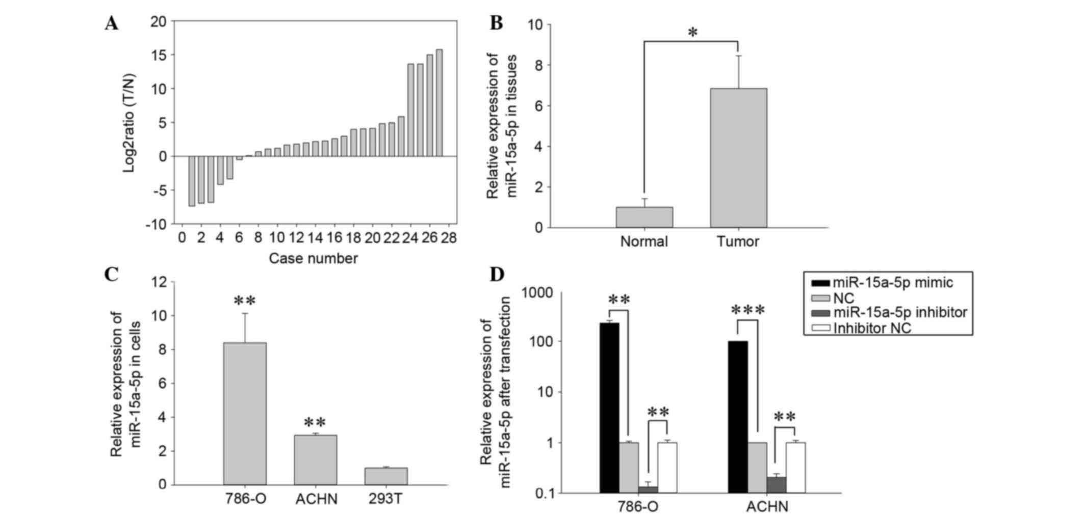
To determine the expression level of miR-15a-5p,
qPCR was performed in 28 paired RCC tissues and adjacent normal
tissues. Relative expression of miR-15a-5p [Log2 (T/N)] was
presented in Fig. 1A. The present
study demonstrated that the expression of miR-15a-5p in RCC tissues
was significantly higher than adjacent normal tissues (P<0.05;
Fig. 1B). The expression levels of
miR-15a-5p in the 786-O and ACHN RCC cell lines and HEK-293T normal
human embryo kidney cell line were also detected and the results
indicated that expression of miR-15a-5p was significantly higher in
786-O and ACHN (P<0.01) compared with 293T cells, which is
consistent with the expression pattern of miR-15a-5p in RCC tissues
(Fig. 1C).
Validation of cell transfection
efficiency
To determine whether the expression level of
miR-15a-5p was changed by transfecting miR-15a-5p mimic or
inhibitor, RT-qPCR was performed. The results indicated that
miR-15a-5p was downregulated by 79.22% in ACHN and 86.54% in 786-O
following transfection with miR-15a-5p inhibitor compared with the
inhibitor NC (P<0.01). In addition, the expression levels of
miR-15a-5p were 228.67 times higher (786-O cells, P<0.05) and
100.08 times higher (ACHN cells, P<0.001) in cells transfected
with miR-15a-5p mimics compared with. negative control (NC) as
presented in Fig. 1D.
miR-15a-5p promoted cell
proliferation
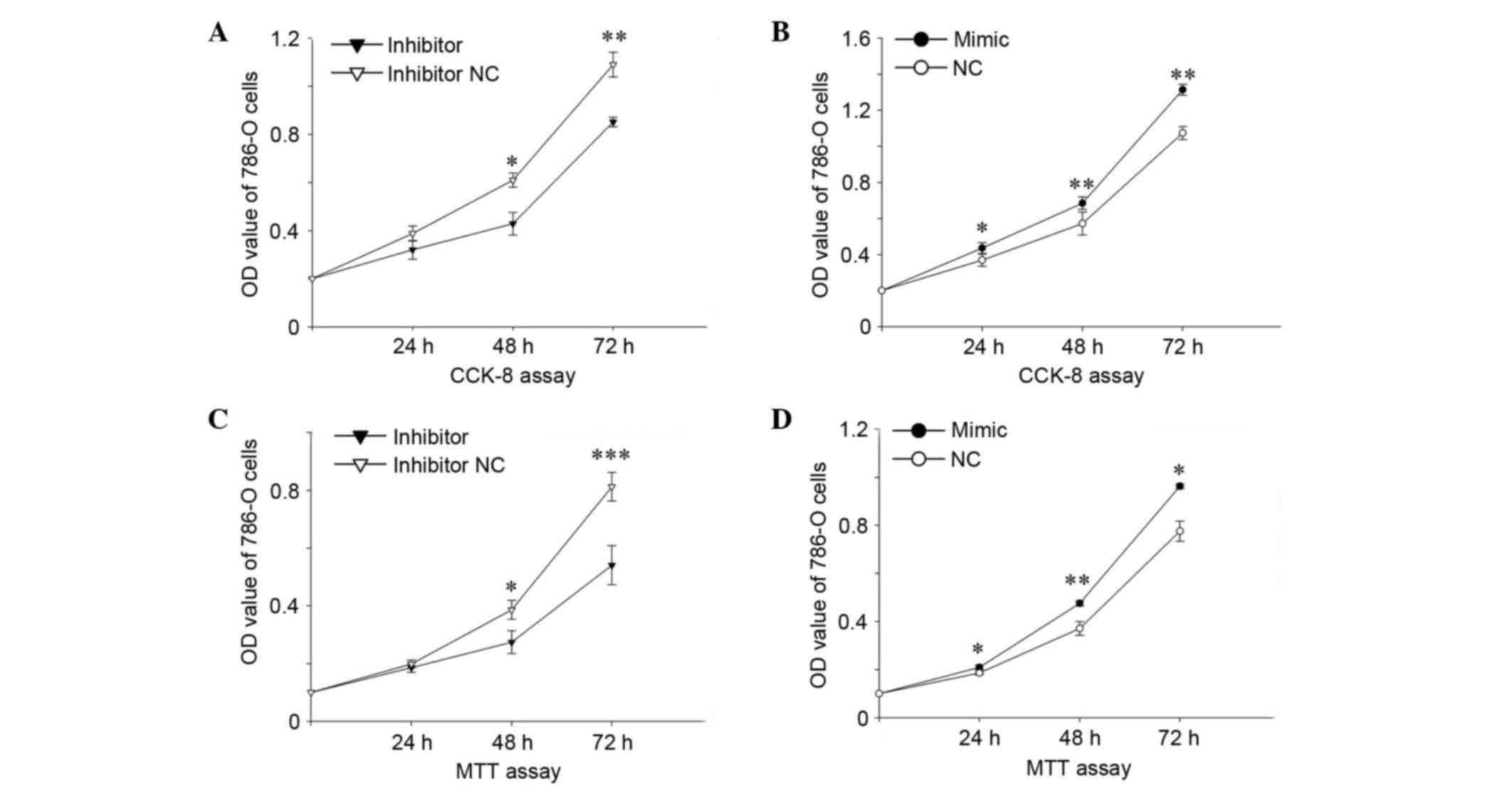
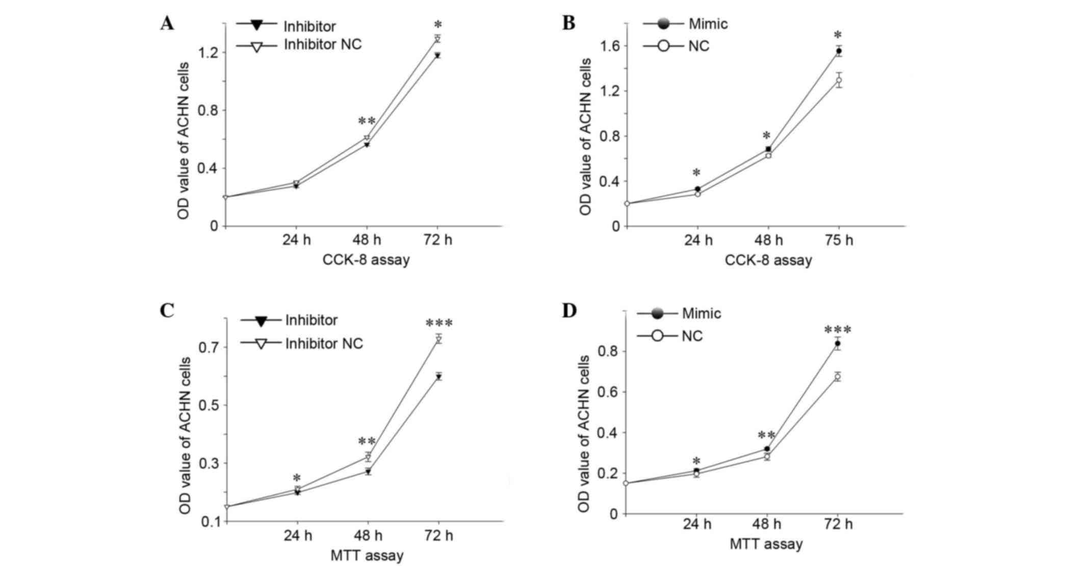
Cell proliferation determined by the MTT assay and
the CCK-8 assay in vitro. The results of the MTT and CCK-8
assays suggested that upregulation of miR-15a-5p promoted cell
proliferation while downregulation of miR-15a-5p inhibited cell
proliferation. The cell proliferation of 786-O (Fig. 2) and ACHN cells (Fig. 3) was reduced by 17.38, 29.73%
(P<0.05), 21.85% (P<0.01;Fig.
2A) and 8.31, 8.13% (P<0.01), 8.87% (P<0.05; Fig. 3A) in CCK-8, 6.30, 29.04%
(P<0.05), 33.44% (P<0.001; Fig.
2C) and 5.47% (P<0.05), 15.61% (P<0.01), 17.80%
(P<0.001; Fig. 3C) as
determined by the MTT assay following transfection with an
miR-15a-5p inhibitor at 24, 48 and 72 h compared with those
transfected with inhibitor NC, respectively. The cell proliferation
of 786-O and ACHN cells was upregulated by 18.19% (P<0.05),
19.78% (P<0.01), 22.32% (P<0.01; Fig. 2B) and 16.58% (P<0.05), 9.69%
(P<0.05), 19.92% (P<0.05; Fig.
3B) in CCK-8, 12.52% (P<0.05), 28.12% (P<0.01), 24.09%
(P<0.05; Fig. 2D) and 8.03%
(P<0.05), 13.51% (P<0.01), 24.17% (P<0.01; Fig. 3D) as demonstrated by the MTT assay
following transfection with miR-15a-5p mimic at 24, 48 and 72 h
compared with those transfected with NC, respectively. The results
demonstrated that miR-15a-5p may promote cell proliferation in
RCC.
miR-15a-5p promoted RCC cell
mobility
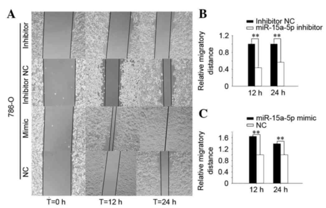
To investigate the effect of miR-15a-5p on cell
mobility in RCC cell lines (786-O and ACHN), a Transwell assay and
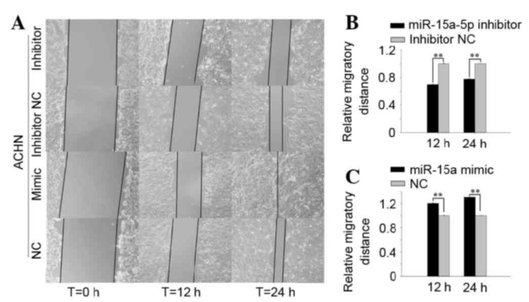
wound scratch assay were performed. As presented in Fig. 4, the results of wound scratch assay
of 786-O indicated that the migratory distance of cells transfected
with miR-15a-5p inhibitor was significantly reduced by 56.27%
(P<0.01) and 43.17% (P<0.01) at 12 and 24 h compared with
cells transfected with inhibitor NC (Fig. 4B). By contrast, upregulation of
miR-15a-5p by transfecting miR-15a-5p mimics promoted migratory
distances by 64.46% (P<0.01) and 38.63% (P<0.05) in 786-O at
12 and 24 h compared cells transfected with NC (Fig. 4C). In ACHN cells (presented in
Fig. 5), the downregulation of
miR-15a-5p reduced the migratory distance by 30.09% (P<0.01) and
22.16% (P<0.01) at 12 and 24 h (Fig. 5B). By contrast, upregulation of
miR-15a-5p promoted migratory distances by 19.94% (P<0.01) and
30.14% (P<0.01) in ACHN cells at 12 and 24 h (Fig. 5C).
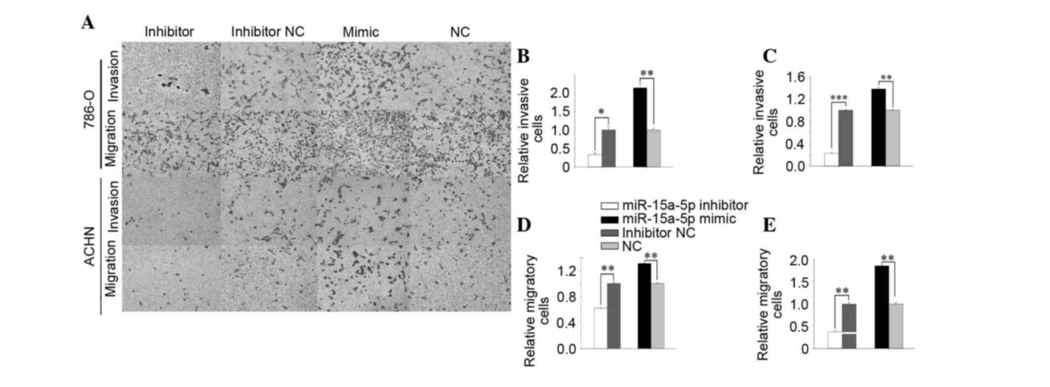
The results of the Transwell assay were presented in
Fig. 6. The results of the
Transwell invasion assay indicated that the invasive ability of
786-O cells was significantly reduced by 66.70% (P<0.05) in the
miR-15a-5p inhibitor group and upregulated by 113.70% (P<0.01)
in the miR-15a-5p mimic group (Fig.
6B), and in ACHN cells invasive ability was reduced by 77.23%
(P<0.001) and upregulated by 38.24% (P<0.01) in miR-15a-5p
mimic group (Fig. 6C).
As presented in Fig.
6D, the migratory ability of 786-O cells transfected with
miR-15a-5p inhibitors was reduced significantly by 37.79%
(P<0.01) and in miR-15a-5p mimic group was increased by 30.75%
(P<0.01). In ACHN cells the migratory ability was reduced by
62.39% (P<0.01) in miR-15a-5p inhibitor group and promoted by
85.99% (P<0.01) in miR-15a-5p mimic group (Fig. 6E). The results indicated that
miR-15a-5p promoted the ability of RCC cell mobility.
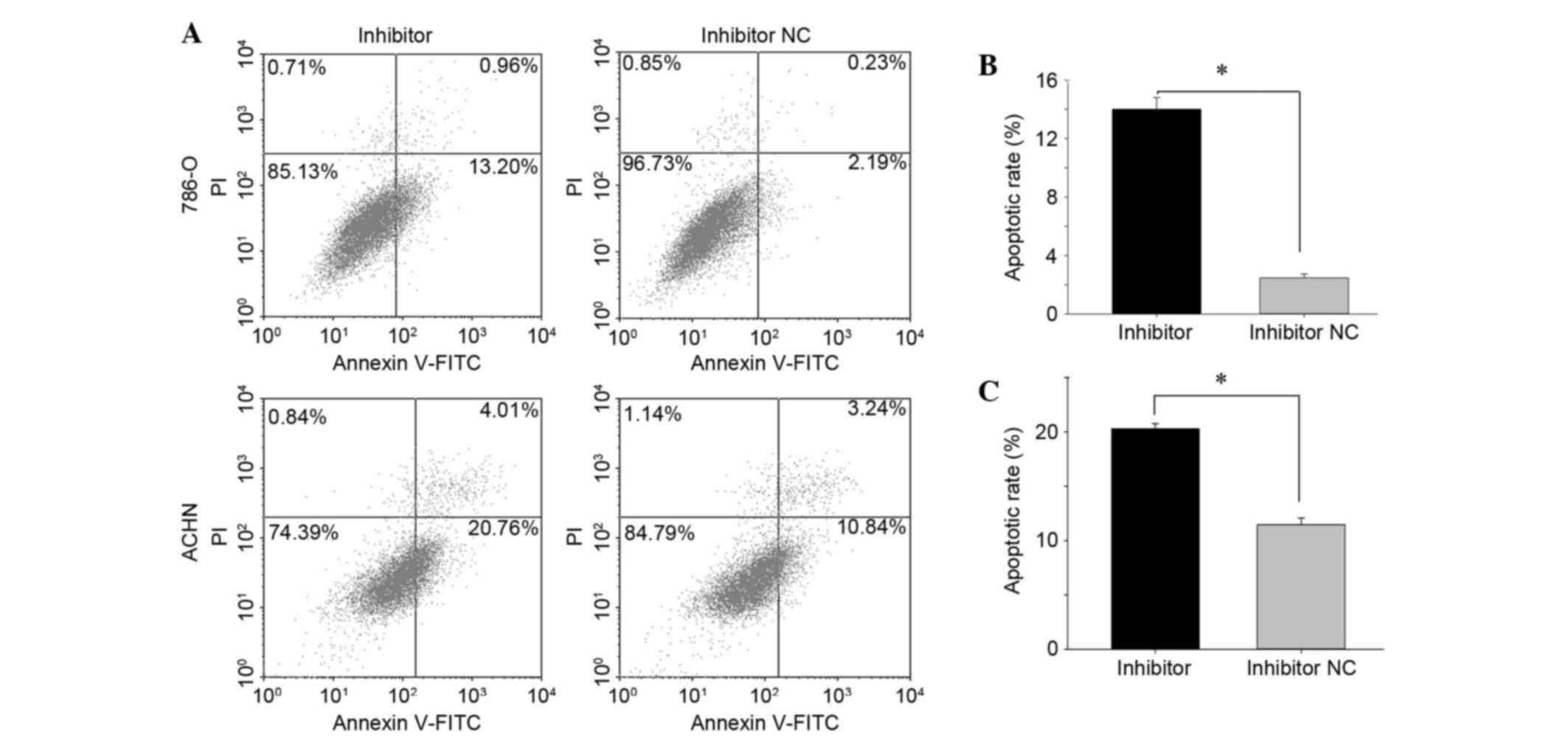
Downregulation of miR-15a-5p induced
cell apoptosis
Apoptotic rate was determined by flow cytometry
(Fig. 7) and Hoechst 33342
staining. The results indicated that downregulation of miR-15a-5p
induced apoptosis of RCC cells. At 48 h after transfection of
miR-15a-5p inhibitor or inhibitor NC, all the cells in a well were
collected for measurement of apoptosis. The results demonstrated
that the early apoptotic rate of 786-O cells transfected with
miR-15a-5p inhibitor or inhibitor NC was 14.01±0.81 vs. 2.47±0.28%
(P<0.05;Fig. 7B) and the
apoptotic rate of ACHN cells was 20.30±0.47 vs. 11.45±0.61%
(P<0.05; Fig. 7C). There was no
difference observed between the mimic and NC group for apoptotic
rates in the two groups (data not shown). The apoptotic ratio in
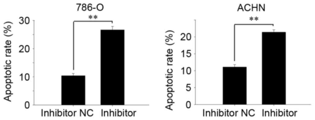
RCC cell lines was also measured by Hoechst 33342 staining. As
presented in Fig. 8, the apoptotic
rate of 786-O cells in the inhibitor group was 27.16±1.75%, with
10.39±0.78% in the inhibitor NC group (P<0.01; Fig. 8A). In the ACHN cells the apoptotic
rate in the inhibitor group or inhibitor NC group was 21.4±0.69 vs.
11.08±0.74% (Fig. 8B; P<0.01).
The results indicated that downregulation of miR-15a-5p induced
cell apoptosis in RCC.
Discussion
Tumorigenesis is a complicated process associated
with activation of various oncogenes or dysfunction of tumor
suppressor genes. miRNAs were described as important in the
pathogenesis of a number of types of cancer (18,19).
Mammalian miRNAs were reported to have the potential to regulate at
least 20–30% of all genes (20),
which suggested that miRNAs are important in oncogenesis and
development of cancer.
Previous studies demonstrated that miR-15a-5p was
downregulated in types of cancer, including prostate cancer
(21), non-small-cell lung cancer
(NSCLC) (22), chronic lymphocytic
leukemia (23) and pancreatic
cancer (14). Previous studies
described the signaling pathways of miR-15a-5p in certain types of
tumor (21–23). However, miR-15a-5p was observed to
be upregulated in RCC tissues compared with paired normal tissues,
which indicated that the underlying mechanism of miR-15a-5p in RCC
is different compared with other tumors. In NSCLC, miR-15a-5p was
demonstrated to induce cell apoptosis and inhibit metastasis by
regulating BCL-2 like protein 2 (2). Xin et al (24) demonstrated that miR-15a-5p promoted
neuroblastoma migration by targeting reversion-inducing
cysteine-rich protein with Kazal motifs and regulating matrix
metalloproteinase-9 expression (24). In a previous study, miR-15a-5p was
observed to suppress cell viability by regulating Wnt family member
3A and fibroblast growth factor 7 in pancreatic cancer (14). In another previous study about
pancreatic ductal adenocarcinoma, miR-15a-5p was observed to
inhibit cell proliferation and epithelial-to-mesenchymal transition
by downregulating polycomb complex protein BMI-1 (6). In addition, miR-15a-5p was also
reported to be downregulated in breast cancer and could affect cell
processes by targeting cyclin E1 (CCNE1) (25). Li et al (26) also described synuclein-γ as a
target of miR-15a-5p in breast cancer (26). In osteosarcoma cells, miR-15a-5p
was reported to regulate cell processes by targeting TNF-α-induced
protein 1 (27). Komabayashi et
al (4) demonstrated that LAMP1
downregulated miR-15a-5p in nasal natural killer/T-cell lymphoma.
In all these types of cancer, restoring the level of miR-15a-5p
induces cell apoptosis or inhibits cell proliferation or
invasion.
The miR-15 family includes miR-15a-5p, miR-15b and
miR-16-1 (previously termed miR-16). As miR-15a-5p and miR-16-1 are
located in the same region of chromosome 13q14 (13), a number of studies have
investigated the role of miR-15a-5p/16 in tumors. Our previous
study determined miR-16 was upregulated in RCC tissues and acts as
an oncogene (28). In addition,
miR-15a-5p/16 was demonstrated to inhibit NSCLC cell progression by
targeting Cripto (29). Lan et
al (30) also demonstrated
that miR-15a-5p/16 enhances radiation sensitivity of NSCLC cells by
targeting the Toll-like receptor 1/nuclear factor-κB signaling
pathway (30). In other previous
studies, CCNE1 (20), Wilms tumor
protein 1 (31) and vascular
endothelial growth factor (32)
are targets of miR-15a-5p and miR-16. miR-15a-5p/16 were also
described as tumor suppressors in the types of cancer
mentioned.
A previous study has demonstrated that the
upregulated miR-15a-5p inversely correlated to protein kinase C α
(PKCα) in RCC (33), however, the
function of miR-15a-5p in RCC remains to be elucidated. In the
present study, it was demonstrated that upregulation of miR-15a-5p
promoted cell proliferation, migration and invasion in 786-O and
ACHN cells. Conversely, downregulation of miR-15a-5p induced cell
apoptosis and inhibited cell proliferation, migration and invasion
in 786-O and ACHN cells. The results indicated that miR-15a-5p acts
as an oncogene in RCC, which is different from the role of
miR-15a-5p in other types of cancer. Similarly, miR-15a-5p-3p was
also described as an oncogene that may contribute to colorectal
adenoma-to-carcinoma progression (34).
In addition to being involved in cancer, miR-15a-5p
was also reported to regulate angiogenesis. Spinetti et al
(35) reported that miR-15a-5p was
increased in the proangiogenic cells (PAC) and serum of patients
with critical limb ischemia, and impaired the function of the
circulating human PACs (35).
miR-15a-5p was described as a direct transcriptional target of
Kruppel-like factor 4, which mediates its anti-proliferative and
anti-angiogenic effects (36).
Previous studies have also reported that miR-15a-5p
could be used as a biomarker for diagnosis and estimation of
prognosis. A high level of miR-15a-5p has been associated with poor
prognosis in multiple myeloma (37). For breast cancer and colorectal
cancer patients low expression levels of miR-15a-5p in the primary
tumor predicted a poor prognosis (9,38).
In addition, in triple-negative patients a low level of miR-15a-5p
expression was significantly associated with shorter disease-free
survival and overall survival (38). There is also a research indicating
that miR-15a-5p is also a potential biomarker to differentiate
between benign and malignant renal tumors in biopsy and urine
samples (33). Thus, suggesting
miR-15a-5p is involved in RCC cellular processes, and PKCα is a
target of miR-15a-5p in RCC. Future research should focus on the
role of miR-15a-5p as a biomarker for use in clinical
situations.
In conclusion, the results of the present study
demonstrated that miR-15a-5p was involved in cellular
proliferation, migration, invasion and apoptosis in renal cancer
cell lines, which indicated that miR-15a-5p is important in RCC.
Furthermore, the results suggested that miR-15a-5p may be used as a
therapeutic target for renal cancer treatment in the future.
Acknowledgements
The present study was supported by the National
Natural Science Foundation of China (grant no. 81101922), the
Science and Technology Development Fund Project of Shenzhen (grant
nos. JCYJ20130402114702124, JCYJ20150403091443304 and
JCYJ20150403091443329) and the Fund of Guangdong Key Medical
Subject.
References
|
1
|
Chen D, Li Y, Yu Z, Li Y, Su Z, Ni L, Yang
S, Gui Y and Lai Y: Downregulated microRNA-510-5p acts as a tumor
suppressor in renal cell carcinoma. Mol Med Rep. 12:3061–3066.
2015.PubMed/NCBI
|
|
2
|
Yang T, Thakur A, Chen T, Yang L, Lei G,
Liang Y, Zhang S, Ren H and Chen M: MicroRNA-15a induces cell
apoptosis and inhibits metastasis by targeting BCL2L2 in non-small
cell lung cancer. Tumour Biol. 36:4357–4365. 2015. View Article : Google Scholar : PubMed/NCBI
|
|
3
|
Su Z, Chen D, Zhang E, Li Y, Yu Z, Shi M,
Jiang Z, Ni L, Yang S, Gui Y, et al: MicroRNA-509-3p inhibits
cancer cell proliferation and migration by targeting the
mitogen-activated protein kinase kinase kinase 8 oncogene in renal
cell carcinoma. Mol Med Rep. 12:1535–1543. 2015.PubMed/NCBI
|
|
4
|
Komabayashi Y, Kishibe K, Nagato T, Ueda
S, Takahara M and Harabuchi Y: Downregulation of miR-15a due to
LMP1 promotes cell proliferation and predicts poor prognosis in
nasal NK/T-cell lymphoma. Am J Hematol. 89:25–33. 2014. View Article : Google Scholar : PubMed/NCBI
|
|
5
|
Wang BS, Liu Z, Xu WX and Sun SL:
Functional polymorphisms in microRNAs and susceptibility to liver
cancer: a meta-analysis and meta-regression, Genetics and molecular
research. GMR. 13:5426–5440. 2014. View Article : Google Scholar : PubMed/NCBI
|
|
6
|
Guo S, Xu X, Tang Y, Zhang C, Li J, Ouyang
Y, Ju J, Bie P and Wang H: miR-15a inhibits cell proliferation and
epithelial to mesenchymal transition in pancreatic ductal
adenocarcinoma by down-regulating Bmi-1 expression. Cancer Lett.
344:40–46. 2014. View Article : Google Scholar : PubMed/NCBI
|
|
7
|
Zitman-Gal T, Green J, Pasmanik-Chor M,
Golan E, Bernheim J and Benchetrit S: Vitamin D manipulates
miR-181c, miR-20b and miR-15a in human umbilical vein endothelial
cells exposed to a diabetic-like environment. Cardiovasc Diabetol.
13:82014. View Article : Google Scholar : PubMed/NCBI
|
|
8
|
Matsushita R, Seki N, Chiyomaru T,
Inoguchi S, Ishihara T, Goto Y, Nishikawa R, Mataki H, Tatarano S,
Itesako T, et al: Tumour-suppressive microRNA-144-5p directly
targets CCNE1/2 as potential prognostic markers in bladder cancer.
British journal of cancer. 2015. View Article : Google Scholar
|
|
9
|
Xiao G, Tang H, Wei W, Li J, Ji L and Ge
J: Aberrant expression of MicroRNA-15a and MicroRNA-16
synergistically associates with tumor progression and prognosis in
patients with colorectal cancer. Gastroenterol Res Pract.
2014:3645492014. View Article : Google Scholar : PubMed/NCBI
|
|
10
|
Rasmussen F: Metastatic renal cell cancer.
Cancer Imaging. 13:374–380. 2013. View Article : Google Scholar : PubMed/NCBI
|
|
11
|
Patel C, Ahmed A and Ellsworth P: Renal
cell carcinoma: A reappraisal. Urol Nurs. 32:182–190; quiz 191.
2012.PubMed/NCBI
|
|
12
|
Alt AL, Boorjian SA, Lohse CM, Costello
BA, Leibovich BC and Blute ML: Survival after complete surgical
resection of multiple metastases from renal cell carcinoma. Cancer.
117:2873–2882. 2011. View Article : Google Scholar : PubMed/NCBI
|
|
13
|
Bottoni A, Piccin D, Tagliati F, Luchin A,
Zatelli MC and Uberti EC degli: miR-15a and miR-16-1
down-regulation in pituitary adenomas. J Cell Physiol. 204:280–285.
2005. View Article : Google Scholar : PubMed/NCBI
|
|
14
|
Zhang XJ, Ye H, Zeng CW, He B, Zhang H and
Chen YQ: Dysregulation of miR-15a and miR-214 in human pancreatic
cancer. J Hematol Oncol. 3:462010. View Article : Google Scholar : PubMed/NCBI
|
|
15
|
Humplikova L, Kollinerova S, Papajik T,
Pikalova Z, Holzerova M, Prochazka V, Divoka M, Modriansky M,
Indrak K and Jarosova M: Expression of miR-15a and miR-16-1 in
patients with chronic lymphocytic leukemia. Biomed Pap Med Fac Univ
Palacky Olomouc Czech Repub. 157:284–293. 2013.PubMed/NCBI
|
|
16
|
Renjie W and Haiqian L: MiR-132, miR-15a
and miR-16 synergistically inhibit pituitary tumor cell
proliferation, invasion and migration by targeting Sox5. Cancer
Lett. 356:568–578. 2015. View Article : Google Scholar : PubMed/NCBI
|
|
17
|
Livak KJ and Schmittgen TD: Analysis of
relative gene expression data using real-time quantitative PCR and
the 2(−Delta Delta C(T)) method. Methods. 25:402–408. 2001.
View Article : Google Scholar : PubMed/NCBI
|
|
18
|
Bhattacharya R, Nicoloso M, Arvizo R, Wang
E, Cortez A, Rossi S, Calin GA and Mukherjee P: MiR-15a and MiR-16
control Bmi-1 expression in ovarian cancer. Cancer Res.
69:9090–9095. 2009. View Article : Google Scholar : PubMed/NCBI
|
|
19
|
Satzger I, Mattern A, Kuettler U,
Weinspach D, Voelker B, Kapp A and Gutzmer R: MicroRNA-15b
represents an independent prognostic parameter and is correlated
with tumor cell proliferation and apoptosis in malignant melanoma.
Int J Cancer. 126:2553–2562. 2010.PubMed/NCBI
|
|
20
|
Ofir M, Hacohen D and Ginsberg D: MiR-15
and miR-16 are direct transcriptional targets of E2F1 that limit
E2F-induced proliferation by targeting cyclin E. Mol Cancer Res.
9:440–447. 2011. View Article : Google Scholar : PubMed/NCBI
|
|
21
|
Musumeci M, Coppola V, Addario A, Patrizii
M, Maugeri-Saccà M, Memeo L, Colarossi C, Francescangeli F, Biffoni
M, Collura D, et al: Control of tumor and microenvironment
cross-talk by miR-15a and miR-16 in prostate cancer. Oncogene.
30:4231–4242. 2011. View Article : Google Scholar : PubMed/NCBI
|
|
22
|
Bandi N, Zbinden S, Gugger M, Arnold M,
Kocher V, Hasan L, Kappeler A, Brunner T and Vassella E: miR-15a
and miR-16 are implicated in cell cycle regulation in a
Rb-dependent manner and are frequently deleted or down-regulated in
non-small cell lung cancer. Cancer Res. 69:5553–5559. 2009.
View Article : Google Scholar : PubMed/NCBI
|
|
23
|
Hanlon K, Rudin CE and Harries LW:
Investigating the targets of MIR-15a and MIR-16-1 in patients with
chronic lymphocytic leukemia (CLL). PLoS One. 4:e71692009.
View Article : Google Scholar : PubMed/NCBI
|
|
24
|
Xin C, Buhe B, Hongting L, Chuanmin Y,
Xiwei H, Hong Z, Lulu H, Qian D and Renjie W: MicroRNA-15a promotes
neuroblastoma migration by targeting reversion-inducing
cysteine-rich protein with Kazal motifs (RECK) and regulating
matrix metalloproteinase-9 expression. FEBS J. 280:855–866.
2013.PubMed/NCBI
|
|
25
|
Luo Q, Li X, Li J, Kong X, Zhang J, Chen
L, Huang Y and Fang L: MiR-15a is underexpressed and inhibits the
cell cycle by targeting CCNE1 in breast cancer. Int J Oncol.
43:1212–1218. 2013.PubMed/NCBI
|
|
26
|
Li P, Xie XB, Chen Q, Pang GL, Luo W, Tu
JC, Zheng F, Liu SM, Han L, Zhang JK, et al: MiRNA-15a mediates
cell cycle arrest and potentiates apoptosis in breast cancer cells
by targeting synuclein-γ. Asian Pac J Cancer Prev. 15:6949–6954.
2014. View Article : Google Scholar : PubMed/NCBI
|
|
27
|
Tian X, Zhang J, Yan L, Dong JM and Guo Q:
MiRNA-15a inhibits proliferation, migration and invasion by
targeting TNFAIP1 in human osteosarcoma cells. Int J Clin Exp
Pathol. 8:6442–6449. 2015.PubMed/NCBI
|
|
28
|
Chen D, Li Y, Yu Z, Su Z, Yu W, Li Y, Yang
S, Gui Y, Ni L and Lai Y: Upregulated microRNA-16 as an oncogene in
renal cell carcinoma. Mol Med Rep. 12:1399–1404. 2015.PubMed/NCBI
|
|
29
|
Chen F, Hou SK, Fan HJ and Liu YF:
MiR-15a-16 represses Cripto and inhibits NSCLC cell progression.
Mol Cell Biochem. 391:11–19. 2014. View Article : Google Scholar : PubMed/NCBI
|
|
30
|
Lan F, Yue X, Ren G, Li H, Ping L, Wang Y
and Xia T: miR-15a/16 enhances radiation sensitivity of non-small
cell lung cancer cells by targeting the TLR1/NF-κB signaling
pathway. Int J Radiat Oncol Biol Phys. 91:73–81. 2015. View Article : Google Scholar : PubMed/NCBI
|
|
31
|
Gao SM, Xing CY, Chen CQ, Lin SS, Dong PH
and Yu FJ: miR-15a and miR-16-1 inhibit the proliferation of
leukemic cells by down-regulating WT1 protein level. J Exp Clin
Cancer Res. 30:1102011. View Article : Google Scholar : PubMed/NCBI
|
|
32
|
Sun CY, She XM, Qin Y, Chu ZB, Chen L, Ai
LS, Zhang L and Hu Y: miR-15a and miR-16 affect the angiogenesis of
multiple myeloma by targeting VEGF. Carcinogenesis. 34:426–435.
2013. View Article : Google Scholar : PubMed/NCBI
|
|
33
|
von Brandenstein M, Pandarakalam JJ, Kroon
L, Loeser H, Herden J, Braun G, Wendland K, Dienes HP, Engelmann U
and Fries JW: MicroRNA 15a, inversely correlated to PKCα, is a
potential marker to differentiate between benign and malignant
renal tumors in biopsy and urine samples. Am J Pathol.
180:1787–1797. 2012. View Article : Google Scholar : PubMed/NCBI
|
|
34
|
de Groen FL, Timmer LM, Menezes RX,
Diosdado B, Hooijberg E, Meijer GA, Steenbergen RD and Carvalho B:
Oncogenic role of miR-15a-3p in 13q amplicon-driven colorectal
adenoma-to-carcinoma progression. PLoS One. 10:e01324952015.
View Article : Google Scholar : PubMed/NCBI
|
|
35
|
Spinetti G, Fortunato O, Caporali A,
Shantikumar S, Marchetti M, Meloni M, Descamps B, Floris I,
Sangalli E, Vono R, et al: MicroRNA-15a and microRNA-16 impair
human circulating proangiogenic cell functions and are increased in
the proangiogenic cells and serum of patients with critical limb
ischemia. Circ Res. 112:335–346. 2013. View Article : Google Scholar : PubMed/NCBI
|
|
36
|
Zheng X, Li A, Zhao L, Zhou T, Shen Q, Cui
Q and Qin X: Key role of microRNA-15a in the KLF4 suppressions of
proliferation and angiogenesis in endothelial and vascular smooth
muscle cells. Biochem Biophys Res Commun. 437:625–631. 2013.
View Article : Google Scholar : PubMed/NCBI
|
|
37
|
Gao X, Zhang R, Qu X, Zhao M, Zhang S, Wu
H, Jianyong L and Chen L: MiR-15a, miR-16-1 and miR-17-92 cluster
expression are linked to poor prognosis in multiple myeloma. Leuk
Res. 36:1505–1509. 2012. View Article : Google Scholar : PubMed/NCBI
|
|
38
|
Shinden Y, Akiyoshi S, Ueo H, Nambara S,
Saito T, Komatsu H, Ueda M, Hirata H, Sakimura S, Uchi R, et al:
Diminished expression of MiR-15a is an independent prognostic
marker for breast cancer cases. Anticancer Res. 35:123–127.
2015.PubMed/NCBI
|