Spandidos Publications Logo
  • About
    • About Spandidos
    • Aims and Scopes
    • Abstracting and Indexing
    • Editorial Policies
    • Reprints and Permissions
    • Job Opportunities
    • Terms and Conditions
    • Contact
  • Journals
    • All Journals
    • Oncology Letters
      • Oncology Letters
      • Information for Authors
      • Editorial Policies
      • Editorial Board
      • Aims and Scope
      • Abstracting and Indexing
      • Bibliographic Information
      • Archive
    • International Journal of Oncology
      • International Journal of Oncology
      • Information for Authors
      • Editorial Policies
      • Editorial Board
      • Aims and Scope
      • Abstracting and Indexing
      • Bibliographic Information
      • Archive
    • Molecular and Clinical Oncology
      • Molecular and Clinical Oncology
      • Information for Authors
      • Editorial Policies
      • Editorial Board
      • Aims and Scope
      • Abstracting and Indexing
      • Bibliographic Information
      • Archive
    • Experimental and Therapeutic Medicine
      • Experimental and Therapeutic Medicine
      • Information for Authors
      • Editorial Policies
      • Editorial Board
      • Aims and Scope
      • Abstracting and Indexing
      • Bibliographic Information
      • Archive
    • International Journal of Molecular Medicine
      • International Journal of Molecular Medicine
      • Information for Authors
      • Editorial Policies
      • Editorial Board
      • Aims and Scope
      • Abstracting and Indexing
      • Bibliographic Information
      • Archive
    • Biomedical Reports
      • Biomedical Reports
      • Information for Authors
      • Editorial Policies
      • Editorial Board
      • Aims and Scope
      • Abstracting and Indexing
      • Bibliographic Information
      • Archive
    • Oncology Reports
      • Oncology Reports
      • Information for Authors
      • Editorial Policies
      • Editorial Board
      • Aims and Scope
      • Abstracting and Indexing
      • Bibliographic Information
      • Archive
    • Molecular Medicine Reports
      • Molecular Medicine Reports
      • Information for Authors
      • Editorial Policies
      • Editorial Board
      • Aims and Scope
      • Abstracting and Indexing
      • Bibliographic Information
      • Archive
    • World Academy of Sciences Journal
      • World Academy of Sciences Journal
      • Information for Authors
      • Editorial Policies
      • Editorial Board
      • Aims and Scope
      • Abstracting and Indexing
      • Bibliographic Information
      • Archive
    • International Journal of Functional Nutrition
      • International Journal of Functional Nutrition
      • Information for Authors
      • Editorial Policies
      • Editorial Board
      • Aims and Scope
      • Abstracting and Indexing
      • Bibliographic Information
      • Archive
    • International Journal of Epigenetics
      • International Journal of Epigenetics
      • Information for Authors
      • Editorial Policies
      • Editorial Board
      • Aims and Scope
      • Abstracting and Indexing
      • Bibliographic Information
      • Archive
    • Medicine International
      • Medicine International
      • Information for Authors
      • Editorial Policies
      • Editorial Board
      • Aims and Scope
      • Abstracting and Indexing
      • Bibliographic Information
      • Archive
  • Articles
  • Information
    • Information for Authors
    • Information for Reviewers
    • Information for Librarians
    • Information for Advertisers
    • Conferences
  • Language Editing
Spandidos Publications Logo
  • About
    • About Spandidos
    • Aims and Scopes
    • Abstracting and Indexing
    • Editorial Policies
    • Reprints and Permissions
    • Job Opportunities
    • Terms and Conditions
    • Contact
  • Journals
    • All Journals
    • Biomedical Reports
      • Information for Authors
      • Editorial Policies
      • Editorial Board
      • Aims and Scope
      • Abstracting and Indexing
      • Bibliographic Information
      • Archive
    • Experimental and Therapeutic Medicine
      • Information for Authors
      • Editorial Policies
      • Editorial Board
      • Aims and Scope
      • Abstracting and Indexing
      • Bibliographic Information
      • Archive
    • International Journal of Epigenetics
      • Information for Authors
      • Editorial Policies
      • Editorial Board
      • Aims and Scope
      • Abstracting and Indexing
      • Bibliographic Information
      • Archive
    • International Journal of Functional Nutrition
      • Information for Authors
      • Editorial Policies
      • Editorial Board
      • Aims and Scope
      • Abstracting and Indexing
      • Bibliographic Information
      • Archive
    • International Journal of Molecular Medicine
      • Information for Authors
      • Editorial Policies
      • Editorial Board
      • Aims and Scope
      • Abstracting and Indexing
      • Bibliographic Information
      • Archive
    • International Journal of Oncology
      • Information for Authors
      • Editorial Policies
      • Editorial Board
      • Aims and Scope
      • Abstracting and Indexing
      • Bibliographic Information
      • Archive
    • Medicine International
      • Information for Authors
      • Editorial Policies
      • Editorial Board
      • Aims and Scope
      • Abstracting and Indexing
      • Bibliographic Information
      • Archive
    • Molecular and Clinical Oncology
      • Information for Authors
      • Editorial Policies
      • Editorial Board
      • Aims and Scope
      • Abstracting and Indexing
      • Bibliographic Information
      • Archive
    • Molecular Medicine Reports
      • Information for Authors
      • Editorial Policies
      • Editorial Board
      • Aims and Scope
      • Abstracting and Indexing
      • Bibliographic Information
      • Archive
    • Oncology Letters
      • Information for Authors
      • Editorial Policies
      • Editorial Board
      • Aims and Scope
      • Abstracting and Indexing
      • Bibliographic Information
      • Archive
    • Oncology Reports
      • Information for Authors
      • Editorial Policies
      • Editorial Board
      • Aims and Scope
      • Abstracting and Indexing
      • Bibliographic Information
      • Archive
    • World Academy of Sciences Journal
      • Information for Authors
      • Editorial Policies
      • Editorial Board
      • Aims and Scope
      • Abstracting and Indexing
      • Bibliographic Information
      • Archive
  • Articles
  • Information
    • For Authors
    • For Reviewers
    • For Librarians
    • For Advertisers
    • Conferences
  • Language Editing
Login Register Submit
  • This site uses cookies
  • You can change your cookie settings at any time by following the instructions in our Cookie Policy. To find out more, you may read our Privacy Policy.

    I agree
Search articles by DOI, keyword, author or affiliation
Search
Advanced Search
presentation
Molecular Medicine Reports
Join Editorial Board Propose a Special Issue
Print ISSN: 1791-2997 Online ISSN: 1791-3004
Journal Cover
August-2017 Volume 16 Issue 2

Full Size Image

Sign up for eToc alerts
Recommend to Library

Journals

International Journal of Molecular Medicine

International Journal of Molecular Medicine

International Journal of Molecular Medicine is an international journal devoted to molecular mechanisms of human disease.

International Journal of Oncology

International Journal of Oncology

International Journal of Oncology is an international journal devoted to oncology research and cancer treatment.

Molecular Medicine Reports

Molecular Medicine Reports

Covers molecular medicine topics such as pharmacology, pathology, genetics, neuroscience, infectious diseases, molecular cardiology, and molecular surgery.

Oncology Reports

Oncology Reports

Oncology Reports is an international journal devoted to fundamental and applied research in Oncology.

Experimental and Therapeutic Medicine

Experimental and Therapeutic Medicine

Experimental and Therapeutic Medicine is an international journal devoted to laboratory and clinical medicine.

Oncology Letters

Oncology Letters

Oncology Letters is an international journal devoted to Experimental and Clinical Oncology.

Biomedical Reports

Biomedical Reports

Explores a wide range of biological and medical fields, including pharmacology, genetics, microbiology, neuroscience, and molecular cardiology.

Molecular and Clinical Oncology

Molecular and Clinical Oncology

International journal addressing all aspects of oncology research, from tumorigenesis and oncogenes to chemotherapy and metastasis.

World Academy of Sciences Journal

World Academy of Sciences Journal

Multidisciplinary open-access journal spanning biochemistry, genetics, neuroscience, environmental health, and synthetic biology.

International Journal of Functional Nutrition

International Journal of Functional Nutrition

Open-access journal combining biochemistry, pharmacology, immunology, and genetics to advance health through functional nutrition.

International Journal of Epigenetics

International Journal of Epigenetics

Publishes open-access research on using epigenetics to advance understanding and treatment of human disease.

Medicine International

Medicine International

An International Open Access Journal Devoted to General Medicine.

Journal Cover
August-2017 Volume 16 Issue 2

Full Size Image

Sign up for eToc alerts
Recommend to Library

  • Article
  • Citations
    • Cite This Article
    • Download Citation
    • Create Citation Alert
    • Remove Citation Alert
    • Cited By
  • Similar Articles
    • Related Articles (in Spandidos Publications)
    • Similar Articles (Google Scholar)
    • Similar Articles (PubMed)
  • Download PDF
  • Download XML
  • View XML
Article

Gossypol induces cell death by activating apoptosis and autophagy in HT-29 cells

  • Authors:
    • Ming‑Dong Lu
    • Li‑Yi Li
    • Pi‑Hong Li
    • Tao You
    • Fei‑Hai Wang
    • Wei‑Jian Sun
    • Zhi‑Qiang Zheng
  • View Affiliations / Copyright

    Affiliations: Department of Gastrointestinal Surgery, The Second Affiliated Hospital of Wenzhou Medical University, Wenzhou, Zhejiang 325000, P.R. China
  • Pages: 2128-2132
    |
    Published online on: June 19, 2017
       https://doi.org/10.3892/mmr.2017.6804
  • Expand metrics +
Metrics: Total Views: 0 (Spandidos Publications: | PMC Statistics: )
Metrics: Total PDF Downloads: 0 (Spandidos Publications: | PMC Statistics: )
Cited By (CrossRef): 0 citations Loading Articles...

This article is mentioned in:


Abstract

Gossypol is a polyphenolic, yellowish compound derived from cottonseed extract. The present study examined the effects of gossypol on the apoptosis and autophagy of HT‑29 cells. A Cell Counting Kit‑8 assay, Annexin V‑FITC, JC‑1 staining and western blotting were used to identify the viability of cells, stages of apoptosis and the expression levels of the signaling proteins. Gossypol promoted apoptosis and induced the loss of mitochondrial membrane potential. Further investigation of the apoptotic mechanism revealed that gossypol increased the ratio of B‑cell lymphoma 2 (Bcl-2)-associated X protein/Bcl‑2 protein levels and upregulated the expression of caspase‑3. Gossypol also enhanced the activity of microtubule‑associated protein light chain 3 LC3‑II and Beclin‑1 and downregulated LC3‑I, in a dose‑dependent manner. Together, these finding suggested that gossypol may be a novel and potential antitumor agent.

Introduction

Colorectal cancer, including cancerous growths in the colon, appendix and rectum, is the third most commonly diagnosed cancer in men and the second most common in women, with an estimated 1,400,000 cases and a mortality rate of 693,900 worldwide in 2012 (1). Current treatment for colon cancer includes surgery, radiation, physical rehabilitation, nutritional therapy and chemotherapy using cytotoxic drugs (2). However, these treatments are not curative. Therefore, effective anticancer drugs for colon cancer are required.

Gossypol is a polyphenolic, yellowish compound derived from cottonseed extract. It is extensively used as a contraceptive agent in men (3,4). Gossypol has been identified as a promising anticancer agent. Moon et al (3) demonstrated that gossypol effectively inhibited tumor necrosis factor-α-induced expression of intercellular adhesion molecule 1 by activating the suppression of nuclear factor-κB, and in vitro invasion and adhesion in human breast cancer cells. Another study demonstrated that gossypol reduced the viability of three prostate cancer cell lines with a half-maximal inhibitory concentration (IC50) of between 3 and 5 µmol/l. In addition, gossypol effectively inhibited prostate tumor-initiating cell-driven tumor growth in a NOD/SCID xenograft model, in addition to inducing DNA damage and activating p53 (5). Knowledge of the molecular mechanisms underlying the anticancer effects of gossypol against HT-29 human colonic cancer cells is limited. Autophagy and apoptosis are two evolutionarily conserved programmed cell death mechanisms, which occur in several physiological conditions (6). Autophagy and apoptosis are dysregulated in cancer cells (6); therefore, the present study examined the effects of gossypol on the apoptosis and autophagy of HT-29 cells.

Materials and methods

Compounds and reagents

Gossypol (>98% pure) was purchased from LKT Laboratories, Inc. (Hanzhou, China) and reconstituted in DMSO (Sigma-Aldrich; Merck Millipore, Darmstadt, Germany). RPMI-1640 medium and fetal bovine serum (FBS) were obtained from Gibco; Thermo Fisher Scientific, Inc. (Waltham, MA, USA). The Cell counting Kit-8 (CCK-8), 5,5′,6,6′-tetra-chloro-1,1′,3,3′-tetra-ethylbenzimidalyl-carbocyanineiodide (JC-1) and Annexin V/PI apoptosis detection kits were purchased from Beyotime Institute of Biotechnology (Nanjing, China). Antibodies against Caspase-3, B-cell lymphoma 2 (Bcl-2), Bcl-2-associated X protein (Bax), microtubule-associated protein light chain 3 (LC3), Beclin-1, Cytochrome c (Cyt-c) and β-actin were commercially available from MBL International Co. (Boston, MA, USA).

Cell culture

The HT-29 human colon cancer cells were purchased from the American Type Culture Collection (Manassas, VA, USA). The cells were cultured in RPMI-1640 medium (Gibco; Thermo Fisher Scientific, Inc.) supplemented with 10% FBS (Gibco; Thermo Fisher Scientific, Inc.), 100 U/ml penicillin and 100 mg/ml streptomycin (Gibco; Thermo Fisher Scientific, Inc.) in a humidified atmosphere in a 5% CO2 incubator at 37°C.

CCK-8 assay

The growth-inhibitory effect of gossypol was determined using a CCK-8 assay. Briefly, the HT-29 human colon cancer cells were seeded at a density of 1×10⁴ cells/100 µl/well in 96-well plates and were cultured overnight for attachment. Following 24 h of incubation, the HT-29 cells were incubated with gossypol (5, 10, 20, 40 or 80 µM/l) in 96-well plates for 24 h at 37°C. Following incubation, 10 µl CCK-8 solution was added to each well and incubated at 37°C for 4 h, and the absorbance was measured using a microplate reader (Bio-Rad Laboratories, Inc., Hercules, CA, USA) at 450 nm.

Analysis of cell apoptosis

Apoptosis kits were used to detect the effect of gossypol on the HT-29 cells. The cells (5×10⁴ cells/well) were treated with gossypol (20 and 40 µM/l) for 24 h at 37°C. The cells were harvested and washed with ice-cold PBS, resuspended with binding buffer, and simultaneously stained with Annexin-V-FITC and PI for 5–15 min. The cells were then analyzed immediately using flow cytometry.

Mitochondrial membrane potential (ΔΨm) assay

JC-1 staining was used to detect changes in ΔΨm. The cells (2×10⁵ cells/well) were seeded in 6-well plates and treated with or without gossypol (20 and 40 µM/l) for 24 h. Following incubation; the collected cells were incubated with 5 µg/ml JC-1 in culture medium for 15 min at 37°C. The staining solution was removed and the cells were washed twice with JC-1 staining buffer. The cell-associated fluorescence was analyzed using a FACSCalibur flow cytometer.

Western blot analysis of Caspase-3, Bax, Bcl-2, LC3, Beclin-1, Cyt-c and β-actin

The HT-29 cells were seeded at a density of 1×10⁵ cells/well in 6-well plates and, following incubation for 24 h, the cells were treated with gossypol (20 and 40 µM/l) for 24 h and then harvested. The expression levels of Bcl-2, Caspase-3, Bax, LC3 and Beclin-1, and the release of mitochondrial Cyt-c were then assessed using western blot analysis. Proteins were extracted from HT-29 cells using a total protein extraction kit (Beyotime Institute of Biotechnology, Shanghai, China) according to the instructions of the manufacturer. Protein concentrations were determined using a BCA protein assay kit (Beyotime Institute of Biotechnology). Aliquots of cell lysates containing 20 µl of protein were separated on a 12% SDS-PAGE gel, and transferred onto PVDF membranes. The membranes were blocked with TBST buffer containing 10 mM Tris-HCl (pH 7.5), 150 mM NaCl and 0.05% Tween-20, containing 5% skimmed milk, and were then incubated with polyclonal antibodies against Bcl-2 (cat. no. 2876; 1:500), Bax (cat. no. sc-493; 1:500), caspase-3 (sc-16647; 1:1,000), Cyt-c (cat. no. 250621; 1:1,000), LC3 (cat. no. 4108; 1:1,000), beclin-1 (cat. no. 4122; 1:500) and β-actin (cat. no. 4967; 1:1,000) overnight at 4°C, respectively. The membrane was then incubated with either HRP-conjugated goat anti-rabbit IgG (cat. no. sc-2005; 1:10,000; Santa Cruz Biotechnology, Inc., Dallas, TX, USA) or goat anti-mouse IgG (cat. no. sc-2004; 1:10,000; Santa Cruz Biotechnology, Inc.) secondary antibodies for 1.5 h at room temperature. Peroxidase activity was detected via ECL visualization of the bands. The images were analyzed and quantified using a Quantity One software version 4.6.2 (Bio-Rad Laboratories, Inc.).

Statistical analysis

Statistical analyses were performed using SPSS version 10.0 (SPSS Inc., Chicago, IL, USA). Data are expressed as the mean ± standard deviation. Statistical comparisons were performed using Student's t-test. P<0.05 was considered significant a statistically significant difference between groups.

Results

Inhibitory effects of gossypol on HT-29 cells

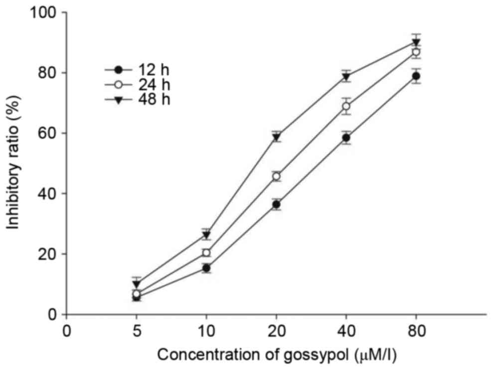
A CCK-8 assay was performed to assess the inhibitory effects of gossypol on the HT-29 cells. The cells were cultured with 5–80 µM/l gossypol for 12, 24 and 48 h. As shown in Fig. 1, gossypol inhibited the growth of HT-29 cells in a time- and dose-dependent manner, with a 12 h IC50 of 31.20 µM/l, 24 h IC50 of 23.60 µM/l and 48 h IC50 of 17.97 µM/l. The two concentrations of 20 and 40 µM/l were selected for the following experiments.

Figure 1.

HT-29 cells were treated with various concentrations of gossypol for 12, 24 and 48 h, and the inhibitory effects were measured using a Cell counting kit-8 assay. Values are presented as the mean ± standard deviation of three independent experiments.

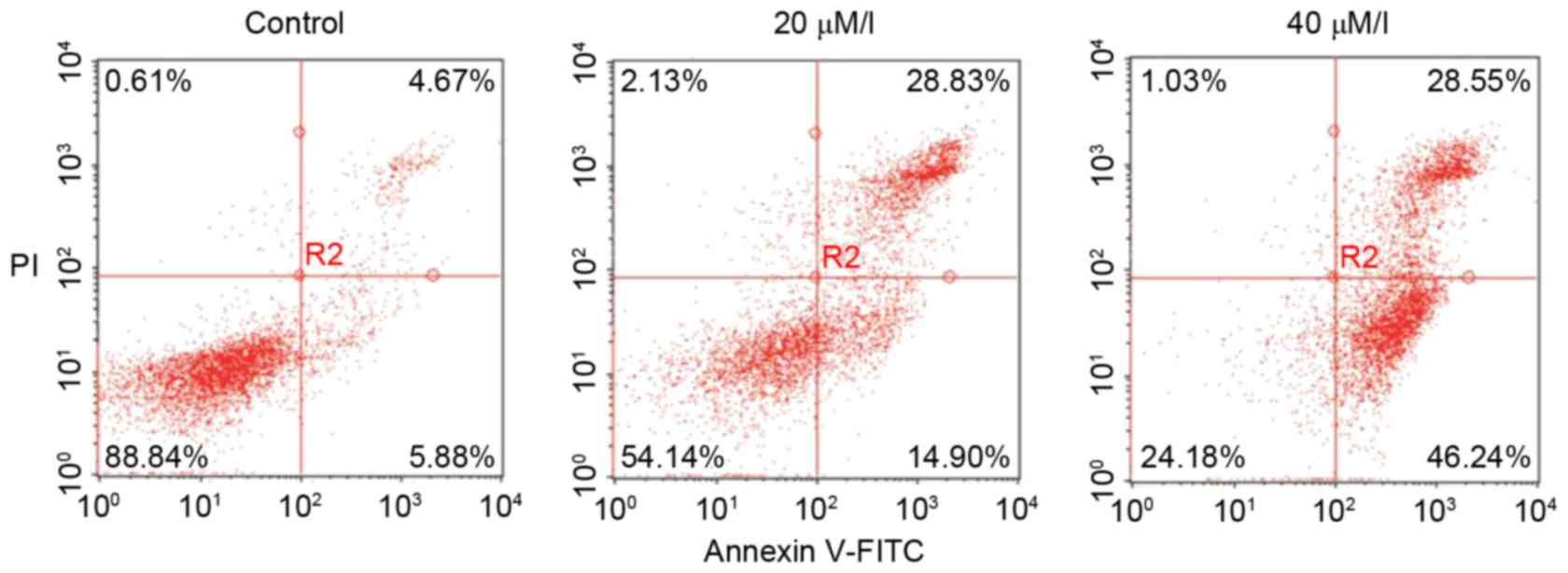
Effect of gossypol on apoptosis

To determine the induction of cell apoptosis by gossypol, an Annexin V and PI staining assay based on flow cytometry was used. As shown in Fig. 2, there were significant changes in the proportions of early and late apoptotic, or necrotic HT-29 cells following exposure to gossypol for 24 h. Compared with the control group, the proportion of early apoptotic cells increased from 5.88 to 46.24% and the proportions of late apoptotic cells increased from 4.67 to 28.55%.

Figure 2.

Apoptotic effects of gossypol on HT-29 cells. Apoptosis of HT-29 cells following treatment with gossypol (20 and 40 µM/l) for 24 h. Representative results from the fluorescence activated cell sorting assays of three independent experiments are presented.

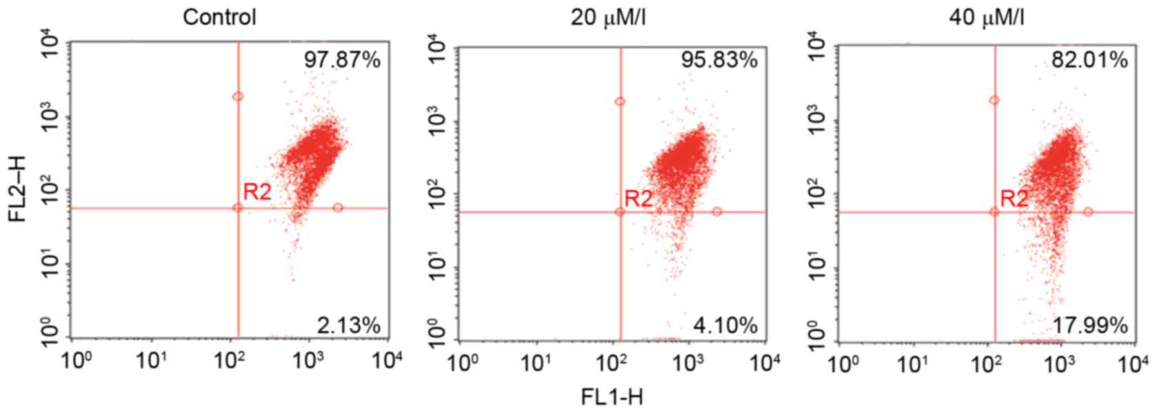
Effects of gossypol on ΔΨm

A loss of ΔΨm was detected by the fluorescent dye, JC-1. As shown in Fig. 3, treatment with gossypol (20 and 40 µM/l) for 24 h induced a loss in the ΔΨm of the HT-29 cells, suggesting damage to the mitochondria.

Figure 3.

Effect of gossypol on the ΔΨm in HT-29 cells when treated for 24 h. The ΔΨm was detected using the fluorescent dye, JC-1, and analyzed using flow cytometry. The results are representatives of three independent experiments. ΔΨm, mitochondrial membrane potential.

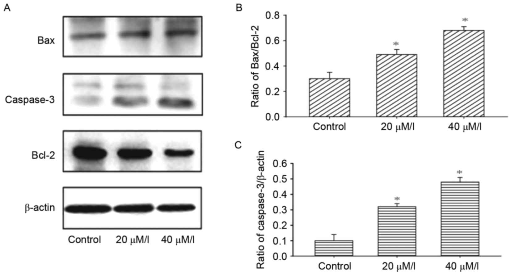
Expression of proteins of the apoptotic pathway

To investigate the mechanism underlying gossypol-induced apoptosis, the effects of gossypol on the modification of apoptosis-associated proteins were examined. The results of the western blot analysis demonstrated that gossypol led to an increase in the protein levels of Bax, and decrease in the levels of Bcl-2 in the HT-29 cells following treatment with gossypol at concentrations of 20 and 40 µM/l (Fig. 4A and B). Gossypol also promoted the activation of caspase-3 (Fig. 4C).

Figure 4.

Effects of gossypol on proteins of the apoptotic pathway. (A) Effects on the protein expression levels of Bax, Bcl-2 and Caspase-3 were detected using western blot analysis following treatment with gossypol for 24 h. (B) Protein expression levels of Bax/Bcl-2 were detected using western blot analysis following treatment with gossypol for 24 h. (C) Protein expression levels of caspase-3 was detected using western blot analysis following treatment with gossypol for 24 h. Results are expressed as the mean ± standard deviation. Each experiment was performed in triplicate (n=3). Comparisons were made between the different concentrations of gossypol and the untreated control. *P<0.05, compared with the control. Bcl-2, B-cell lymphoma 2; Bax, Bcl-2-associated X protein.

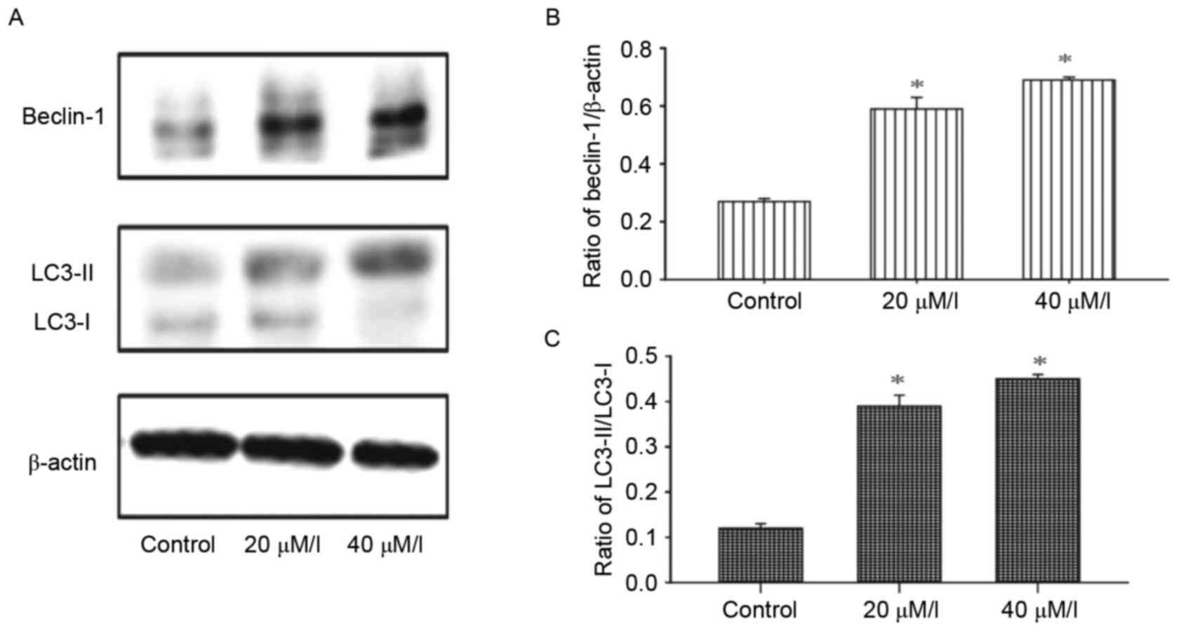
Autophagy is induced by gossypol in HT-29 cells

The activity of autophagy in HT-29 cells was evaluated following treatment with gossypol. Alterations in the levels of autophagy marker proteins, LC3-I and LC3-II, in HT-29 cells were detected using western blot analysis. As shown in Fig. 5, the ratio of LC3-II/LC3-I and Beclin-1 were enhanced in a dose-dependent manner following exposure to gossypol (20 and 40 µM/l) for 24 h.

Figure 5.

Effects of gosspol on the expression of markers of autophagy. (A) Effects on the protein expression levels of Beclin-1 and LC3 were detected using western blot analysis following treatment with gossypol for 24 h. (B) Protein expression level of Beclin-1 was detected using western blot analysis following treatment with gossypol for 24 h. (C) Protein expression levels of LC3-II/LC3-I were detected using western blot analysis following treatment with gossypol for 24 h. Results are expressed as the mean ± standard deviation. Each experiment was performed in triplicate (n=3). Comparisons were made between different concentrations of gossypol and the untreated group. *P<0.05, compared with the control. LC3, microtubule-associated protein light chain 3.

Discussion

The aims of the present study were to evaluate the antitumor activities of gossypol and the possible underlying molecular mechanism. Gossypol inhibited the proliferation of HT-29 cells in a concentration-dependent manner. The loss of ΔΨm is a hallmark of the early-stage of apoptosis (7). Mitochondrial involvement in gossypol-induced apoptosis was assessed using JC-1 staining, and Annexin V and PI double-staining was used to differentiate early and late apoptosis. The proportions of early and late apoptotic cells were higher, compared with those in the control group.

The signal of apoptosis in several human cancer cells in response to antitumor agents is upregulation of the mitochondrial apoptotic pathway triggered by an alteration in the ratio of Bax/Bcl-2 and the activation of caspases (8,9). In the present study, proteins associated with mitochondria-dependent apoptosis were measured in the HT-29 cells. The expression of Bcl-2 was significantly decreased following gossypol exposure (20 and 40 µM/l) for 24 h, whereas the expression of Bax was markedly increased. Therefore, the ratio of Bax/Bcl-2 was increased. A high Bax/Bcl-2 ratio leads to the release of Cyt-c and activates caspase 3, which is the final step in apoptosis (10,11). In the present study, caspase-3 was significantly increased following gossypol exposure (20 and 40 µM/l) for 24 h. These results suggested that gossypol induced apoptosis via the mitochondrial apoptotic pathway.

In addition to apoptosis, the present study investigated the autophagic effects of gossypol on the proliferation of HT-29 cells. A number of studies have reported that autophagy eliminates cancer cells, thus autophagy is important in the fight against cancer. It has been found that autophagy suppresses the development of carcinogenesis (12,13). LC3 and Beclin 1 are the most well-known markers of autophagy. During autophagy, LC3 is transformed from LC3-I to LC3-II for movement onto isolated membranes and autophagosomes (14,15). In the present study, the ratio of LC3-II/LC3-I was increased and the expression of Beclin 1 was upregulated following treatment with gossypol. Therefore, apoptosis and autophagy were induced in the HT-29 cells following gossypol treatment. Apoptosis is involved in eliminating damaged cells and tumor cells.

Although the molecular processes of apoptosis and autophagy differ, the end result of their actions is to remove unnecessary cells (16). The combinatorial use of anticancer agents, which induce autophagy and apoptosis, can be an effective therapeutic strategy and be used in treatment against cancer. The data presented in the present study demonstrated that gossypol induced apoptosis and autophagy, and can offer potential as a promising anticancer agent for the treatment of colorectal cancer due to its specific antitumor activity.

References

1 

Torre LA, Bray F, Siegel RL, Ferlay J, Lortet-Tieulent J and Jemal A: Global cancer statistics, 2012. CA Cancer J Clin. 65:87–108. 2012. View Article : Google Scholar

2 

Lee SY, Debnath T, Kim SK and Lim BO: Anti-cancer effect and apoptosis induction of cordycepin through DR3 pathway in the human colonic cancer cell HT-29. Food Chem Toxicol. 60:439–447. 2013. View Article : Google Scholar : PubMed/NCBI

3 

Moon DO, Choi YH, Moon SK, Kim WJ and Kim GY: Gossypol decreases tumor necrosis factor-α-induced intercellular adhesion molecule-1 expression via suppression of NF-κB activity. Food Chem Toxicol. 49:999–1005. 2011. View Article : Google Scholar : PubMed/NCBI

4 

Balakrishnan K, Wierda WG, Keating MJ and Gandhi V: Gossypol, a BH3 mimetic, induces apoptosis in chronic lymphocytic leukemia cells. Blood. 112:1971–1980. 2008. View Article : Google Scholar : PubMed/NCBI

5 

Volate SR, Kawasaki BT, Hurt EM, Milner JA, Kim YS, White J and Farrar WL: Gossypol induces apoptosis by activating p53 in prostate cancer cells and prostate tumor-initiating cells. Mol Cancer Ther. 9:461–470. 2010. View Article : Google Scholar : PubMed/NCBI

6 

Lisiak N, Paszel-Jaworska A, Bednarczyk-Cwynar B, Zaprutko L, Kaczmarek M and Rybczyńska M: Methyl 3-hydroxyimino-11-oxoolean-12-en-28-oate (HIMOXOL), a synthetic oleanolic acid derivative, induces both apoptosis and autophagy in MDA-MB-231 breast cancer cells. Chem Biol Interact. 208:47–57. 2014. View Article : Google Scholar : PubMed/NCBI

7 

Zhu J, Zhou Y, Wang GN, Tai G and Ye XS: Cell cycle arrest, apoptosis and autophagy induced by iminosugars on K562 cells. Eur J Pharmacol. 731:65–72. 2014. View Article : Google Scholar : PubMed/NCBI

8 

Elmore S: Apoptosis: A review of programmed cell death. Toxicol Pathol. 35:495–516. 2007. View Article : Google Scholar : PubMed/NCBI

9 

Yang Z and Klionsky DJ: Mammalian autophagy: Core molecular machinery and signaling regulation. Curr Opin Cell Biol. 22:124–131. 2010. View Article : Google Scholar : PubMed/NCBI

10 

Chen Y, Zhou Y, Wang X, Qian W and Han X: Microcystin-LR induces autophagy and apoptosis in rat Sertoli cells in vitro. Toxicon. 76:84–93. 2013. View Article : Google Scholar : PubMed/NCBI

11 

Sawada M, Nakashima S, Banno Y, Yamakawa H, Hayashi K, Takenaka K, Nishimura Y, Sakai N and Nozawa Y: Ordering of ceramide formation, caspase activation, and Bax/Bcl-2 expression during etoposide-induced apoptosis in C6 glioma cells. Cell Death Differ. 7:761–772. 2000. View Article : Google Scholar : PubMed/NCBI

12 

Berardi DE, Campodónico PB, Díaz Bessone MI, Urtreger AJ and Todaro LB: Autophagy: Friend or foe in breast cancer development, progression, and treatment. Int J Breast Cancer. 2011:5950922011. View Article : Google Scholar : PubMed/NCBI

13 

Carew JS, Kelly KR and Nawrocki ST: Autophagy as a target for cancer therapy: New developments. Cancer Manag Res. 4:357–365. 2012.PubMed/NCBI

14 

Backer JM: The regulation and function of Class III PI3Ks: Novel roles for Vps34. Biochem J. 410:1–17. 2008. View Article : Google Scholar : PubMed/NCBI

15 

Petiot A, Ogier-Denis E, Blommaart EF, Meijer AJ and Codogno P: Distinct classes of phosphatidylinositol 3′-kinases are involved in signaling pathways that control macroautophagy in HT-29 cells. J Biol Chem. 275:992–998. 2000. View Article : Google Scholar : PubMed/NCBI

16 

Maiuri M, Zalckvar E, Kimchi A and Kroemer G: Self-eating and self-killing: Crosstalk between autophagy and apoptosis. Nat Rev Mol Cell Biol. 8:741–752. 2007. View Article : Google Scholar : PubMed/NCBI

Related Articles

  • Abstract
  • View
  • Download
  • Twitter
Copy and paste a formatted citation
Spandidos Publications style
Lu MD, Li LY, Li PH, You T, Wang FH, Sun WJ and Zheng ZQ: Gossypol induces cell death by activating apoptosis and autophagy in HT-29 cells. Mol Med Rep 16: 2128-2132, 2017.
APA
Lu, M., Li, L., Li, P., You, T., Wang, F., Sun, W., & Zheng, Z. (2017). Gossypol induces cell death by activating apoptosis and autophagy in HT-29 cells. Molecular Medicine Reports, 16, 2128-2132. https://doi.org/10.3892/mmr.2017.6804
MLA
Lu, M., Li, L., Li, P., You, T., Wang, F., Sun, W., Zheng, Z."Gossypol induces cell death by activating apoptosis and autophagy in HT-29 cells". Molecular Medicine Reports 16.2 (2017): 2128-2132.
Chicago
Lu, M., Li, L., Li, P., You, T., Wang, F., Sun, W., Zheng, Z."Gossypol induces cell death by activating apoptosis and autophagy in HT-29 cells". Molecular Medicine Reports 16, no. 2 (2017): 2128-2132. https://doi.org/10.3892/mmr.2017.6804
Copy and paste a formatted citation
x
Spandidos Publications style
Lu MD, Li LY, Li PH, You T, Wang FH, Sun WJ and Zheng ZQ: Gossypol induces cell death by activating apoptosis and autophagy in HT-29 cells. Mol Med Rep 16: 2128-2132, 2017.
APA
Lu, M., Li, L., Li, P., You, T., Wang, F., Sun, W., & Zheng, Z. (2017). Gossypol induces cell death by activating apoptosis and autophagy in HT-29 cells. Molecular Medicine Reports, 16, 2128-2132. https://doi.org/10.3892/mmr.2017.6804
MLA
Lu, M., Li, L., Li, P., You, T., Wang, F., Sun, W., Zheng, Z."Gossypol induces cell death by activating apoptosis and autophagy in HT-29 cells". Molecular Medicine Reports 16.2 (2017): 2128-2132.
Chicago
Lu, M., Li, L., Li, P., You, T., Wang, F., Sun, W., Zheng, Z."Gossypol induces cell death by activating apoptosis and autophagy in HT-29 cells". Molecular Medicine Reports 16, no. 2 (2017): 2128-2132. https://doi.org/10.3892/mmr.2017.6804
Follow us
  • Twitter
  • LinkedIn
  • Facebook
About
  • Spandidos Publications
  • Careers
  • Cookie Policy
  • Privacy Policy
How can we help?
  • Help
  • Live Chat
  • Contact
  • Email to our Support Team