Introduction
Gastric cancer is one of the most common types of
malignant tumors worldwide (1).
According to the data recently issued by World Health Organization
International Agency for Research on Cancer, gastric cancer is
still a serious health threat in many parts of the world,
especially in some Asian countries, such as China, due to high
incidence and low early detection rates (2–4). Due
to symptomatic delay, many gastric cancer cases have developed into
advanced stage with obvious invasion and migration of cancer cells
by the time of diagnosis (5).
Degradation of extracellular matrix (ECM) is
recognized to be a key step in the complex processes of invasion
and migration of cancer cells (6,7).
Many studies have demonstrated that matrix metaloproteinases
(MMPs), a large family of proteolytic enzymes, serve an important
role in resolving constituents of the ECM (8–10).
MMP-2 and MMP-9 are proven to act as chief members in the family
for their special abilities to degrade type IV collagen, the major
component of basement membrane in ECM (11–13).
It has been verified that the metastasis of various malignant
tumors, including gastric cancer, are associated with an increase
in MMP-2 and MMP-9 expression levels (14–17).
Thus, expression levels of MMP-2 and MMP-9 are often used as
important markers to evaluate the invasion and migration of cancer
cells (18–20).
Paeonol (Pae, 2-hydroxy-4-methoxyacetophenone) is
the principal bioactive component in the root bark of Paeonia
suffruticosa Andr. (Ranunculaceae) and the root of Cynanchum
paniculatum (Bunge) K. Schum, which are herbal medicines widely
used in China (21,22). A number of studies have
demonstrated that Pae has inhibitory effects, including inhibition
of cell proliferation and induction of apoptosis, on various
malignant tumors (23–26). Anti-metastatic activities of Pae on
human fibrosarcoma and chondrosarcoma cells have been reported
(27,28). Therefore, Pae may exert
anti-metastatic activities in highly metastatic gastric cancer.
Therefore, the present study aimed to investigate the effects of
Pae on the growth, invasion and migration of poorly differentiated
BGC823 gastric cancer cells with strong invasive and metastatic
abilities.
In the present study, the anti-proliferative and
pro-apoptotic effects of Pae on BGC823 cells were verified; it was
also demonstrated that Pae could inhibit the invasion and migration
of BGC823 cells. Furthermore, the expression levels of MMP-2 and
MMP-9 were assessed, and their dose-dependent downregulation by Pae
may be one of the potential mechanisms responsible for the
promising anti-cancer effects of Pae.
Materials and methods
Materials
Pae (solid, 20 mg/bottle, purity >98% by high
performance liquid chromatography) was obtained from Dalian Meilun
Biotechnology Co., Ltd. (Dalian, China; cat. no. MB1762-S). The
BGC823 human gastric cancer cell line was obtained from CHI
Scientific, Inc. (Jiangyin, China). RPMI-1640 medium and fetal
bovine serum (FBS) were purchased from Hyclone; GE Healthcare Life
Sciences (Logan, UT, USA). A Cell Counting kit-8 (CCK-8) was
purchased from Beyotime Institute of Biotechnology (Shanghai,
China). An Annexin V-fluorescein isothiocyanate/propidium iodine
(FITC/PI) apoptosis detection kit was obtained from BD Biosciences
(San Jose, CA, USA). Cell lysis buffer, antibiotic (penicillin and
streptomycin) solution and dimethyl sulfoxide (DMSO) were from
Beijing Solarbio Science & Technology Co., Ltd. (Beijing,
China). A Transwell chamber and Matrigel were from Corning
Incorporated (Corning, NY, USA). An Enhanced Chemiluminescence
(ECL) kit was from EMD Millipore (Billerica, MA, USA). A β-actin
antibody, and horseradish peroxidase-labeled goat anti-mouse and
anti-rabbit secondary antibodies (cat. nos. TA-09, ZB-2305 and
ZB-2301 respectively) were from Beijing Zhongshan Golden Bridge
Biotechnology Co., Ltd. (Beijing, China). MMP-2 (cat. no. YT2798)
and MMP-9 (cat. no. YT1892) antibodies were from ImmunoWay
Biotechnology Company (Plano, TX, USA).
Cell culture and preparation of Pae
solution
BGC823 human gastric cancer cells were routinely
cultured in RPMI-1640 medium supplemented with 10% FBS, 100 U/ml
penicillin and 100 µg/ml streptomycin, in a humidified incubator
(37°C, 5% CO2). DMSO was used as a solvent to dissolve
Pae in serial concentrations (0.05, 0.1, 0.2, 0.4, 0.6 and 0.8
mg/µl). Corresponding concentrations of Pae in culture solution
(0.05, 0.1, 0.2, 0.4, 0.6 and 0.8 mg/ml) were obtained for
experiments by mixing 1 µl Pae/DMSO solution into 1 ml culture
solution.
CCK-8 assay
Cell viability was detected by CCK-8 assay. First,
BGC823 cells were seeded into 96-well plates (200 µl,
5×10³ cells/well). During logarithmic cell growth, the
medium was replaced with fresh medium containing Pae of serial
concentrations (0, 0.05, 0.1, 0.2, 0.4, 0.6 and 0.8 mg/ml). Three
wells were set for each concentration group. Culture medium
containing equivalent DMSO without Pae was applied in control
wells. CCK-8 reagent (10 µl) was added into each well under
protection from light at 24 and 48 h of treatment. After 4 h, the
absorbance (A) was measured at a wavelength of 490 nm using a
microplate autoreader (Bio-Rad Laboratories, Inc., Hercules, CA,
USA). The formula for calculation of relative inhibition rate (RIR)
was as follows: RIR (%) = (the mean A value of control group-the
mean A value of each concentration Pae group) /the mean A value of
control group × 100%.
Based on the results of the CCK-8 assay, BGC823
cells were divided into four groups for subsequent experiments:
Control group without Pae (0 mg/ml), low concentration Pae (LP, 0.1
mg/ml) group, moderate concentration Pae (MP, 0.2 mg/ml) group and
high concentration Pae (HP, 0.4 mg/ml) group.
Apoptosis analysis by flow
cytometry
An Annexin V-FITC/PI assay was used to examine cell
apoptosis of the four groups. BGC823 cells were seeded into 6-well
plates (2 ml, 0.5×10⁵ cells/well) and treated with the
corresponding concentration of Pae (0, 0.1, 0.2 and 0.4 mg/ml).
After 24 h, cells in each group were harvested with 0.25% trypsin
(Gibco; Thermo Fisher Scientific, Inc., Waltham, MA, USA), washed
in cold phosphate-buffered saline (PBS) and centrifuged at 168 × g
and at 4°C for 3 min. The supernatant was removed and the cells
were resuspended with 400 ml binding buffer from BD Biosciences
(San Jose, CA, USA), containing 0.1 M Hepes/NaOH (pH 7.4), 1.4 M
NaCl and 25 µM CaCl2. Subsequently, 5 µl AnnexinV-FITC
and 5 µl PI were added and mixed gently with the resuspended cells
in the dark at room temperature. After a 15-min incubation, cell
apoptosis was detected within 1 h of staining by flow cytometry (BD
Biosciences). Apoptosis data was analyzed using FlowJo software
version 7.6 (FlowJo LLC., Ashland, OR, USA).
Cell scratch-wound healing assay
BGC823 cells were cultured in 6-well plates. Once
cells had grown to 70–80% confluence as a monolayer, the monolayer
was scratched in a straight line using 200 µl pipette tips along
the bottom middle of the well to establish wound areas.
Subsequently, the original culture solution was discarded, and the
cells were washed gently twice with PBS. Subsequently, BGC823 cells
in each group were cultured in new medium containing the
corresponding concentration of Pae. At the set treatment time
points (0, 24 and 48 h), cell wound areas were imaged under an
inverted optical microscope (Olympus Corporation, Tokyo, Japan).
The width of the cell wound was measured by Photoshop CS5 software
version 12.0.2 (Adobe Systems Europe, Ltd., Maidenhead, UK).
Healing rate (HR) was calculated as follows: HR (%) = (width of
wound at 0 h - width of wound at 24 or 48 h) /width of wound at 0 h
× 100%.
Invasion assay
Transwell invasion assay was performed to determine
cell invasion. BGC823 cells grown by routine culture were harvested
and resuspended with culture solution containing the appropriate
concentration of Pae for each group. RPMI-1640 medium supplemented
with 20% FBS (500 µl) was added in advance into Transwell lower
chambers in a 24-well plate. Following this, the resuspended BGC823
cells were seeded onto the artificial basement membrane (Matrigel)
in Transwell upper chambers with 8 µm pores, and incubated for 24
h. Cells which had passed through the artificial basement membrane
were fixed with 4% paraformaldehyde for 30 min, stained with
crystal violet for 20 min at room temperature and air-dried.
Meanwhile, the cells on the upside of the artificial basement
membrane were cleared out. Subsequently, the stained cells were
imaged under an inverted optical microscope. The numbers of stained
cells were measured in three different randomly selected
fields.
Western blot analysis
The expression levels of MMP-2 and MMP-9 were
detected by western blotting. Briefly, BGC823 cells of each
treatment group were incubated for 48 h with exposure to
corresponding concentrations of Pae. Subsequently, the culture
solution was removed, cells were washed twice with PBS, lysed with
an ice-cold mixture of lysis buffer and protease inhibitors, and
centrifuged for 15 min at 19,700 × g at 4°C. The protein
concentrations in the collected supernatant was determined using a
Bicinchoninic Acid kit, and then the protein lysates were mixed
with loading buffer and heated for 10 min at 98°C. Sodium dodecyl
sulfate (SDS)-polyacrylamide gel (8% separating gel and 5% stacking
gel) electrophoresis (PAGE) was used to separate the proteins, and
10 µl of each protein sample was loaded onto the corresponding lane
of the gel. Following separation by SDS-PAGE, the proteins were
transferred to a polyvinylidene fluoride membrane and blocked in a
mixture of Tris-buffered saline with Tween-20 (TBST) with non-fat
milk (50 mg/ml) for 1.5 h at room temperature. After being washed
with TBST, the membranes were incubated at 4°C for 16 h with
appropriate primary antibodies (1:1,000 dilution for MMP-2, MMP-9
and β-actin). Subsequently, the membranes were taken out, washed
with TBST and incubated at room temperature for 1.5 h with the
appropriate secondary antibody (1:5,000 dilutions for both
anti-mouse and anti-rabbit). After being washed with TBST, the
protein bands on the membranes were visualized using an ECL kit,
processed by Image Lab Software 5.1 (Bio-Rad Laboratories, Inc.)
and quantified by Image J software version 1.46r (National
Institutes of Health, Bethesda, MD, USA).
Statistical analysis
Data were analyzed using SPSS 20.0 software (IBM
Corp., Armonk, NY, USA). All data are presented as the mean ±
standard deviation. One-way analysis of variance and Student's
t-tests were used to analyze statistical data. The post hoc test
was performed using Tukey's method in conjunction with the analysis
of variance to find means that were significantly different from
each other. P<0.05 was considered to indicate statistically
significant difference.
Results
Inhibitory effect of Pae on the
proliferation of BGC823 cells
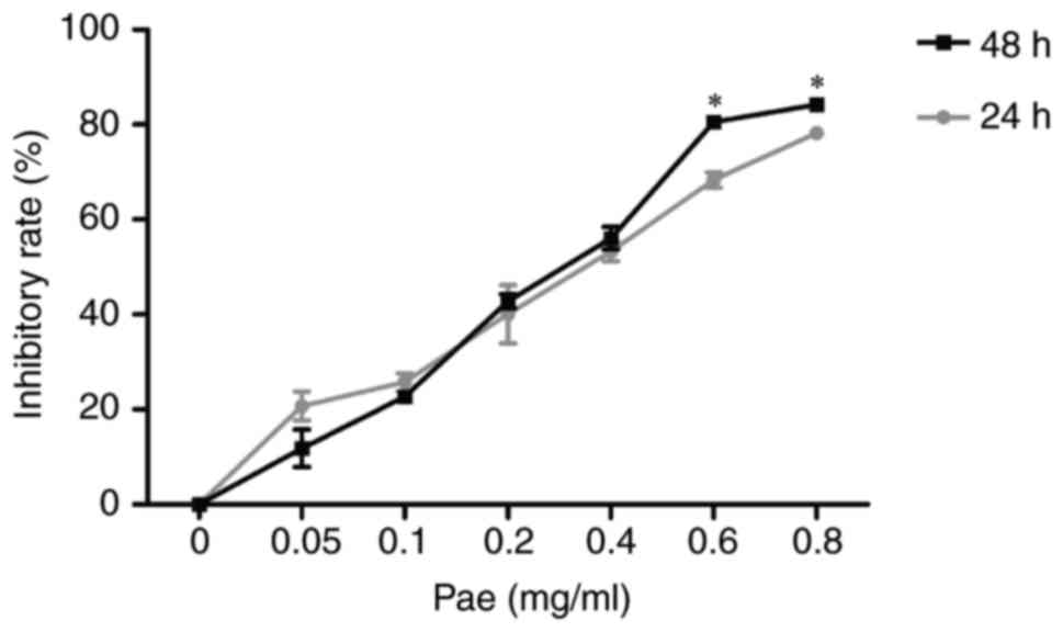
To verify the inhibitory effect of Pae on the
proliferation of gastric cancer cells, CCK-8 assays were used. The
results (Fig. 1) demonstrated that
at the 24 and 48 h time points, inhibitory rates of Pae on BGC823
cells increased gradually with the rise of concentration (0, 0.05,
0.1, 0.2, 0.4, 0.6 and 0.8 mg/ml). When the concentration of Pae
was 0.4 mg/ml, the 24 and 48 h inhibitory rates were (53.3±2.1) and
(56.1±2.4)%, respectively. However, when the concentration reached
0.6 mg/ml, the 24 and 48 h inhibitory rates were (68.3±1.6) and
(80.1±6.0)%, respectively, revealing obvious cytotoxicity.
According to the results of CCK-8 assays, low, moderate and high
concentrations of Pae were established as 0.1, 0.2 and 0.4 mg/ml,
respectively.
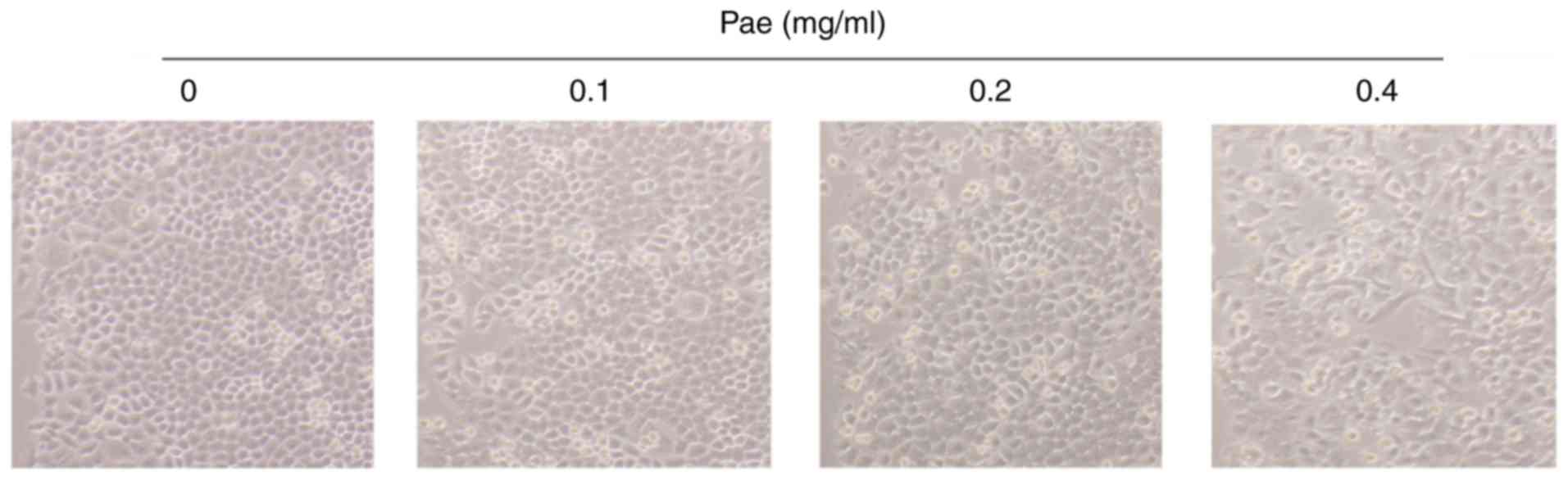
Cell morphological observation
Observed under an optical microscope after 24 h of
treatment (Fig. 2), BGC823 cells
in the control group exhibited a clear shape and orderly
arrangement. However, compared with the control group, the LP, MP
and HP groups demonstrated obvious morphological changes.
Furthermore, the HP group altered the most obviously with an
unclear shape, disorderly arrangement, fewer cells and more cell
death.
Pae induces apoptosis of BGC823
cells
The pro-apototic effect of Pae on BGC823 cells was
verified using an AnnexinV-FITC/PI assay. The results demonstrated
that cell apoptosis rates of the LP, MP and HP groups, (12.7±2.2),
(15.1±2.0) and (18.3±1.7)%, respectively, increased in a
significant concentration-dependent effect and were markedly higher
compared with the control group (P<0.05 or P<0.01; Fig. 3).
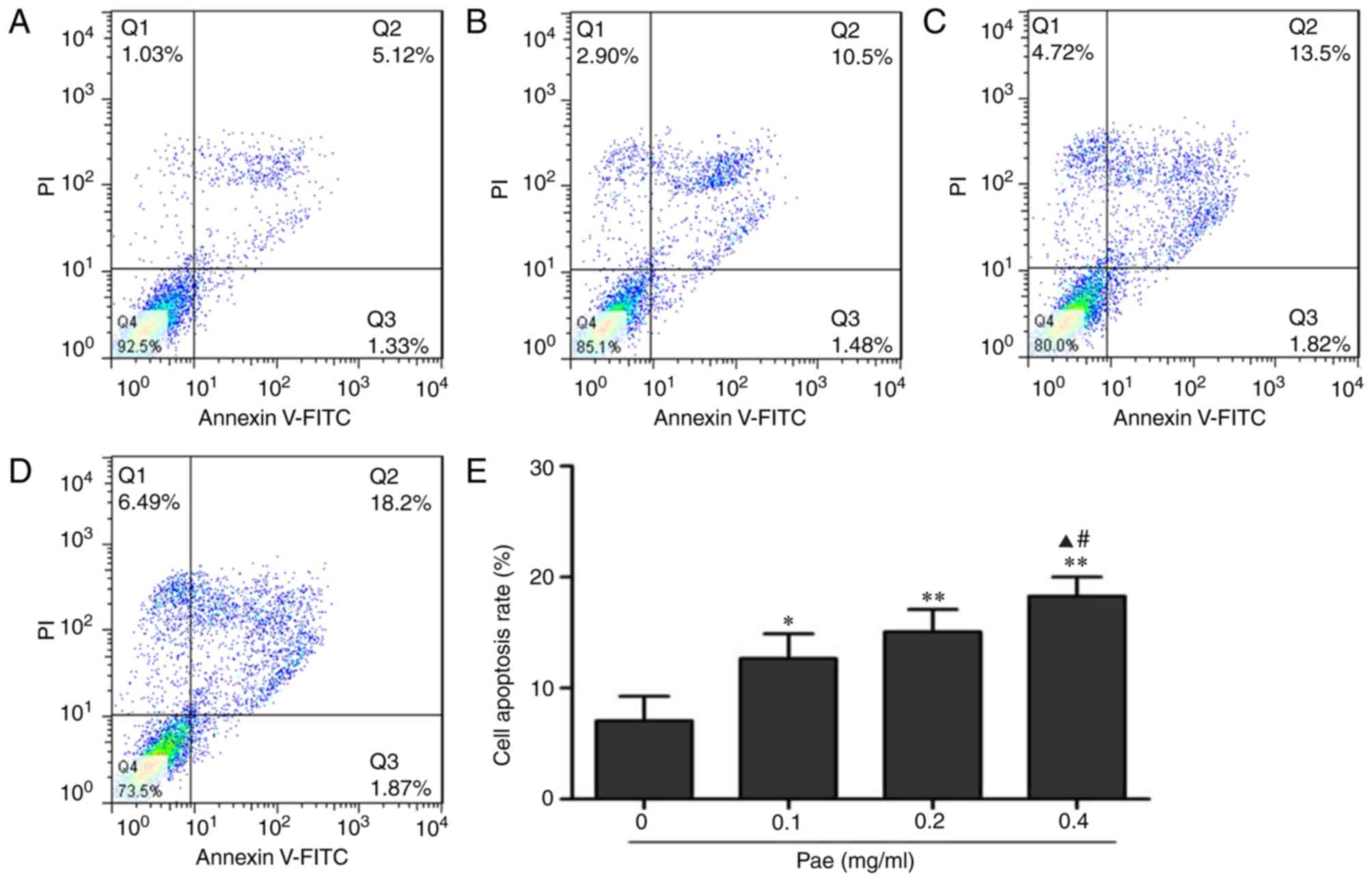 | Figure 3.Effect of Pae on the apoptosis of
BGC823 cells. Cell apoptosis data were analyzed in each group after
48 h treatment with corresponding concentration of Pae at (A) 0
mg/ml, (B) 0.1 mg/ml, (C) 0.2 mg/ml and (D) 0.4 mg/ml. (E)
Quantitative comparison of the apoptosis rates among groups. Data
are expressed as the mean ± standard deviation (n=4). *P<0.05,
**P<0.01 vs. 0; ▲P<0.05 vs. 0.1;
#P<0.05 vs. 0.2. Pae 0 mg/ml, control group; 0.1
mg/ml, low concentration Pae group; 0.2 mg/ml, moderate
concentration Pae group; 0.4 mg/ml, high concentration Pae group.
FITC, fluorescein isothiocyanate; PI, propidium iodide; Pae,
paeonol. n, the number of independent experiments performed. |
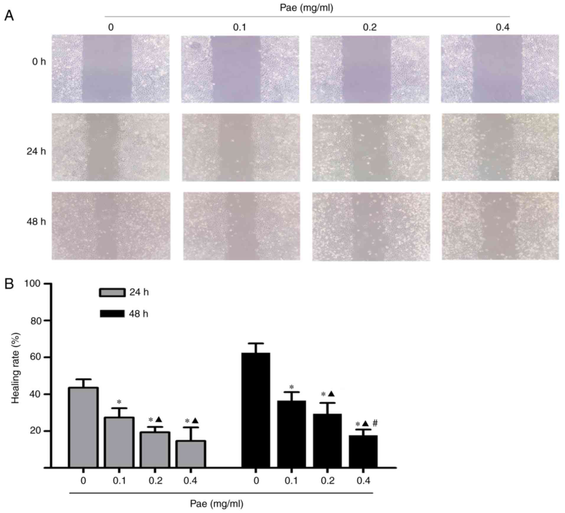
Effects of Pae on the migration and
invasion of BGC823 cells
To explore the effects of Pae on migration and
invasion of gastric cancer cells, the cell scratch-wound healing
and Transwell methods were applied, respectively. As presented in
Fig. 4, the 24 h wound healing
rates of the LP, MP and HP groups were (27.3±5.1), (19.3±2.9) and
(14.7±7.3)%, and the 48 h wound healing rates were (36.5±4.6),
(29.3±6.0) and (17.7±3.1)%, respectively, which were markedly
reduced compared with the 24 and 48 h healing rates [(43.5±4.6) and
(62.5±5.0)%] of the control group (P<0.05 or P<0.01).
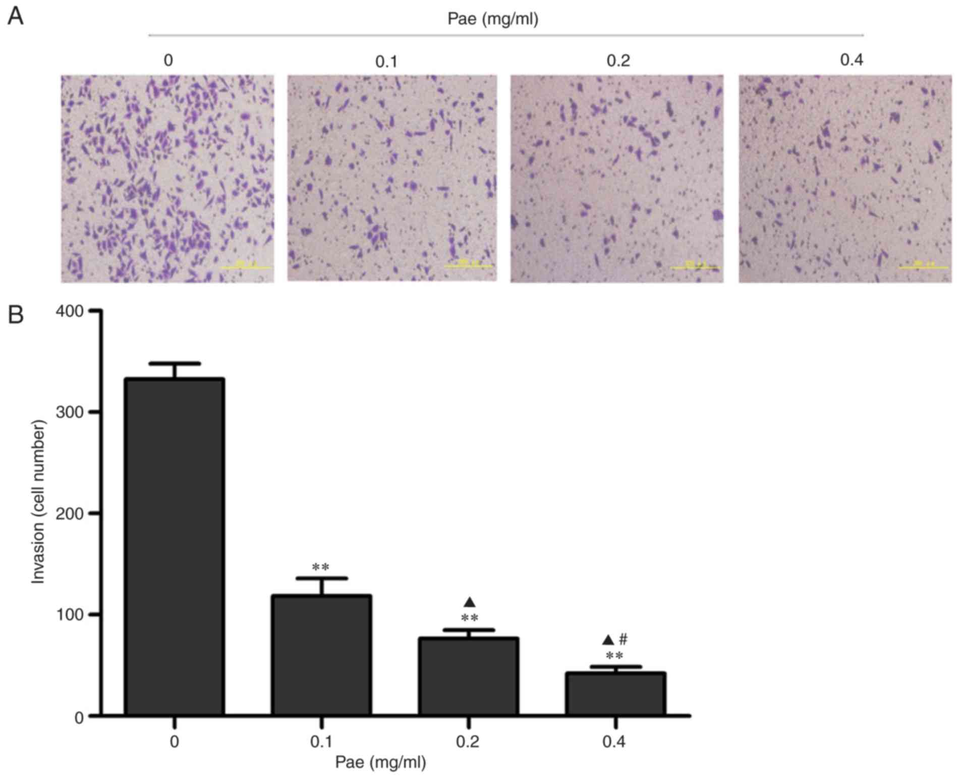
The Transwell invasion assay demonstrated that the
cell numbers passing the artificial basement membrane in the LP, MP
and HP groups (118±17, 76±9, 42±7 and, respectively) were
significantly reduced compared with the number of invading cells
(332±16) observed in the control group (P<0.01; Fig. 5). Together, the cell scratch-wound
healing and Transwell assays demonstrated that Pae could inhibit
the migration and invasion of BGC823 cells with a significant
concentration-dependent effect.
Pae downregulated the expression
levels of MMP-2 and MMP-9 in BGC823 cells
In order to further investigate possible mechanisms
of the inhibitory effects of Pae on the migration and invasion of
gastric cancer cells, the expression levels of MMP-2 and MMP-9 in
BGC823 cells were detected by western blotting. The results
demonstrated that the expression levels of MMP-2 and MMP-9 in
BGC823 cells of the LP, MP and HP groups were significantly reduced
compared with the control group (Fig.
6). Furthermore, with the concentration increase of Pae from
0–0.4 mg/ml, the expression levels of MMP-2 and MMP-9 demonstrated
a gradual concentration-dependent decrease (P<0.05 or
P<0.01). Thus, the mechanism that Pae exerts an inhibitory
effect the migration and invasion of BGC823 cells may, at least in
part, be attributable to the downregulation of MMP-2 and MMP-9
protein expression.
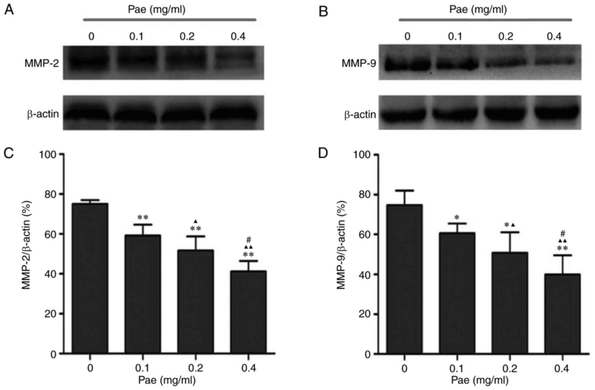 | Figure 6.Effect of Pae on the expression
levels of MMP-2 and MMP-9 of BGC823 cells at 48 h of treatment.
Representative western blot images of (A) MMP-2 and (B) MMP-9
protein expression levels. Quantitative comparison of the
expression levels of (C) MMP-2 and (D) MMP-9 among groups, with
β-actin as a loading control. Data are expressed as the mean ±
standard deviation (n=5). *P<0.05, **P<0.01 vs. 0;
▲P<0.05, ▲▲P<0.01 vs. 0.1;
#P<0.05 vs. 0.2. Pae 0 mg/ml, control group; 0.1
mg/ml, low concentration Pae group; 0.2 mg/ml, moderate
concentration Pae group; 0.4 mg/ml, high concentration Pae group.
MMP, matrix metalloproteinase; Pae, paeonol. n, the number of
independent experiments performed. |
Discussion
Pae is a natural product, with a white needle
crystal structure and melting point of 51–52°C that is extracted
from the root bark of Paeonia suffruticosa and the root of
Cynanchum paniculatum, and is widely used in traditional
Chinese medicine (TCM) (22,29).
Medicinally, Pae is extensively implemented for its various
pharmacological and physiological effects such as analgesia,
antipyresis, sedation, immunoregulation, anti-inflammation and
tumor suppression (30,31). In clinical TCM anti-tumor
treatment, Cynanchum paniculatum is usually prescribed for
pain reduction. Our previous clinical observations indicated that
the recipes with Cynanchum paniculatum could improve
chemotherapy effects of gastric cancer patients at advanced stages
of disease progression. Furthermore, Pae, the major bioactive
component of Cynanchum paniculatum, was previously reported
to enhance apoptotic induction effect of cisplatin on human
hepatoma cells and reverse paclitaxel resistance in human breast
cancer cells (32,33).
The anti-cancer effects of Pae with respect to
proliferation inhibition and apoptosis induction on cancer cells
are well established in the scientific literature. However, the
effect of Pae on tumor metastasis lacks conclusive evidence.
Therefore, the present study aimed to investigate the effects of
Pae with an emphasis on the invasion and migration of BGC823
gastric cancer cells with strong invasive and metastatic abilities.
The results indicated that Pae could inhibit proliferation and
induce apoptosis of BGC823 cells, which is in agreement with
existing reports (34).
Furthermore, the present study presents, to the best of our
knowledge, the first evidence of the inhibitory influence of Pae
upon invasion and migration of BGC823 cells using cell
scratch-wound healing and Transwell invasion assays.
In order to further investigate the possible
mechanism of inhibitory activities of Pae on the invasion and
migration of gastric cancer cells, the expression levels of MMP-2
and MMP-9 in BGC823 cells were determined after treatment with
gradient concentrations of Pae. The results demonstrated that Pae
could downregulate the expression levels of MMP-2 and MMP-9 in
BGC823 cells in a concentration-dependent manner, which may account
for anti-metastatic activities of Pae and represent a potential
target mechanism for clinical development of its promising
anti-cancer effects. Further studies are needed to investigate the
anti-cancer effects of Pae and elucidate its more definite
mechanisms of action.
Cell over-proliferation, inhibition of apoptosis and
metastasis are basic biological characteristics of malignant
tumors. For gastric cancer patients, cancer metastasis is often the
main cause leading to disease aggravation and mortality (35,36).
Previous research has demonstrated that there are higher MMP-2 and
MMP-9 expression levels in gastric tumors, especially in metastatic
tumors, than in normal mucosa (37,38).
MMPs could be used as diagnostic markers in body fluid, and MMP-2
might be a prognostic marker in ascites of advanced gastric cancer
patients with disseminated metastasis (39). Additionally, MMPs have been
selected as promising targets for cancer treatment based on their
upregulation in malignant tumors and their ability to enhance
cancer metastasis (9). Therefore,
along with the anti-proliferative and pro-apoptotic effects, the
anti-metastatic activities exerted through downregulation of MMPs
may support Pae to become a promising therapeutic candidate for the
gastric cancer therapy.
Acknowledgements
The present study was supported by the projects of
the Traditional Chinese Medical Science and Technology Development
Program in Shandong Province (grant no. 2013-27) and the
Fundamental Research Funds of Shandong University (grant no.
2014QLKY07).
References
|
1
|
de Martel C, Forman D and Plummer M:
Gastric cancer: Epidemiology and risk factors. Gastroenterol Clin
North Am. 42:219–240. 2013. View Article : Google Scholar : PubMed/NCBI
|
|
2
|
Ferlay J, Soerjomataram I, Dikshit R, Eser
S, Mathers C, Rebelo M, Parkin DM, Forman D and Bray F: Cancer
incidence and mortality worldwide: Sources, methods and major
patterns in GLOBOCAN 2012. Int J Cancer. 136:E359–E386. 2015.
View Article : Google Scholar : PubMed/NCBI
|
|
3
|
Jemal A, Bray F, Center MM, Ferlay J, Ward
E and Forman D: Global cancer statistics. CA Cancer J Clin.
61:69–90. 2011. View Article : Google Scholar : PubMed/NCBI
|
|
4
|
Ohtsu A, Yoshida S and Saijo N:
Disparities in gastric cancer chemotherapy between the East and
West. J Clin Oneol. 24:2188–2196. 2006. View Article : Google Scholar
|
|
5
|
de Vries AC and Kuipers EJ: Epidemiology
of premalignant gastric lesions: Implications for the development
of screening and surveillance strategies. Helicobacter. 12 Suppl
2:S22–S31. 2007. View Article : Google Scholar
|
|
6
|
Kalluri R: Basement membranes: Structure,
assembly and role in tumour angiogenesis. Nat Rev Cancer.
3:422–433. 2003. View
Article : Google Scholar : PubMed/NCBI
|
|
7
|
Liotta LA and Kohn EC: The
microenvironment of the tumour-host interface. Nature. 411:375–379.
2001. View
Article : Google Scholar : PubMed/NCBI
|
|
8
|
Kessenbrock K, Plaks V and Werb Z: Matrix
metalloproteinases: Regulators of the tumor microenvironment. Cell.
141:52–67. 2010. View Article : Google Scholar : PubMed/NCBI
|
|
9
|
Hua H, Li M, Luo T, Yin Y and Jiang Y:
Matrix metalloproteinases in tumorigenesis: An evolving paradigm.
Cell Mol Life Sci. 68:3853–3868. 2011. View Article : Google Scholar : PubMed/NCBI
|
|
10
|
Curran S and Morray GI: Matrix
metalloproteitases in tumour invasion and metastasis. J Pathol.
189:300–308. 1999. View Article : Google Scholar : PubMed/NCBI
|
|
11
|
Zeng ZS, Cohen AM and Guillem JG: Loss of
basement membrane type IV collagen is associated with increased
expression of metalloproteinases 2 and 9 (MMP-2 and MMP-9) during
human colorectal tumorigenesis. Carcinogenesis. 20:749–755. 1999.
View Article : Google Scholar : PubMed/NCBI
|
|
12
|
Heikinheimo K and Salo T: Expression of
basement membrane type IV collagen and type IV collagenases (MMP-2
and MMP-9) in human fetal teeth. J Dent Res. 74:1226–1234. 1995.
View Article : Google Scholar : PubMed/NCBI
|
|
13
|
Roach DM, Fitridge RA, Laws PE, Millard
SH, Varelias A and Cowled PA: Up-regulation of MMP-2 and MMP-9
leads to degradation of type IV collagen during skeletal muscle
reperfusion injury; protection by the MMP inhibitor, doxycycline.
Eur J Vasc Endovasc Surg. 23:260–269. 2002. View Article : Google Scholar : PubMed/NCBI
|
|
14
|
Zhao Y, Zhou FL, Li WP, Wang J and Wang
LJ: Slit2-Robo1 signaling promotes the adhesion, invasion and
migration of tongue carcinoma cells via upregulating matrix
metalloproteinases 2 and 9 and downregulating E-cadherin. Mol Med
Rep. 14:1901–1906. 2016. View Article : Google Scholar : PubMed/NCBI
|
|
15
|
Liu C: Pathological and prognostic
significance of matrix metalloproteinase-2 expression in ovarian
cancer: A meta-analysis. Clin Exp Med. 16:375–382. 2016. View Article : Google Scholar : PubMed/NCBI
|
|
16
|
Pazzaglia L, Ponticelli F, Magagnoli G,
Magagnoli G, Gamberi G, Ragazzini P, Balladelli A, Picci P and
Benassi M: Activation of metalloproteinases-2 and −9 by
interleukin-1alpha in S100A4-positive liposarcoma cell line:
Correlation with cell invasiveness. Anticancer Res. 24:967–972.
2004.PubMed/NCBI
|
|
17
|
Hwang TL, Changchien TT, Wang CC and Wu
CM: Claudin-4 expression in gastric cancer cells enhances the
invasion and is associated with the increased level of matrix
metalloproteinase-2 and −9 expression. Oncol Lett. 8:1367–1371.
2014.PubMed/NCBI
|
|
18
|
Chen SJ, Yao XD, Peng BO, Xu YF, Wang GC,
Huang J, Liu M and Zheng JH: Epigallocatechin-3-gallate inhibits
migration and invasion of human renal carcinoma cells by
downregulating matrix metalloproteinase-2 and matrix
metalloproteinase-9. Exp Ther Med. 11:1243–1248. 2016. View Article : Google Scholar : PubMed/NCBI
|
|
19
|
Sun W, Liu DB, Li WW, Zhang LL, Long GX,
Wang JF, Mei Q and Hu GQ: Interleukin-6 promotes the migration and
invasion of nasopharyngeal carcinoma cell lines and upregulates the
expression of MMP-2 and MMP-9. Int J Oncol. 44:1551–1560. 2014.
View Article : Google Scholar : PubMed/NCBI
|
|
20
|
Lee KR, Lee JS, Song JE, Ha SJ and Hong
EK: Inonotus obliquus-derived polysaccharide inhibits the migration
and invasion of human non-small cell lung carcinoma cells via
suppression of MMP-2 and MMP-9. Int J Oncol. 45:2533–2540. 2014.
View Article : Google Scholar : PubMed/NCBI
|
|
21
|
Riley CM and Ren TC: Simple method for the
determination of paeonol in human and rabbit plasma by
high-performance liquid chromatography using solid-phase extraction
and ultraviolet detection. J Chromatogr. 489:432–437. 1989.
View Article : Google Scholar : PubMed/NCBI
|
|
22
|
Jiang SP and Chen YX: Advances in the
research and its clinical application of Cynanchum paniculatum
(Bge.) Kitag. Zhongguo Zhong Yao Za Zhi. 19:311–314. 1994.(In
Chinese). PubMed/NCBI
|
|
23
|
Anh Hle T, Cuc NT, Tai BH, Yen PH, Nhiem
NX, do T Thao, Nam NH, Van Minh C, Van Kiem P and Kim YH: Synthesis
of chromonylthiazolidines and their cytotoxicity to human cancer
cell lines. Molecules. 20:1151–1160. 2015. View Article : Google Scholar : PubMed/NCBI
|
|
24
|
Xu SP, Sun GP, Shen YX, Wei W, Peng WR and
Wang H: Antiproliferation and apoptosis induction of paeonol in
HepG2 cells. World J Gastroenterol. 13:250–256. 2007. View Article : Google Scholar : PubMed/NCBI
|
|
25
|
Li M, Tan SY and Wang XF: Paeonol exerts
an anticancer effect on human colorectal cancer cells through
inhibition of PGE2 synthesis and COX-2 expression. Oncol
Rep. 32:2845–2853. 2014. View Article : Google Scholar : PubMed/NCBI
|
|
26
|
Yin J, Wu N, Zeng F, Cheng C, Kang K and
Yang H: Paeonol induces apoptosis in human ovarian cancer cells.
Acta Histochem. 115:835–839. 2013. View Article : Google Scholar : PubMed/NCBI
|
|
27
|
Horng CT, Shieh PC, Tan TW, Yang WH and
Tang CH: Paeonol suppresses chondrosarcoma metastasis through
up-regulation of miR-141 by modulating PKCδ and c-Src signaling
pathway. Int J Mol Sci. 15:11760–11772. 2014. View Article : Google Scholar : PubMed/NCBI
|
|
28
|
Kim SA, Lee HJ, Ahn KS, Lee HJ, Lee EO,
Ahn KS, Choi SH, Jung SJ, Kim JY, Baek N and Kim SH: Paeonol exerts
anti-angiogenic and anti-metastatic activities through
downmodulation of Akt activation and inactivation of matrix
metalloproteinases. Biol Pharm Bull. 32:1142–1147. 2009. View Article : Google Scholar : PubMed/NCBI
|
|
29
|
Katakai M, Akamaru T and Tani T: An
analysis of appearance frequency of formulations and crude drugs in
Jin-Kui-Yao-Lue. Yakushigaku Zasshi. 38:1–10. 2003.(In Japanese).
PubMed/NCBI
|
|
30
|
Fan L, Song B, Sun G, Ma T, Zhong F and
Wei W: Endoplasmic reticulum stress-induced resistance to
doxorubicin is reversed by paeonol treatment in human
hepatocellular carcinoma cells. PLoS One. 8:e626272013. View Article : Google Scholar : PubMed/NCBI
|
|
31
|
Chou TC: Anti-inflammatory and analgesic
effects of paeonol in carrageenan-evoked thermal hyperalgesia. Br J
Pharmacol. 139:1146–1152. 2003. View Article : Google Scholar : PubMed/NCBI
|
|
32
|
Xu SP, Sun GP, Shen YX, Peng WR, Wang H
and Wei W: Synergistic effect of combining paeonol and cisplatin on
apoptotic induction of human hepatoma cell lines. Acta Pharmacol
Sin. 28:869–878. 2007. View Article : Google Scholar : PubMed/NCBI
|
|
33
|
Cai J, Chen S, Zhang W, Hu S, Lu J, Xing J
and Dong Y: Paeonol reverses paclitaxel resistance in human breast
cancer cells by regulating the expression of transgelin 2.
Phytomedicine. 21:984–991. 2014. View Article : Google Scholar : PubMed/NCBI
|
|
34
|
Li N, Fan LL, Sun GP, Wan XA, Wang ZG, Wu
Q and Wang H: Paeonol inhibits tumor growth in gastric cancer in
vitro and in vivo. World J Gastroenterol. 16:4483–4490. 2010.
View Article : Google Scholar : PubMed/NCBI
|
|
35
|
Hohenberger P and Gretschel S: Gastric
cancer. Lancet. 362:305–315. 2003. View Article : Google Scholar : PubMed/NCBI
|
|
36
|
Krejs GJ: Gastric cancer: Epidemiology and
risk factors. Dig Dis. 28:600–603. 2010. View Article : Google Scholar : PubMed/NCBI
|
|
37
|
Parsons SL, Watson SA, Collins HM, Griffin
NR, Clarke PA and Steele RJ: Gelatinase (MMP-2 and −9) expression
in gastrointestinal malignancy. Br J Cancer. 78:1495–1502. 1998.
View Article : Google Scholar : PubMed/NCBI
|
|
38
|
Lim SC: Expression of matrix
metalloproteinases and its inhibitor in gastric adenocarcinoma.
Cancer Res Treat. 33:199–206. 2001. View Article : Google Scholar : PubMed/NCBI
|
|
39
|
Noh S, Jung JJ, Jung M, Kim TS, Park CH,
Lim SJ, Jeung HC, Cheol H, Chung HC and Rha SY: MMP-2 as a putative
biomarker for carcinomatosis in gastric cancer.
Hepatogastroenterology. 58:2015–2019. 2011. View Article : Google Scholar : PubMed/NCBI
|