Introduction
Osteoporosis is a common bone metabolic disease
characterized by systemic bone loss, impaired bone microstructure
and reduced bone strength (1,2).
Clinically, it typically manifests as chronic pain and increased
susceptibility to low-traumatic fractures, resulting in compromised
life quality and increased patient mortality (3–5).
Postmenopausal osteoporosis is the primary form of osteoporosis
which results from estrogen deficiency, leading to excessive bone
absorption and inadequate bone formation (6). Bone mineral density (BMD) measurement
using dual energy X-ray absorptiometry is currently the gold
standard for confirmative diagnosis of osteoporosis. However, the
early stage of osteoporosis is usually asymptomatic and >50% of
low-traumatic patients do not meet the diagnostic criteria of
osteoporosis, according to BMD value (7), therefore, the search for more
sensitive markers for osteoporosis is essential and will be
beneficial for evaluation and treatment for osteoporosis patients,
particularly at the early stages.
Previously, investigations using osteoporotic serum
have suggested carboxy-terminal crosslinking telopeptide of type I
collagen and procollagen type I N propeptide as biomarkers of bone
resorption and bone formation (8).
The serum cathepsin K has been explored as a biomarker of
osteoclast activity, whereas fibroblast growth factor-23 and
sclerostin were considered osteocyte factors (9–11).
In addition, with the rapid development of omics technologies,
various osteoporosis associated biomarkers have been discovered.
Micro (mi)RNA-133a and miR-422a in circulating monocytes have been
demonstrated to act as potential nucleic biomarkers for
postmenopausal osteoporosis (12,13).
However, all these candidates are not fully specific or sensitive
as early-stage markers of osteoporosis. Recently developed protein
array technology is sensitive and specific in detecting trace
amounts of proteins in body fluid and has been successfully applied
in the discovery of various disease associated biomarkers (14–16).
Therefore, the present study hypothesized that protein array
screening in combination with osteoporosis models may provide
useful information in identifying biomarkers for early osteoporosis
diagnosis.
The present study successfully established the
ovariectomized rat as a postmenopausal osteoporosis model, which
demonstrated progressive bone loss starting from four weeks
following surgery. In the protein array screening, B7-2, β-nerve
growth factor (β-NGF), Fractalkine, interferon-γ (IFN-γ), tissue
inhibitor of metalloproteinases-1 (TIMP-1) and monocyte chemotactic
protein-1 (MCP-1) were observed to be increased with the
development of osteoporosis in the rat serum. Validation in human
samples demonstrated that Fractalkine, TIMP-1 and MCP-1 were
increased in serum of patients suffering from osteoporosis or
decreased bone density compared with that of healthy people with
normal bone mineral density. Overall, the present study identified
a novel panel of serum protein markers that may successfully
predict the development and severity of postmenopausal
osteoporosis.
Materials and methods
Animals and experimental
procedures
A total of 10 female Sprague-Dawley rats, (age, 3
months; weight, 239±17.5 g), were obtained from the Experimental
Animal Center at the Fourth Military Medical University (Xi'an,
China), and were housed under specific pathogen-free conditions
(20°C, 12-h light/dark cycle and 50–55% humidity) with free access
to food and water. They were randomly divided into two groups, an
ovariectomy group (OVX, n=5) and a sham group (Sham, n=5). A total
of 5 rats of OVX group underwent an ovariectomy to establish the
postmenopausal osteoporosis model. The rats were anesthetized
intraperitoneally with pentobarbital at a dose of 40 mg/kg. The
ovariectomy surgery was performed using the dorsal approach
(17). All 5 rats in the sham
group underwent the same operation with the exception of the ovary
ablation. Blood samples were obtained from the angular veins on
each animal at 2-week intervals for 2 months following the
operations (2, 4, 6 and 8 weeks). Serum samples were collected by
centrifugation and stored at −80°C for further analysis. There was
no significant difference in total body weight between the 2 groups
at the differing time points. All experimental procedures conducted
using animals were approved by the Ethics in Animal Research
Committee of the Fourth Military Medical University (permission
code 20110405-5).
Patients
A total of 24 women aged 57–68 from Xijing Hospital
(Xi'An, China) were recruited as human subjects and signed
informed-consent documents, prior to being enrolled in the present
study. Subjects were excluded if they had a history of cancer,
cardiovascular disease or diabetes mellitus. None of the subjects
had been diagnosed with metabolic bone diseases such as
osteoarthritis and rheumatoid arthritis or had been treated with
medication known to impact upon bone metabolism, such as hormone
therapy, bisphosphonates or calcitonin. Patients had normal
hepatorenal function and were not suffering from any endocrine
disturbances, hypercalcemia or urolithiasis. The human
investigations were approved by the ethics committee of the Fourth
Military Medical University. A total of 8 subjects had normal BMD
(Tm ≥-1.0) and 8 had low BMD (−2.5< Tm <-1.0). The other 8
subjects were postmenopausal osteoporosis patients (Tm ≤-2.5). BMD
of all subjects for the lumbar spine (L1-4) and total hip (femoral
neck, trochanter and intertrochanteric region) were measured using
dual energy X-ray absorptiometry (DXA) scanners. Blood samples were
collected from subjects and centrifuged at 1,500 × g for 10 min at
room temperature. Supernatant sera were obtained and stored at
−80°C for further analysis.
Micro-computed tomography (micro-CT)
assessment
Micro-CT scanning was performed in vivo on
each animal at 2 week intervals for 2 months following the
ovariectomy or sham operations (2, 4, 6, and 8 weeks). Rats were
anesthetized with pentobarbital at a dose of 40 mg/kg during each
measurement. The distal femurs were scanned using Pre-Clinical
Inveon Micro-CT (Siemens Healthineers, Erlangen, Germany) with an 8
mm resolution, a 50 kV tube voltage and a 0.1 mA tube current.
Reconstruction and three-dimensional quantitative analyses were
determined using the software provided by a desktop micro-CT system
(Inveon Research Workplace 2.2; Siemens Healthineers). The scanning
regions were confined to the distal metaphysis and extended 2.0 mm
proximally from the proximal tip of the primary spongiosa. The
following three-dimensional indices in the defined region of
interest were analyzed: BMD, trabecular number (Tb.N), trabecular
thickness (Tb.Th), trabecular separation (Tb.Sp) and relative bone
volume over the total volume (BV/TV, %). The operator who conducted
the scan analyses was blinded to the procedure associated with the
subjects.
Cytokine antibody array analysis
Rat serum samples were assessed for the presence of
27 cytokines using Quantibody Rat Cytokine Array 3 kit (RayBiotech
Inc., Norcross, GA, USA). Antibody array membranes were first
blocked with Tris-buffered saline containing 0.05% Tween-20,
supplemented with 5% skimmed milk at room temperature for 1 h, to
which rat sera were subsequently added for a final 10-fold
dilution, following the manufacturer's protocol. The cytokine
expression levels were detected and quantified on a fluorescent
scanner (Axon GenePix; Molecular Devices, LLC, Sunnyvale, CA,
USA).
ELISA assay
Human serum Fractalkine, TIMP-1 and MCP-1 expression
levels were measured using sandwich ELISA assay kits (P78423,
P01033 and P13500; RayBiotech Inc.) according to the manufacturer's
protocol. Total protein concentration was calculated using the
Bradford protein assay method.
Statistical analysis
Statistical analyses were performed using SPSS
software, version 15.0 (SPSS Inc., Chicago, IL, USA). Quantitative
data are presented as the mean ± standard deviation. Statistical
tests were two-sided; the differences between two groups were
assessed using Student's t-tests, and analysis of variance followed
by the Bonferroni post-hoc test was conducted to analyze multiple
comparisons among groups. Pearson correlation was employed to
determine the linear relationship between two variables. P<0.05
was considered to indicate a statistically significant
difference.
Results
Establishment and confirmation of
early osteoporosis in ovariectomized rat model
To determine when early postmenopausal osteoporosis
occurs, the present study measured BMD and other osteoporosis
parameters starting from two weeks following surgery. Distal femurs
of the rats were scanned using micro-CT and trabecular bone
structures were reconstructed based on the images (Fig. 1A). The results revealed that there
was no difference in BMD between the OVX and the sham group 2 weeks
following the ovariectomy. A gradual reduction of BMD in the
ovariectomized rats was observed 4 weeks post-surgery and reached
>25% reduction 8 weeks following surgery (Fig. 1B). No observable BMD alteration was
detected in the sham group during this time course. Consistent with
the decreased BMD values, quantitative analysis of further
osteoporosis parameters revealed a significant decrease in BV/TV
and Tb.Th with a similar pattern to BMD from 4 weeks post-surgery
(P<0.01). Tb.N, as a marker of trabecule number, decreased later
and revealed a significant reduction from 6 weeks following surgery
(P<0.05 and P<0.01). A prominent increase in trabecular space
marker, Tb.Sp, was also observed from 4 weeks following surgery
(P<0.05 and P<0.01). These data suggested that initiation of
rat postmenopausal osteoporosis occurred 4 weeks post-surgery.
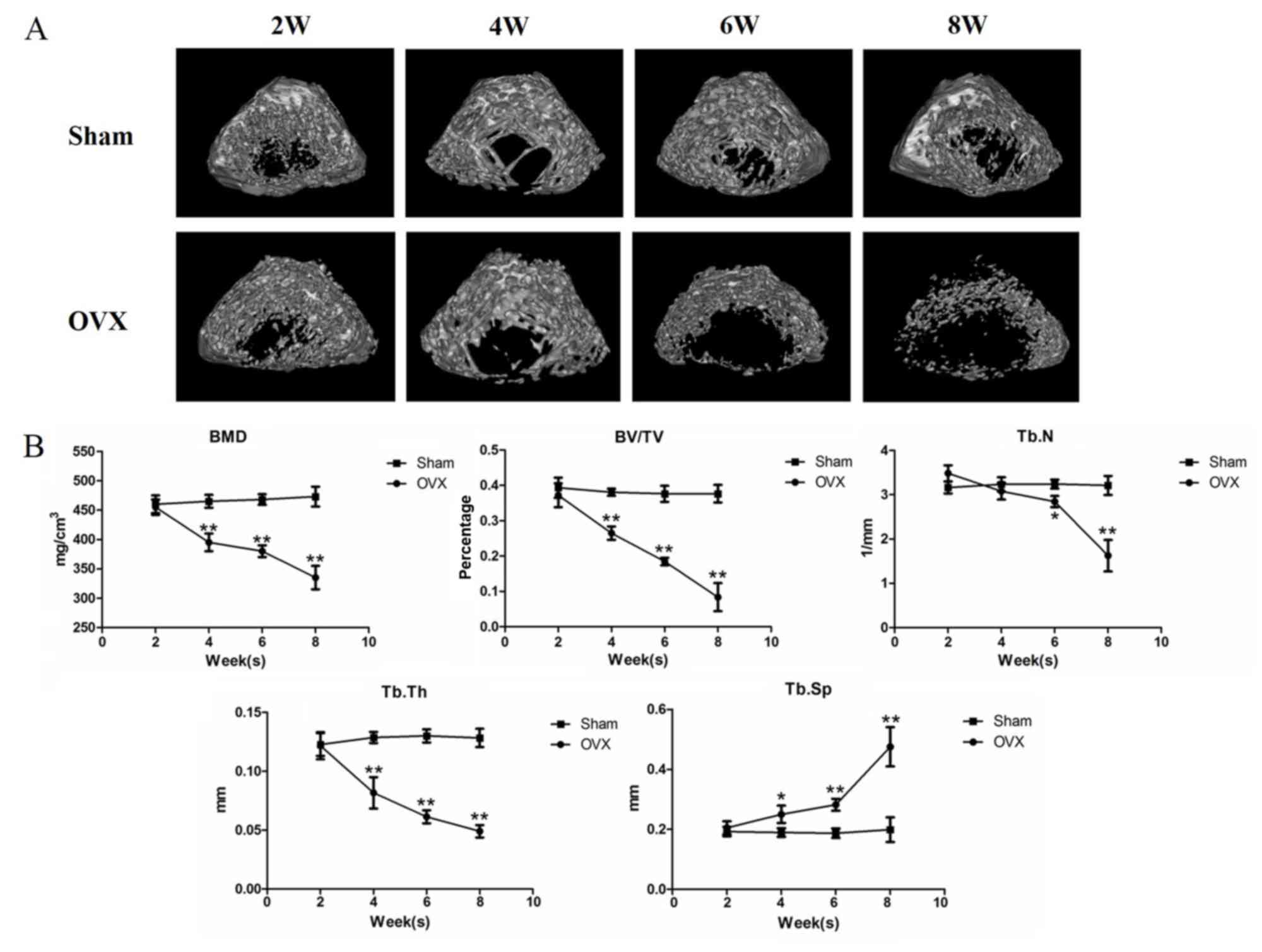 | Figure 1.Establishment and confirmation of
early osteoporosis in ovariectomized rat models. (A) Micro-CT
analysis within the distal metaphyseal femur region at 2, 4, 6 and
8 weeks following sham or ovariectomy operations. Magnification ×40
of the original section. (B) Dynamic alterations of BMD, BV/TV,
Tb.N, Tb.Th and Tb.Sp in rats that received either sham or
ovariectomy operations. *P<0.05 and **P<0.01 vs. sham group
(n=5). OVX, ovariectomy; BMD, bone mineral density; BV/TV, relative
bone volume over the total volume; Tb.N, trabecular number; Tb.Th,
trabecular thickness; Tb.Sp, trabecular separation; OVX,
ovariectomy group. |
Screening of postmenopausal
osteoporosis-associated biomarkers using protein array
A total of 27 proteins, B7-2, β-NGF,
cytokine-induced neutrophil chemoattractant (CINC)-1, CINC-2,
CINC-3, ciliary neurotrophic factor, Fractalkine,
granulocyte-macrophage colony-stimulating factor, intercellular
adhesion molecule-1, IFN-γ, interleukin (IL)-1α, IL-1β, IL-2, IL-4,
IL-6, IL-10, IL-13, chemokine (C-X-C motif) ligand 5, L-selectin,
MCP-1, platelet derived growth factor subunit A, Prolactin R,
receptor for advanced glycation end products, calmodulin-binding
kinesin-like protein-1, TIMP-1, tumor necrosis factor-α and
vascular endothelial growth factor, were included in the protein
array screening assay, based on previous literature. By analyzing
the fluorescence intensities, the results demonstrated that the
serum levels of B7-2, Fractalkine, IFN-γ, β-NGF, MCP-1 and TIMP-1
increased following ovariectomy (Fig.
2). Further statistical study revealed that TIMP-1
significantly increased at 4 weeks following the ovariectomy,
almost in parallel with the aforementioned significant alterations
in bone mineral density at 4 weeks. Other biomarkers increased
significantly at 6 weeks post-surgery, two weeks later than when
the alterations in bone mineral density were observed (P<0.05
and P<0.01). The levels of other candidate proteins did not
significantly alter in the screening during this time course.
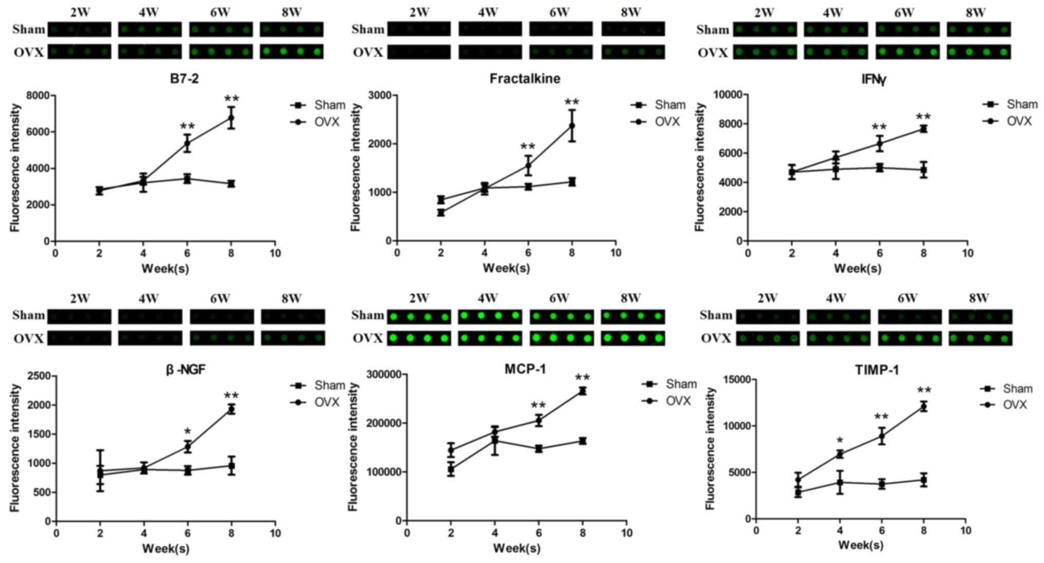 | Figure 2.Screening results of postmenopausal
osteoporosis-associated biomarkers using protein array. Upper
panel: fluorescence intensity diagram of B7-2, Fractalkine, IFN-γ,
β-NGF, MCP-1 and TIMP-1 in sera obtained from sham or
ovariectomized rats at 2, 4, 6 and 8 weeks following surgery in the
protein array screening. Each sample repeated in four slots. Lower
panel: dynamic alterations of serum biomarkers in the sham or
ovariectomized rats post-surgery. *P<0.05, **P<0.01 vs. sham
group. n=5. IFN-γ, interferon-γ; β-NGF, β-nerve growth factor;
MCP-1, monocyte chemotactic protein-1; TIMP-1, tissue inhibitor of
metalloproteinases-1; OVX, ovariectomy group. |
Fractalkine, IFN-γ, MCP-1 and TIMP-1
correlate with the severity of bone loss in the ovariectomized rat
model
In the postmenopausal osteoporosis rat model, eight
weeks following ovariectomy is usually defined as the standard
period for the presentation of obvious bone loss. Six biomarkers
were observed to be elevated in the serum during the early stage of
osteoporosis in our model. However, whether they were also
correlated with osteoporosis progression remained unclear. To
verify the association of these six proteins with the progression
of postmenopausal osteoporosis, the present study analyzed the
correlation between the protein levels and the severity of bone
loss of the ovariectomized rats. As presented in Fig. 3 and Table I, Fractalkine, IFN-γ, MCP-1 and
TIMP-1 were negatively correlated with the BMD of the
ovariectomized rats (P<0.05), whereas the results for B7-2 and
β-NGF were not significant.
 | Figure 3.Correlation between serum B7-2,
Fractalkine, IFN-γ, β-NGF, MCP-1 and TIMP-1 expression levels and
bone loss. The fluorescence intensity of B7-2, Fractalkine, IFN-γ,
β-NGF, MCP-1 and TIMP-1 in the protein array screening was plotted
against the BMD of the rats following surgery. BMD, bone mineral
density; IFN-γ, interferon-γ; β-NGF, β-nerve growth factor; MCP-1,
monocyte chemotactic protein-1; TIMP-1, tissue inhibitor of
metalloproteinases-1. |
 | Table I.Correlation between potential early
postmenopausal osteoporosis biomarker and bone mineral density in
ovariectomized rats. |
Table I.
Correlation between potential early
postmenopausal osteoporosis biomarker and bone mineral density in
ovariectomized rats.
| Biomarker | r | P-value |
|---|
| B7-2 | −0.914 | 0.086 |
| Fractalkine | −0.971 |
0.029a |
| IFNγ | −0.976 |
0.024a |
| β-NGF | −0.888 | 0.112 |
| MCP1 | −0.979 |
0.021a |
| TIMP1 | −0.971 |
0.029a |
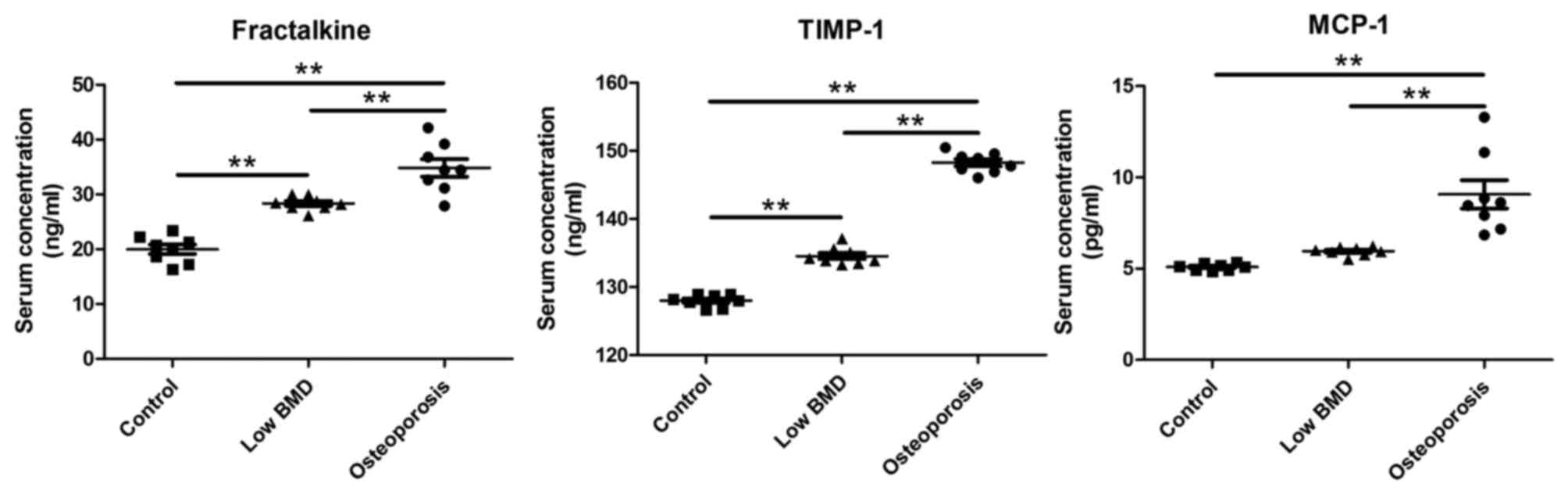
Validation of early postmenopausal
osteoporosis-associated biomarkers in human samples
To further verify the clinical significance of the
potential early postmenopausal osteoporosis serum biomarkers, the
present study detected the serum levels of Fractalkine, TIMP-1 and
MCP-1 using commercially available ELISA kits in patients. The
different serum protein levels among people with normal BMD
(Tm≥-1.0, n=8), patients with reduced BMD that did not reach the
osteoporosis diagnosis criteria (−2.5<Tm<-1.0, n=8) and
patients suffering from postmenopausal osteoporosis (Tm≤-2.5, n=8)
were compared and analyzed. The results demonstrated that these
three candidates, Fractalkine, TIMP-1 and MCP-1, increased as the
BMD decreased from normal people to patients with reduced BMD and
postmenopausal osteoporosis with significant differences (Fig. 4).
Discussion
Postmenopausal osteoporosis is one of the most
common skeletal diseases lacking practical methods for early
diagnosis and intervention to reduce the possibility of
low-traumatic fracture. In the present study, through a protein
array screening in the ovariectomy osteoporosis rat model, a total
of 6 serum protein markers were identified with expression levels
that altered in parallel with the development of early
postmenopausal osteoporosis. Of the six potential markers,
Fractalkine, TIMP-1 and MCP-1 were further validated to be
increased in serum at the early stage of osteoporosis in human
samples.
Postmenopausal osteoporosis is gradually acquired
following the menopause and there is currently no consent on
parameters that define the early stage of this disease. The World
Health Organization classification of osteoporosis using the BMD T
score (normal if BMD ≥-1.0; low bone mass or alternatively
osteopenia if −2.5< BMD <-1.0; osteoporosis if BMD ≤-2.5;
severe or established osteoporosis if BMD ≤-2.5 with history of
fragility fracture) is useful for clinically diagnosing and
evaluating the response to treatment of osteoporosis patients
(7). However, the BMD T score is
not sensitive in identifying members of the population that are at
a high risk of developing osteoporosis, prior to the prominent
occurrence of bone loss. To establish the early stage of
postmenopausal osteoporosis, the BMD value of the rat femur was
measured post ovariectomy. The majority of parameters were still
comparable 2 weeks following the surgery, slight alterations
appeared 4 weeks following surgery and the trends extended to the
end of the observation period, at 8 weeks following the
ovariectomy. Therefore, the time window of 2–4 weeks following
ovariectomy in the rat may be considered as the early stage of
postmenopausal osteoporosis development, and serum biochemical
alterations may be explored for discovery of biomarkers predicting
the upcoming osteoporosis. This time window for early
postmenopausal osteoporosis may not be strict, however it does
provide helpful information for screening early postmenopausal
osteoporosis associated markers.
Following the establishment of the 2–4-week period
following ovariectomy as the early stage of postmenopausal
osteoporosis genesis, the present study focused on investigation of
the biomarkers that demonstrated increased trends during this
period. A total of 6 protein markers were observed to exhibit this
trend, and the elevation of Fractalkine, TIMP-1 and MCP-1 were
further verified with investigations using serum from osteopenia
and osteoporosis patients.
Fractalkine, additionally termed C-X3-C motif ligand
1, is the only member of the CX3C chemokine subfamily that
functions through binding with the CX3C receptor 1 (18). Fractalkine is a membrane-bound
protein that may be released into serum by metalloproteinase, thus
it acts as an adhesion molecule and potential chemoattractant
(19,20). Certain studies have previously
linked Fractalkine with bone metabolism. In mouse, ionizing
irradiation augments Fractalkine production in skeletal vascular
endothelium, which promotes recruitment of osteoclast precursor
cells resulting in enhanced osteoclastogenesis and bone absorption
(21). The membrane-bound form of
Fractalkine has been demonstrated to be critical for the
osteoblast-osteoclast interaction and the osteoblast-induced
osteoclast differentiation (22,23).
Furthermore, it has been reported that circulating Fractalkine
levels are associated with the severity of postmenopausal
osteoporosis (24). The present
study demonstrated that serum Fractalkine concentration was
elevated not only in postmenopausal osteoporosis patients, however
additionally in osteopenia patients, compared with the healthy
control. The study further provided evidence that demonstrated
Fractalkine progressively increased from osteopenia to
osteoporosis, acting as an potential warning marker for early
postmenopausal osteoporosis.
TIMP-1 belongs to the inhibitor of
metalloproteinases (TIMPs) family and works together with the
metalloproteinases (MMPs) to remodel the extracellular matrices
(25). Coordination degradation of
bone matrices by MMPs and TIMPs is critical in bone formation and
absorption. It has been reported that TIMP-1 levels are elevated in
chronic obstructive pulmonary disease (COPD) and early rheumatoid
arthritis (RA) patients with bone loss (26,27).
COPD and RA are associated with systemic chronic inflammation and
it has previously been demonstrated that dysregulation of immunity
contributes to postmenopausal osteoporosis (28). The present study revealed that
serum TIMP-1 was increased in osteopenia and osteoporosis patients.
In a previous study (29), the
researchers excluded TIMP-1 as a potential biomarker for
osteoporosis diagnosis. However, the authors may have
misinterpreted their data, as a negative correlation exists between
TIMP-1 level and BMD, in addition to the other two serum biomarkers
of osteoporosis in their study.
MCP-1 is one of the most important chemokines that
recruits different kinds of immune cells during inflammation and
infection. Gene polymorphism analysis demonstrated that the MCP-1
A2518G polymorphism is correlated with osteopenia and osteoporosis
risk in postmenopausal women (30). To the best of the author's
knowledge, the present study, for the first time, observed that
MCP-1 was elevated in the serum of postmenopausal osteoporosis
patients and prominently increased during the progression of
osteopenia to osteoporosis, suggesting that MCP-1 may serve as a
potential biomarker for early postmenopausal osteoporosis
diagnosis.
In conclusion, through protein array screening in
ovariectomy postmenopausal osteoporosis models and validation in
human samples, the present study identified Fractalkine, TIMP-1 and
MCP-1 as potential candidate biomarkers for early postmenopausal
osteoporosis diagnosis. Further verification of these biomarkers in
clinical large-data trials will help to enable establishment of
novel therapeutic strategies for readily evaluating and diagnosing
postmenopausal osteoporosis at an early stage.
Acknowledgements
The present study was supported by the National
Natural Science Foundation of China (grant no. 81572192) and the
Natural Science Foundation of Shannxi Province (grant no.
606308942,029).
References
|
1
|
Kanis JA, Melton LJ III, Christiansen C,
Johnston CC and Khaltaev N: The diagnosis of osteoporosis. J Bone
Miner Res. 9:1137–1141. 1994. View Article : Google Scholar : PubMed/NCBI
|
|
2
|
Consensus development conference:
Diagnosis, prophylaxis and treatment of osteoporosis. Am J Med.
94:646–650. 1993. View Article : Google Scholar : PubMed/NCBI
|
|
3
|
Mediati RD, Vellucci R and Dodaro L:
Pathogenesis and clinical aspects of pain in patients with
osteoporosis. Clin Cases Miner Bone Metab. 11:169–172.
2014.PubMed/NCBI
|
|
4
|
Nagae M, Hiraga T, Wakabayashi H, Wang L,
Iwata K and Yoneda T: Osteoclasts play a part in pain due to the
inflammation adjacent to bone. Bone. 39:1107–1115. 2006. View Article : Google Scholar : PubMed/NCBI
|
|
5
|
Roth T, Kammerlander C, Gosch M, Luger TJ
and Blauth M: Outcome in geriatric fracture patients and how it can
be improved. Osteoporos Int. 21 Suppl 4:S615–S619. 2010. View Article : Google Scholar : PubMed/NCBI
|
|
6
|
Rosen CJ: Clinical practice.
Postmenopausal osteoporosis. N Engl J Med. 353:595–603. 2005.
View Article : Google Scholar : PubMed/NCBI
|
|
7
|
McCormick RK: Osteoporosis: Integrating
biomarkers and other diagnostic correlates into the management of
bone fragility. Altern Med Rev. 12:113–145. 2007.PubMed/NCBI
|
|
8
|
Vasikaran S, Eastell R, Bruyère O, Foldes
AJ, Garnero P, Griesmacher A, McClung M, Morris HA, Silverman S,
Trenti T, et al: Markers of bone turnover for the prediction of
fracture risk and monitoring of osteoporosis treatment: A need for
international reference standards. Osteoporos Int. 22:391–420.
2011. View Article : Google Scholar : PubMed/NCBI
|
|
9
|
Kassahun K, McIntosh I, Koeplinger K, Sun
L, Talaty JE, Miller DL, Dixon R, Zajic S and Stoch SA: Disposition
and metabolism of the cathepsin K inhibitor odanacatib in humans.
Drug Metab Dispos. 42:818–827. 2014. View Article : Google Scholar : PubMed/NCBI
|
|
10
|
Li X, Ominsky MS, Niu QT, Sun N, Daugherty
B, D'Agostin D, Kurahara C, Gao Y, Cao J, Gong J, et al: Targeted
deletion of the sclerostin gene in mice results in increased bone
formation and bone strength. J Bone Miner Res. 23:860–869. 2008.
View Article : Google Scholar : PubMed/NCBI
|
|
11
|
Wang H, Yoshiko Y, Yamamoto R, Minamizaki
T, Kozai K, Tanne K, Aubin JE and Maeda N: Overexpression of
fibroblast growth factor 23 suppresses osteoblast differentiation
and matrix mineralization in vitro. J Bone Miner Res. 23:939–948.
2008. View Article : Google Scholar : PubMed/NCBI
|
|
12
|
Wang Y, Li L, Moore BT, Peng XH, Fang X,
Lappe JM, Recker RR and Xiao P: MiR-133a in human circulating
monocytes: A potential biomarker associated with postmenopausal
osteoporosis. PLoS One. 7:e346412012. View Article : Google Scholar : PubMed/NCBI
|
|
13
|
Cao Z, Moore BT, Wang Y, Peng XH, Lappe
JM, Recker RR and Xiao P: MiR-422a as a potential cellular microRNA
biomarker for postmenopausal osteoporosis. PLoS One. 9:e970982014.
View Article : Google Scholar : PubMed/NCBI
|
|
14
|
Cahill DJ: Protein and antibody arrays and
their medical applications. J Immunol Methods. 250:81–91. 2001.
View Article : Google Scholar : PubMed/NCBI
|
|
15
|
Senior K: Fingerprinting disease with
protein chip arrays. Mol Med Today. 5:326–327. 1999. View Article : Google Scholar : PubMed/NCBI
|
|
16
|
Austen BM, Frears ER and Davies H: The use
of seldi proteinchip arrays to monitor production of Alzheimer's
betaamyloid in transfected cells. J Pept Sci. 6:459–469. 2000.
View Article : Google Scholar : PubMed/NCBI
|
|
17
|
Park SB, Lee YJ and Chung CK: Bone Mineral
Density Changes after Ovariectomy in Rats as an Osteopenic Model:
Stepwise Description of Double Dorso-Lateral Approach. J Korean
Neurosurg Soc. 48:309–312. 2010. View Article : Google Scholar : PubMed/NCBI
|
|
18
|
Yoshie O, Imai T and Nomiyama H:
Chemokines in immunity. Adv Immunol. 78:57–110. 2001. View Article : Google Scholar : PubMed/NCBI
|
|
19
|
Imai T, Hieshima K, Haskell C, Baba M,
Nagira M, Nishimura M, Kakizaki M, Takagi S, Nomiyama H, Schall TJ
and Yoshie O: Identification and molecular characterization of
fractalkine receptor CX3CR1, which mediates both leukocyte
migration and adhesion. Cell. 91:521–530. 1997. View Article : Google Scholar : PubMed/NCBI
|
|
20
|
Fong AM, Robinson LA, Steeber DA, Tedder
TF, Yoshie O, Imai T and Patel DD: Fractalkine and CX3CR1 mediate a
novel mechanism of leukocyte capture, firm adhesion and activation
under physiologic flow. J Exp Med. 188:1413–1419. 1998. View Article : Google Scholar : PubMed/NCBI
|
|
21
|
Han KH, Ryu JW, Lim KE, Lee SH, Kim Y,
Hwang CS, Choi JY and Han KO: Vascular expression of the chemokine
CX3CL1 promotes osteoclast recruitment and exacerbates bone
resorption in an irradiated murine model. Bone. 61:91–101. 2014.
View Article : Google Scholar : PubMed/NCBI
|
|
22
|
Koizumi K, Saitoh Y, Minami T, Takeno N,
Tsuneyama K, Miyahara T, Nakayama T, Sakurai H, Takano Y and
Nishimura M: Role of CX3CL1/fractalkine in osteoclast
differentiation and bone resorption. J Immunol. 183:7825–7831.
2009. View Article : Google Scholar : PubMed/NCBI
|
|
23
|
Hoshino A, Ueha S, Hanada S, Imai T, Ito
M, Yamamoto K, Matsushima K, Yamaguchi A and Iimura T: Roles of
chemokine receptor CX3CR1 in maintaining murine bone homeostasis
through the regulation of both osteoblasts and osteoclasts. J Cell
Sci. 126:1032–1045. 2013. View Article : Google Scholar : PubMed/NCBI
|
|
24
|
Chen YD, Huang CY, Liu HY, Yao WF, Wu WG,
Lu YL and Wang W: Serum CX3CL1/fractalkine concentrations are
positively associated with disease severity in postmenopausal
osteoporotic patients. Br J Biomed Sci. 73:121–128. 2016.
View Article : Google Scholar : PubMed/NCBI
|
|
25
|
Rifas L, Fausto A, Scott MJ, Avioli LV and
Welgus HG: Expression of metalloproteinases and tissue inhibitors
of metalloproteinases in human osteoblast-like cells:
Differentiation is associated with repression of metalloproteinase
biosynthesis. Endocrinology. 134:213–221. 1994. View Article : Google Scholar : PubMed/NCBI
|
|
26
|
Murphy E, Roux-Lombard P, Rooney T,
Fitzgerald O, Dayer JM and Bresnihan B: Serum levels of tissue
inhibitor of metalloproteinase-1 and periarticular bone loss in
early rheumatoid arthritis. Clin Rheumatol. 28:285–291. 2009.
View Article : Google Scholar : PubMed/NCBI
|
|
27
|
Stanojkovic I, Kotur-Stevuljevic J, Spasic
S, Milenkovic B, Vujic T, Stefanovic A and Ivanisevic J:
Relationship between bone resorption, oxidative stress and
inflammation in severe COPD exacerbation. Clin Biochem.
46:1678–1682. 2013. View Article : Google Scholar : PubMed/NCBI
|
|
28
|
Ginaldi L, Di Benedetto MC and De Martinis
M: Osteoporosis, inflammation and ageing. Immun Ageing. 2:142005.
View Article : Google Scholar : PubMed/NCBI
|
|
29
|
Luo XH, Guo LJ, Shan PF, Xie H, Wu XP,
Zhang H, Cao XZ, Yuan LQ and Liao EY: Relationship of circulating
MMP-2, MMP-1, and TIMP-1 levels with bone biochemical markers and
bone mineral density in postmenopausal Chinese women. Osteoporos
Int. 17:521–526. 2006. View Article : Google Scholar : PubMed/NCBI
|
|
30
|
Eraltan H, Cacina C, Kahraman OT, Kurt O,
Aydogan HY, Uyar M, Can A and Cakmakoğlu B: MCP-1 and CCR2 gene
variants and the risk for osteoporosis and osteopenia. Genet Test
Mol Biomarkers. 16:229–233. 2012. View Article : Google Scholar : PubMed/NCBI
|