Introduction
Adipose tissue was originally thought to only store
fat; however, it is now known to act as an active endocrine organ,
which secretes various types of bioactive molecules, including
leptin, adiponectin (APN), tumor necrosis factor α (TNF-α) and
interleukin (IL)-6 (1–4). Ridker and Silvertown (5) reported that aberrant secretion of
endocrine substances by epicardial adipose tissue (EAT) results in
the induction of inflammation (6),
which is closely associated with the occurrence and development of
coronary artery disease (CAD), particularly atherosclerosis
(7,8). In addition, there is a close
association between APN and atherosclerosis. It has been reported
that APN protects against atherosclerosis by inhibiting neointimal
thickening, proliferation and migration of smooth muscle cells, and
the expression of vascular adhesion molecules, as well as reducing
the lipid plaque area (9–11). However, the specific underlying
mechanisms remain unclear.
In a preliminary study, quantitative polymerase
chain reaction (qPCR) was used to detect peroxisome
proliferator-activated receptor γ (PPARγ) and preadipocyte factor-1
(Pref-1) gene expression in EAT from patients with atherosclerosis;
compared with the gene expression in individuals without CAD,
Pref-1 was significantly increased and PPARγ was decreased. It is
well known that PPARγ and Pref-1 are closely associated with
proliferation and adipocyte differentiation (12,13).
However, the association between the proliferation and
differentiation of adipocytes and atherosclerosis remains to be
determined, as does the role of APN in atherosclerosis. Therefore,
the present study simulated the inflammatory physiological
environment in the EAT of patients with atherosclerosis by
stimulating mature adiopocytes with lipopolysaccharide (LPS);
subsequently, the effects of APN on preadiopocyte differentiation
in this environment were determined.
Materials and methods
Reagents
LPS was obtained from Sigma-Aldrich (Merck KGaA,
Darmstadt, Germany) and was diluted in pyrogen-free 0.9% saline.
Insulin was also purchased from Sigma-Aldrich (Merck KGaA),
T0070907 was purchased from Selleck Chemicals (Houston, TX, USA).
TRIzol reagent was obtained from Invitrogen (Thermo Fisher
Scientific, Inc., Waltham, MA, USA). Transcriptor First Strand cDNA
Synthesis kit was purchased from Roche Diagnostics (Indianapolis,
IN, USA). Anti-APN (ab22554), anti-PPARγ (ab45036), anti-Pref-1
(ab21682) and anti-CCAAT/enhancer binding protein α (C/EBPα)
(ab40764) antibodies were purchased from Abcam (Cambridge, MA,
USA); anti-β-actin antibody (KL002) was obtained from the Nanjing
Jiancheng Bioengineering Institute (Nanjing, China); and
horseradish peroxidase-labeled immunoglobulin G secondary antibody
(goat anti-mouse lgG; SA00001-1 and goat anti-rabbit lgG;
SA00001-2) was purchased from ProteinTech Group, Inc. (Chicago, IL,
USA).
EAT acquisition and detection
Patients provided written informed consent prior to
tissue collection, and the present study was approved by the Ethics
Committee of Human Investigation of Union Hospital, Huazhong
University of Science and Technology (Wuhan, China), and conformed
to the Helsinki Declaration. Donation procedures complied with the
laws of China, and the specimens obtained were registered with the
relevant governmental authorities of Hubei (China). Between March
2013 and August 2013, a total of 34 patients (40–75 years old, 25
male and 9 female) who had been planning cardiac surgery initially
underwent selective coronary angiography (CAG). The patients were
then divided into a CAD group (n=23) and a non-CAD control group
(n=11), according to the results of CAG. EAT biopsy samples
(average weight, 0.5–1.0 g) were collected from aortic root near
the right coronary artery and were shock-frozen and immediately
stored in liquid nitrogen for total RNA extraction. The mRNA
expression levels of PPARγ, Pref-1 and C/EBPα were detected by
qPCR.
Cell culture and differentiation
The 3T3-L1 murine preadipocyte cell line was
purchased from the Type Culture Collection of the Chinese Academy
of Sciences (Shanghai, China). 3T3-L1 preadipocytes were cultured
in Dulbecco's modified Eagle's medium (DMEM; (Tianjin Hao Yang
Biological Products Technology Co., Ltd., Tianjin, China)
supplemented with 10% fetal bovine serum (FBS) (TBD) and penicillin
(100 IU/ml), and streptomycin (100 mg/ml) (Wuhan Boster Biological
Technology, Ltd., Wuhan, China) under an atmosphere of 5%
CO2 in air (v/v) at 37°C. The medium was replaced every
2–3 days. Differentiation was induced by replacing the medium with
DMEM supplemented with 10% FBS, and 0.5 mM
3-isobutyl-1-methylaxanthine, 0.5 mM dexamethasone and 5 mg/ml
insulin (MDI; Sigma-Aldrich; Merck KGaA). After 48 h of MDI
induction, differentiation medium was replaced with DMEM
supplemented with 10% FBS and 5 mg/ml insulin. The medium was then
replaced every other day with DMEM containing 10% FBS until day 8.
On day 8, the preadipocytes became mature adipocytes (14).
Construction of recombinant adenovirus
for human APN overexpression
Cloning of human adiponectin gene from human adipose
tissue and an adenovirus expressing apM1 (Ad-apM1) was assembled
using pAxCAwt [Cyagen Biosciences (Guangzhou) Inc., Guangzhou,
China]. The sequence was co-transfected into 293 cells (the Cell
Bank of Type Culture Collection of Chinese Academy of Sciences,
Shanghai, China) alongside Ad5DNA-TPC [Cyagen Biosciences
(Guangzhou) Inc.] using Lipofectamine 2000 (Invitrogen; Thermo
Fisher Scientific, Inc.) according to manufacturer's protocol. The
recombinant adenovirus was produced through homologous
recombination. After 48 h, the sample was centrifuged for 20 min at
3,960 × g at 4°C and the viral supernatant was collected. High
speed centrifugation was used to concentrate virus particles by
ultra-speed centrifuge (Beckman, Fullerton, CA, USA) at 5,000 × g
for 2.5 h. Virus was packed and stored at −80°C. Tissue culture
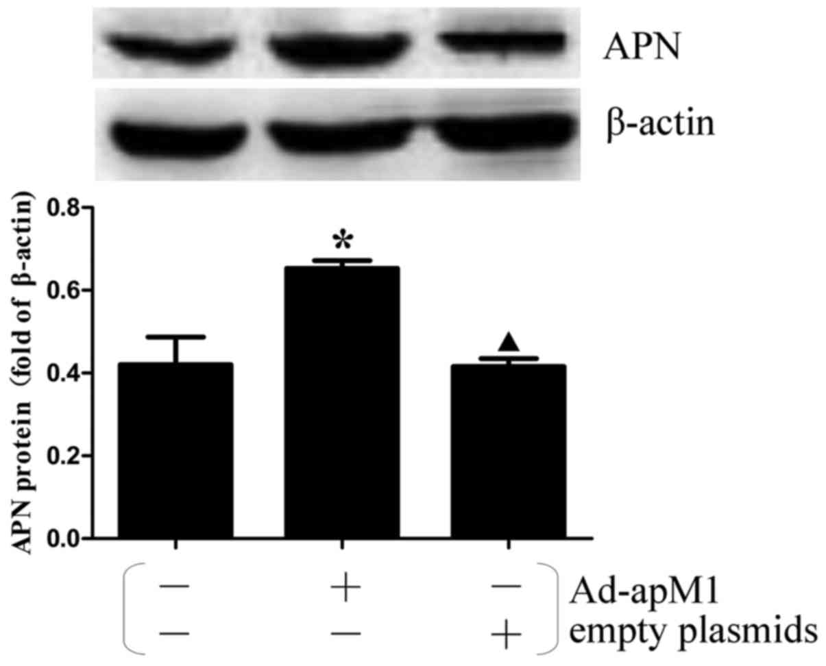
infective dose 50 was used to determine virus titer. Western
blotting was performed to verify the effects of APN overexpression
on protein expression levels following infection of 3T3-L1
preadipocytes with the adenovirus (Fig. 1).
Experimental groups and
treatments
In a 6 wells Transwell plate (Corning, Corning, NY,
USA), 3T3-L1 preadipocytes (1×10⁵ cells) in the inner
chamber, were co-cultured with mature adipocytes (2×10⁵
cells) in outer chamber, which were differentiated from 3T3-L1
preadipocytes. The cells were divided into the following
experimental groups: i) The control group, in which 3T3-L1
preadipocytes were induced to differentiate into adipocytes in the
inner chamber, as aforementioned. Mature adipocytes were cultured
in the outer chamber, with the addition of PBS, equal in volume to
LPS in group ii. ii) The LPS stimulation group, in which mature
adipocytes were stimulated with LPS (1 µg/ml, 18 h) to induce
inflammation, as described in previous studies (15,16).
The mature adipocytes in the outer chamber were co-cultured with
3T3-L1 preadipocytes in the inner chamber, which were induced to
differentiate into mature adipocytes using MDI, as aforementioned.
iii) The human APN recombinant adenovirus group (LPS + Ad-apM1), in
which preadipocytes were infected with Ad-apM1 [multiplicity of
infection (MOI), 100], as previously described (17). After a 48-h infection with the
adenovirus preadipocytes were induced into mature adipocytes and
were co-cultured with LPS-stimulated mature adipocytes. iv) The
negative control (NC) group, in which preadipocytes were infected
with an adenovirus containing empty plasmids (MOI, 100) as an NC;
after 48 h, the cells were induced to differentiate into mature
adipocytes and were co-cultured with mature adipocytes stimulated
with LPS. v) The PPARγ-inhibited group (LPS + Ad-apM1 + T0070907),
in which preadipocytes were infected with Ad-apM1 as aforementioned
and were treated with 10 µM T0070907 24 h prior to being induced to
differentiate into mature adipocytes via co-culturing with
LPS-treated mature adipocytes. The time point and titer of
adenoviruses and the dose of T0070907 used in the present study
were determined during preliminary experiments. At day 8,
subsequent experiments on the 3T3-L1 cells in the inner chamber
were performed.
MTT assay
Cell viability of preadipocytes was determined
following differentiation in the inner chamber using an MTT assay
(Promega Corporation, Madison, WI, USA). MTT was directly dissolved
in cell culture medium and was then incubated with live cells.
Briefly, cells in the exponential growth phase were plated at
1×10⁴ cells/well in a 96-plate, and were incubated at
37°C with 20 µl MTT (5 mg/ml) in 100 µl cell culture medium for 4
h. After 4 h, the supernatant was discarded and 150 µl of DMSO was
added to each well. The vortex was rotated for about 10 min to
allow the crystals to dissolve. The absorbance of each well was
measured at a wavelength of 490 nm using a Spectra Max Paradigm
microplate reader (Molecular Devices, LLC, Sunnyvale, CA, USA).
Oil Red O staining
Oil Red O (Sigma-Aldrich; Merck KGaA) staining was
performed according to the manufacturer's protocol. A total of 8
days after the induction of adipocyte differentiation, adipocytes
in the inner chamber were washed three times with PBS and were
fixed with 10% formalin for 1 h at room temperature. The dishes
were washed once with 60% isopropanol and were then left to dry
completely. Subsequently, the cells were stained with 2 ml Oil Red
O for 2 h at 37°C, rinsed with 60% isopropanol, and were thoroughly
washed four times with PBS. Finally, images were captured using an
inverted microscope (magnification, ×400). After extracting Oil Red
O with 100% isopropanol, the absorbance of the extracted dye was
determined spectrophotometrically at 490 nm wavelength using a
Spectra Max Paradigm microplate reader (Molecular Devices, LLC)
(18).
RNA preparation and reverse
transcription (RT)-qPCR analysis
TRIzol reagent (Invitrogen; Thermo Fisher
Scientific, Inc.) was used to extract total RNA from EAT and
adipocytes in the inner chamber after 8 days of differentiation
according to the manufacturer's protocol. RT was conducted using
the Transcriptor First Strand cDNA Synthesis kit (Roche
Diagnostics) according to the manufacturer's protocol. The
oligonucleotide primer sequences, which were designed using Premier
Primer 5.0 software (Premier Biosoft International, Palo Alto, CA,
USA), are presented in Table I.
β-actin was used as an internal control. The synthesized
first-strand cDNA samples were subjected to qPCR using SYBR-Green
PCR Master mix (Toyobo Life Science, Osaka, Japan) and PCR was
performed using an ABI Prism 7700 Sequence Detector (Applied
Biosystems; Thermo Fisher Scientific, Inc.). Thermocycling
conditions were as follows: 2 min at 95°C followed by 40 cycles of
15 sec at 95°C, 15 sec at 60°C, 20 sec at 72°C, with a final
extension step of 60°C for 30 min. Integrity of the PCR products
was confirmed by dissociation curve analysis using 7500 Software
version 2.0.4 (Applied Biosystems; Thermo Fisher Scientific, Inc.).
The quantification cycle (Cq) values were determined and the
2−ΔΔCq method (19) was
used to calculate relative gene expression.
 | Table I.Quantitative polymerase chain
reaction primers. |
Table I.
Quantitative polymerase chain
reaction primers.
| Gene | Primer
sequence |
|---|
| β-actin | Forward:
TTACAGGAAGTCCCTCACCCTC |
|
| Reverse:
TCAGGGCATGGACGCGA |
| MCP-1 | Forward:
AACTGCATCTGCCCTAAGGT |
|
| Reverse:
ACTGTCACACTGGTCACTCC |
| IL-6 | Forward:
ACAAAGCCAGAGTCCTTCAGAG |
|
| Reverse:
GTGACTCCAGCTTATCTCTTGGT |
| IL-8 | Forward:
GCACTTGGGAAGTTAACGCA |
|
| Reverse:
GCACTTGGGAAGTTAACGCA |
| TNF-α | Forward:
AGCCGATGGGTTGTACCTTG |
|
| Reverse:
ATAGCAAATCGGCTGACGGT |
Western blotting
Following 8 days of differentiation, cells harvested
from the inner chamber were lysed with radioimmunoprecipitation
assay lysis buffer containing 50 mM Tris-HCl (pH 7.4), 150 mM NaCl,
1% sodium deoxycholate, 1% NP-40, 1 mM phenylmethylsulfonyl
fluoride and 1 mM EDTA. The extracts were then centrifuged at
10,140 × g for 15 min at 4°C to remove insoluble material. Total
protein concentrations were determined using a bicinchoninic acid
protein assay kit (Beyotime Institute of Biotechnology, Haimen,
China) according to the manufacturer's protocol. An equivalent
amount of total protein (40 µg) per well was diluted with sample
buffer containing 100 mM dithiothreitol and was heated at 98°C for
5 min. Subsequently, proteins were separated by 10–15% SDS-PAGE
using gel apparatus (Bio-Rad Laboratories, Inc., Hercules, CA,
USA), and were transferred to polyvinylidene fluoride membranes.
The membranes were then soaked in 5% non-fat dry milk for 2 h at
room temperature and were incubated overnight at 4°C with primary
antibodies (1:500) against β-actin, APN, PPARγ, Pref-1and C/EBPα.
Subsequently, the membranes were washed with Tris-buffered saline
containing 0.05% Tween (TBS-T) and were incubated with horseradish
peroxidase-conjugated secondary antibody (1:2,000) with agitation
at room temperature for 1 h. The membranes were washed three times
with TBS-T (20 min/wash) and immune complexes were visualized by
enhanced chemiluminescence (Tiangen Biotech Co., Ltd., Beijing,
China); band intensity was finally measured and semi-quantified
(20). The resulting images were
analyzed with Quantity One version 4.62 (Bio-Rad Laboratories,
Inc.).
Statistical analysis
Data are presented as the mean ± standard error of
the mean. The variance homogeneity test and one-way analysis of
variance were performed by SPSS version 20.0 (IBM Corp., Armonk,
NY, USA). The least significant difference method was used to
compare between groups. P<0.05 was considered to indicate a
statistically significant difference.
Results
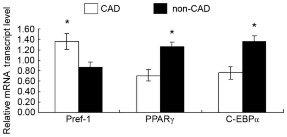
Expression levels of PPARγ, Pref-1 and
C/EBPα in EAT
The mRNA expression levels of PPARγ, Pref-1 and
C/EBPα in EAT were detected by qPCR. As presented in Fig. 2, compared with in the non-CAD
control group, the mRNA expression levels of PPARγ and C/EBPα were
significantly downregulated in the EAT from patients with CAD,
whereas Pref-1 was upregulated. These results suggested that PPARγ,
Pref-1 and C/EBPα may be involved in the occurrence and development
of atherosclerosis.
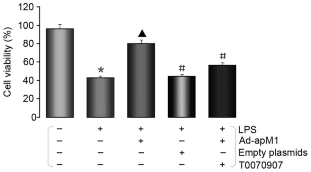
APN increases preadipocyte
viability
In order to determine the effects of APN on the
viability of 3T3-L1 cells co-cultured with LPS-stimulated
adipocytes, an MTT assay was conducted. As presented in Fig. 3, APN overexpression significantly
increased 3T3-L1 cell viability, whereas it was decreased in the
LPS group. Conversely, the effects of APN on cell viability were
reversed by T0070907, which is a selective inhibitor of PPARγ.
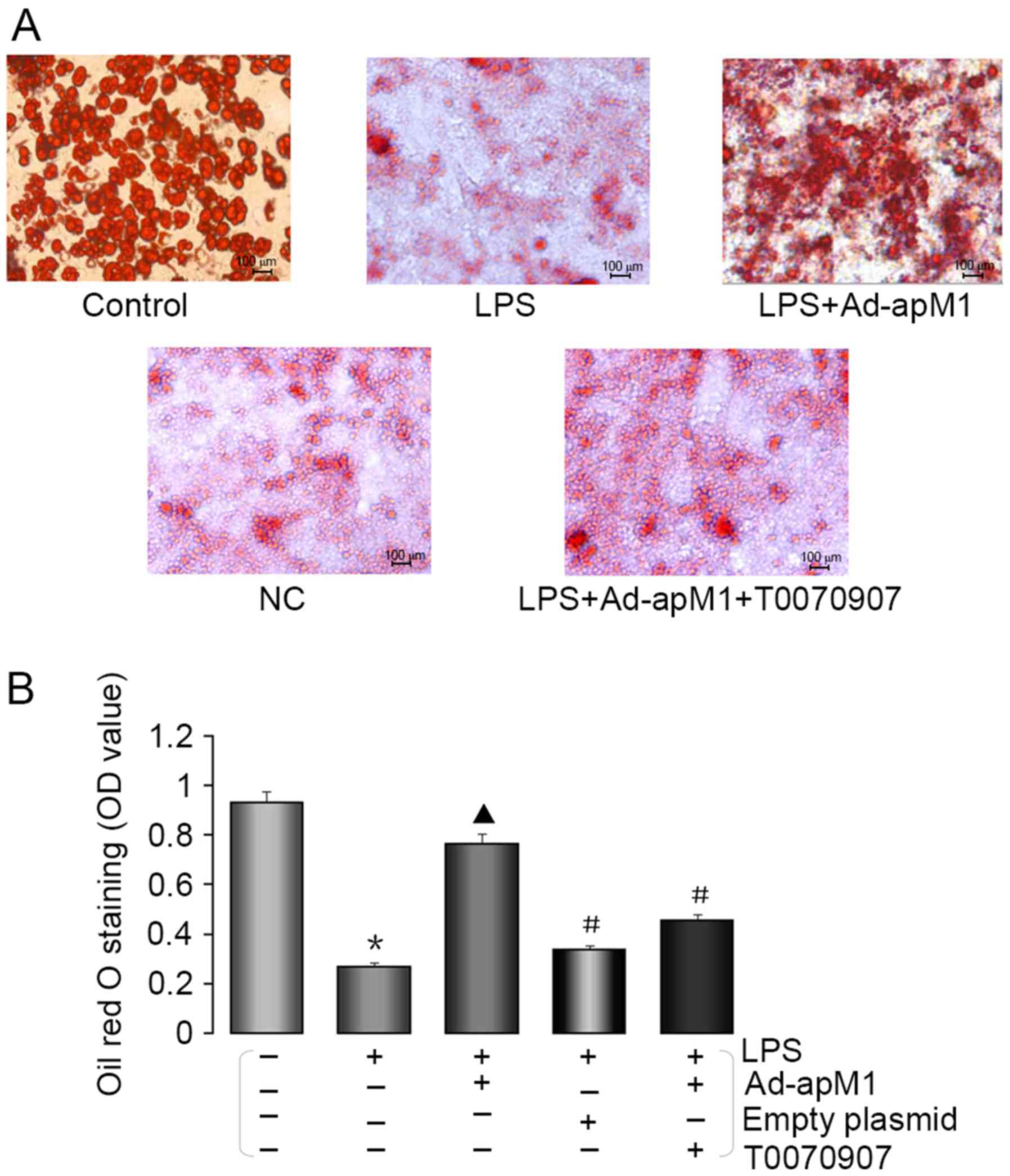
APN promotes preadipocyte
differentiation
The differentiation of preadipocytes was examined by
Oil Red O staining. As shown in Fig.
4A, cell volume and the number of lipid droplets was reduced in
3T3-L1 cells on day 8 following MDI-induced differentiation and
co-culture with LPS-stimulated adipocytes compared with in the
control group. However, this situation was markedly reversed in the
LPS + Ad-apM1 group. As expected, the effects of APN were markedly
attenuated in the NC and LPS + Ad-apM1 + T0070907 groups.
Following extraction of Oil Red O with 100%
isopropanol, absorbance of the extracted dye was determined
spectrophotometrically. As shown in Fig. 4B, the optical density (OD) value
was markedly decreased in the LPS group compared with in the
control group (P<0.05), whereas it was significantly increased
in the LPS + Ad-apM1 group compared with in the LPS group
(P<0.05). Conversely, the OD values were significantly decreased
in the NC and LPS + Ad-apM1 + T0070907 groups.
APN suppresses the expression of
inflammatory factors
The mRNA expression levels of MCP-1, IL-6, IL-8 and
TNF-α were detected in 3T3-L1 preadipocytes by qPCR. As shown in
Fig. 5, the mRNA expression levels
of MCP-1, IL-6, IL-8 and TNF-α were significantly increased in the
LPS group compared with in the control group (P<0.05).
Conversely, the mRNA expression levels of MCP-1, IL-6, IL-8 and
TNF-α were decreased in the APN overexpression group compared with
in the LPS group (P<0.05). However, the effects of APN
overexpression were markedly attenuated in the NC and LPS + Ad-apM1
+ T0070907 groups.
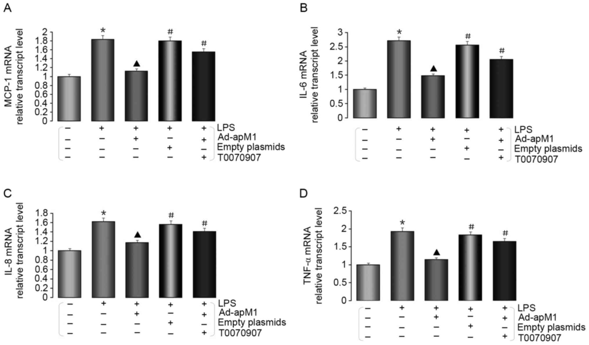 | Figure 5.APN suppresses the expression of
MCP-1, IL-6, IL-8 and TNF-α in preadipocytes 8 days after
co-culturing with LPS-stimulated mature adipocytes. Relative mRNA
expression levels of (A) MCP-1, (B) IL-6, (C) IL-8 and (D) TNF-α
were detected using quantitative polymerase chain reaction. Data
are presented as the mean ± standard error of the mean for six
independent experiments. *P<0.05 vs. the control group;
▲P<0.05 vs. the LPS group; #P<0.05 vs.
the LPS + Ad-apM1 group. Ad-apM1, adenovirus containing the apM1
gene; APN, adiponectin; IL, interleukin; LPS, lipopolysaccharide;
MCP-1, monocyte chemoattractant protein-1; TNF-α, tumor necrosis
factor-α. |
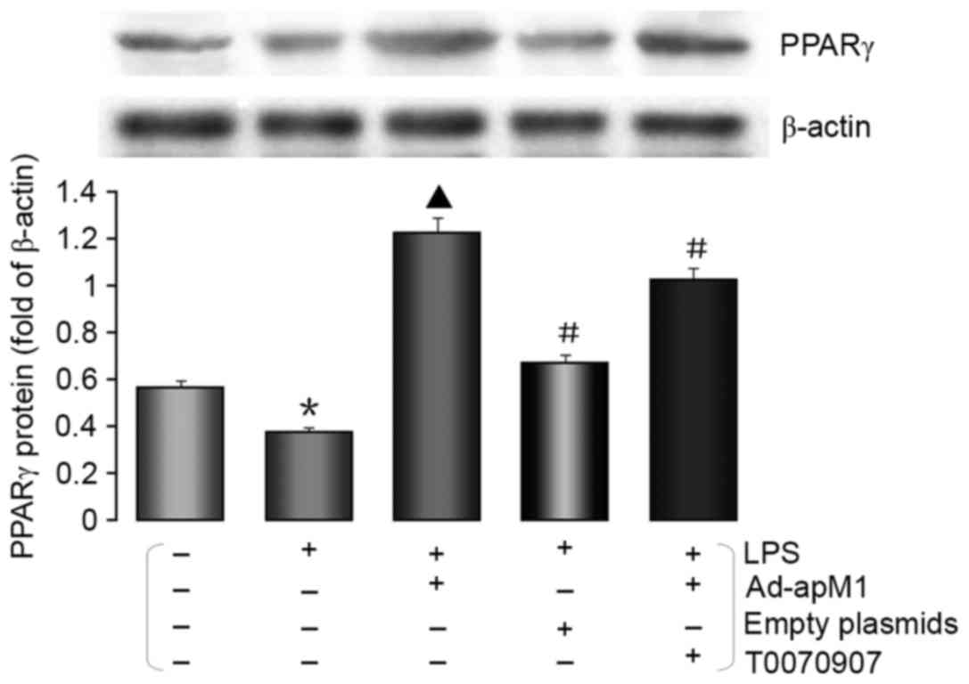
APN increases the protein expression
levels of PPARγ
To further investigate whether PPARγ expression was
altered in response to APN overexpression, the protein expression
levels of PPARγ were detected by western blotting. As shown in
Fig. 6, the protein expression
levels of PPARγ were significantly increased in the LPS + Ad-apM1
group compared with in the LPS group (P<0.05). However, there
was a marked decrease in the expression of PPARγ in the NC and LPS
+ Ad-apM1 + T0070907 groups. These results suggested that APN may
promote the differentiation of preadipocytes co-cultured with
LPS-stimulated mature adipocytes via the upregulation of PPARγ.
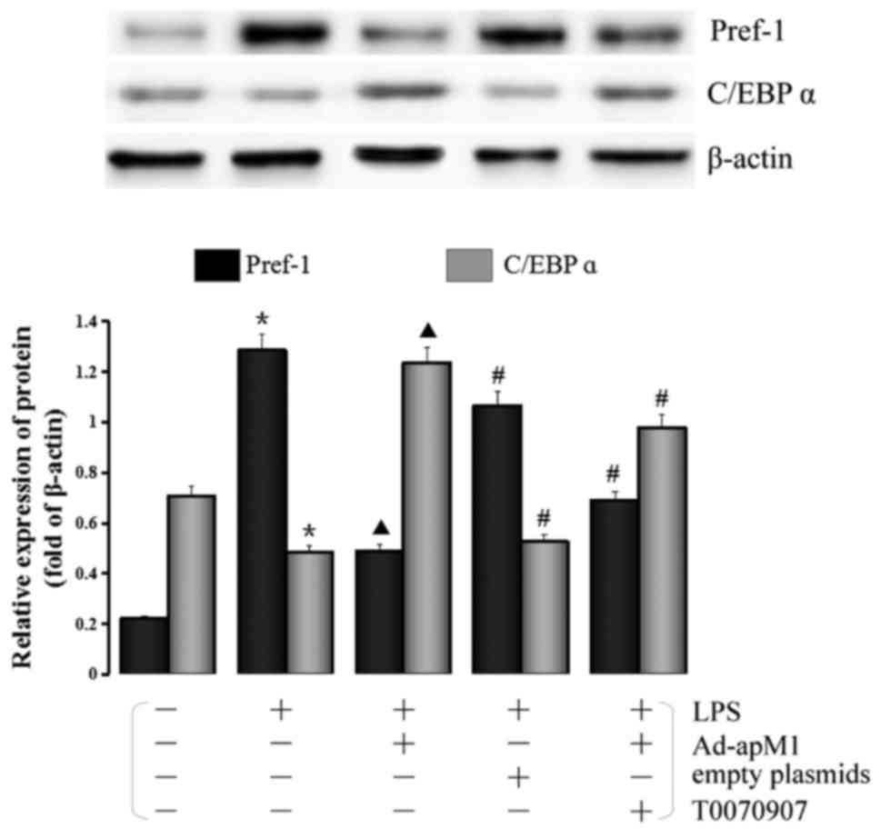
Effects of APN overexpression on the
protein expression levels of Pref-1 and C/EBPα
As shown in Fig. 7,
the inflammatory environment markedly increased Pref-1 expression
compared with in the control group (P<0.05), whereas APN
overexpression decreased the expression levels of Pref-1 compared
with in the LPS group. Conversely, the expression levels of Pref-1
were significantly increased in the NC and LPS + Ad-apM1+T0070907
groups (P<0.05). Furthermore, the expression levels of C/EBPα
were significantly downregulated in the LPS group compared with in
the control groups (P<0.05), whereas its expression was
increased in the LPS + Ad-apM1 group compared with in the LPS group
(P<0.05). However, the expression levels of C/EBP α were
markedly decreased in the NC and LPS + Ad-apM1 + T0070907
groups.
Discussion
It has previously been reported that elevated
proinflammatory cytokine levels mediate insulin resistance, type II
diabetes and cardiovascular disease (21). Inflammatory cytokines, including
MCP-1, IL-6, IL-8 and TNF-α, which are secreted by adipose tissue,
are able to increase infiltration of local immune cells and
aggravate chronic inflammation in adipose tissue, thus leading to
adipose tissue dysfunction and metabolic disorders (22,23).
As adipose tissue, EAT is able to secrete numerous inflammatory
factors, which induce inflammation and atherosclerosis (24). In the present study, the
differentiation of preadipocytes into mature adipocytes was
significantly inhibited, and the expression of inflammatory factors
was markedly increased, under inflammatory stimulation.
There are numerous causes of metabolic disorders, of
which adipocyte hypertrophy is one of the major risk factors. Under
normal conditions, adipose tissue remains metabolically healthy
through the constant production of newer and smaller fat cells;
preadipocyte differentiation serves an important role in this
process (25). APN, which is
secreted by adipose tissue, is an adipokine hormone that is closely
associated with lipid and carbohydrate metabolism, and is composed
of an N-terminal collagenous domain and a C-terminal globular
domain (26,27). The effects of APN on adipocyte
differentiation remain controversial. It has previously been
suggested that APN may promote adipocyte differentiation, insulin
sensitivity and lipid accumulation (28). However, Bauche et al
(29) observed that in a mouse
model of obesity, APN overexpression resulted in a marked reduction
in energy expenditure and impairment in the differentiation of
adipocytes. In the present study, the overexpression of APN
significantly improved preadipocyte differentiation under
inflammatory conditions, which is consistent with the findings of
Fu et al (28). Previous
studies have reported that APN could suppress the growth of
myelomonocytic progenitors and the function of mature macrophages,
inhibit macrophage-to-foam cell transformation and promote the
secretion of anti-inflammatory cytokines from macrophages (30–32).
Furthermore, in adipocytes, APN may suppress LPS-induced nuclear
factor-κB activation, which is closely associated with the
downregulation of inflammatory responses, and it has been suggested
that there is an inverse relationship between TNF-α and APN
(33–37). In the present study, the results of
a qPCR analysis indicated that the expression levels of MCP-1,
IL-6, IL-8 and TNF-α were suppressed by APN overexpression, thus
indicating that the effects of APN on the promotion of preadipocyte
differentiation may be associated with suppression of inflammatory
factor secretion by adipocytes. These findings suggested that APN
may act as an anti-inflammatory factor in adipose tissue
metabolism.
The regulatory mechanism underlying adipocyte
differentiation is complex. A regulated transcriptional cascade is
known to control adipocyte differentiation through activating or
suppressing the expression of transcription factors in a sequential
fashion (38). PPARγ is a member
of the nuclear receptor superfamily of ligand-activated
transcription factors (39). In
vivo and in vitro studies have reported that PPARγ is an
essential regulator of adipogenesis (40). PPARγ is an important transcription
factor that initiates the expression of genes that are required to
convert precursor cells into mature adipocytes; the activation of
PPARγ is necessary and sufficient for adipocyte differentiation
(41). There are two isoforms of
PPARγ, PPARγ1 and PPARγ2, which are generated from alternate
promoter usage and splicing; at the amino-terminus 30 additional
amino acids are contained in PPARγ2 (42). Both isoforms are specifically
activated in the process of adipocyte differentiation; however,
only PPARγ2 is strictly expressed in adipose tissues. In addition,
it has been reported that PPARγ2 serves a more central role in
adipocyte differentiation (43). A
previous study has demonstrated that cytokines that regulating
adipocyte differentiation exercise their effects by regulating
PPARg expression or activity (44). The present study demonstrated that
APN overexpression may increase the protein expression levels of
PPARγ in preadipocytes following MDI-induced differentiation and
co-culture with LPS-stimulated mature adipocytes. Conversely, the
positive effects of APN on preadipocyte differentiation could be
antagonized following treatment with T0070907, which is a specific
inhibitor of PPARγ (45). These
results suggested that the effects of APN on preadipocytes in an
inflammatory environment may be involved with the PPARγ signaling
pathway.
C/EBPs are a basic leucine zipper family of
transcription factors, which are crucial for adipogenesis. C/EBPα
is a member of the C/EBP family that is specifically required for
adipogenesis (38). A previous
report indicated that PPARγ and C/EBPα may promote adipogenesis
through modulating the expression of each other. C/EBPα cooperates
with PPARγ by inducing the expression of multiple subsets of
adipocyte-specific genes during adipocyte differentiation (46). C/EBPβ and δ, which are two other
members of the C/EBP family, are also associated with PPARγ and
C/EBPα transcriptional induction (38). The majority of induced genes in the
process of adipogenesis are bound by PPARγ and C/EBPα, thus
indicating that the two master regulators may cooperatively
upregulate the expression of adipogenic genes (47). The present study demonstrated that
APN overexpression could upregulate the protein expression levels
of C/EBPα in 3T3-L1 cells following MDI-induced differentiation
under inflammatory conditions, whereas its expression was inhibited
by T0070907. Furthermore, the expression of Pref-1 was evaluated;
Pref-1 is an epidermal growth factor repeat-containing
transmembrane protein. The overexpression of Pref-1, or the
treatment of preadipocytes with soluble Pref-1, leads to the
inhibition of adipocyte differentiation (48). The results of the present study
demonstrated that an increase in the expression of inflammatory
factors may promote overexpression of Pref-1, which could suppress
the differentiation of preadipocytes. Conversely, APN may inhibit
Pref-1 expression and promote preadipocyte differentiation, which
was attenuated by T0070907. Taken together, these results suggested
that APN promotes preadipocyte differentiation under inflammatory
conditions, which may be due to upregulation of PPARγ expression,
and the regulation of C/EBPα, Pref-1 and inflammatory factors
expression. However, the specific mechanisms remain to be further
elucidated.
In conclusion, the present study demonstrated that
APN attenuates inflammation-induced inhibition of preadipocyte
differentiation, potentially via the PPARγ signaling pathway.
Therefore, it may be hypothesized that APN promotes the
differentiation of adipocytes in response to inflammatory
stimulation, accelerates the metabolism of visceral adipose tissue
and reduces the secretion of inflammatory cytokines in EAT. As a
result, atherosclerosis may be improved. However, in the present
study, the inflammatory environment of EAT was simulated using
LPS-stimulated 3T3-L1 mature adipocytes, which differs from the
actual situation in human EAT, which is the most marked limitation
of the present research. Although numerous studies have suggested
that APN is beneficial to atherosclerosis (9–11),
other studies have reported a lack of association between APN
levels and atherosclerosis in preclinical rodent models (49). Further research is required in
vivo and in vitro to explore the role of APN in
improving atherosclerosis.
Acknowledgements
The present study was supported by the Science and
Technology Planning Project of Jiangxi Provincial Health Department
(grant no. 20133019).
References
|
1
|
Zhou Y, Wei Y, Wang L, Wang X, Du X, Sun
Z, Dong NG and Chen XZ: Decreased adiponectin and increased
inflammation expression in epicardial adipose tissue in coronary
artery disease. Cardiovasc Diabetol. 10:22011. View Article : Google Scholar : PubMed/NCBI
|
|
2
|
Barton M: Obesity and aging: Determinants
of endothelial cell dysfunction and atherosclerosis. Pflugers Arch.
460:825–837. 2010. View Article : Google Scholar : PubMed/NCBI
|
|
3
|
Ragbir S and Farmer JA: Dysfunctional
high-density lipoprotein and atherosclerosis. Curr Atheroscler Rep.
12:343–348. 2010. View Article : Google Scholar : PubMed/NCBI
|
|
4
|
Caccamo G, Bonura F, Bonura F, Vitale G,
Novo G, Evola S, Evola G, Grisanti MR and Novo S: Insulin
resistance and acute coronary syndrome. Atherosclerosis.
211:672–675. 2010. View Article : Google Scholar : PubMed/NCBI
|
|
5
|
Ridker PM and Silvertown JD: Inflammation,
C-reactive protein, and atherothrombosis. J Periodontol.
79:1544–1551. 2008. View Article : Google Scholar : PubMed/NCBI
|
|
6
|
Stamatelopoulos KS, Kitas GD, Papamichael
CM, Chryssohoou E, Kyrkou K, Georgiopoulos G, Protogerou A,
Panoulas VF, Sandoo A, Tentolouris N, et al: Atherosclerosis in
rheumatoid arthritis versus diabetes: A comparative study.
Arterioscler Thromb Vasc Biol. 29:1702–1708. 2009. View Article : Google Scholar : PubMed/NCBI
|
|
7
|
Iacobellis G, Corradi D and Sharma AM:
Epicardial adipose tissue: Anatomic, biomolecular and clinical
relationships with the heart. Nat Clin Pract Cardiovasc Med.
2:536–543. 2005. View Article : Google Scholar : PubMed/NCBI
|
|
8
|
Ruisi P, Makaryus JN, Ruisi M and Makaryus
AN: Inflammatory bowel disease as a risk factor for premature
coronary artery disease. J Clin Med Res. 7:257–261. 2015.
View Article : Google Scholar : PubMed/NCBI
|
|
9
|
Carneiro FS, Webb RC and Tostes RC:
Emerging role for TNF-α in erectile dysfunction. J Sex Med.
7:3823–3834. 2010. View Article : Google Scholar : PubMed/NCBI
|
|
10
|
Saarikoski LA, Juonala M, Huupponen R,
Viikari JS, Lehtimäki T, Jokinen E, Hutri-Kähönen N, Taittonen L,
Laitinen T and Raitakari OT: Low serum adiponectin levels in
childhood and adolescence predict increased intima-media thickness
in adulthood. The cardiovascular risk in young finns study. Ann
Med. 49:42–50. 2017. View Article : Google Scholar : PubMed/NCBI
|
|
11
|
Gasbarrino K, Gorgui J, Nauche B, Côté R
and Daskalopoulou SS: Circulating adiponectin and carotid
intima-media thickness: A systematic review and meta-analysis.
Metabolism. 65:968–986. 2016. View Article : Google Scholar : PubMed/NCBI
|
|
12
|
Liu X, Malki A, Cao Y, Li Y, Qian Y, Wang
X and Chen X: Glucose-and triglyceride-lowering dietary
penta-o-galloyl-α-d-glucose reduces expression of PPARγ and C/EBPα,
induces p21-mediated G1 phase cell cycle arrest and inhibits
adipogenesis in 3T3-L1 preadipocytes. Exp Clin Endocrinol Diabetes.
123:308–316. 2015. View Article : Google Scholar : PubMed/NCBI
|
|
13
|
Lv S, Wu L, Cheng P, Yu J, Zhang A, Zha J,
Liu J, Wang L, DI W, Hu M, et al: Correlation of obesity and
osteoporosis: Effect of free fatty acids on bone marrow-derived
mesenchymal stem cell differentiation. Exp Ther Med. 1:603–610.
2010. View Article : Google Scholar : PubMed/NCBI
|
|
14
|
Fukuhara A, Matsuda M, Nishizawa M, Segawa
K, Tanaka M, Kishimoto K, Matsuki Y, Murakami M, Ichisaka T,
Murakami H, et al: Visfatin: A protein secreted by visceral fat
that mimics the effects of insulin. Science. 307:426–430. 2005.
View Article : Google Scholar : PubMed/NCBI
|
|
15
|
Jensen MD: Role of body fat distribution
and the metabolic complications of obesity. J Clin Endocrinol
Metab. 93 11 Suppl 1:S57–S63. 2008. View Article : Google Scholar : PubMed/NCBI
|
|
16
|
Antuna-Puente B, Feve B, Fellahi S and
Bastard JP: Adipokines: The missing link between insulin resistance
and obesity. Diabetes Metab. 34:2–11. 2008. View Article : Google Scholar : PubMed/NCBI
|
|
17
|
Dubuisson O, Dhurandhar EJ, Krishnapuram
R, Kirk-Ballard H, Gupta AK, Hegde V, Floyd E, Gimble JM and
Dhurandhar NV: PPARgamma-independent increase in glucose uptake and
adiponectin abundance in fat cells. Endocrinology. 152:3648–3660.
2011. View Article : Google Scholar : PubMed/NCBI
|
|
18
|
Chen X, Luo Y, Huang Z, Jia G, Liu G and
Zhao H: Role of phosphotyrosine interaction domain containing 1 in
porcine intramuscular preadipocyte proliferation and
differentiation. Anim Biotechnol. 27:287–294. 2016. View Article : Google Scholar : PubMed/NCBI
|
|
19
|
Livak KJ and Schmittgen TD: Analysis of
relative gene expression data using real-time quantitative PCR and
the 2(-Delta Delta C(T)) method. Methods. 25:402–408. 2001.
View Article : Google Scholar : PubMed/NCBI
|
|
20
|
Chen C, He H, Luo Y, Zhou M, Yin D and He
M: Involvement of Bcl-2 signal pathway in the protective effects of
apigenin on anoxia/reoxygenation-induced myocardium injury. J
Cardiovasc Pharmacol. 67:152–163. 2016. View Article : Google Scholar : PubMed/NCBI
|
|
21
|
Guo W, Li Y, Liang W, Wong S, Apovian C,
Kirkland JL and Corkey BE: Beta-mecaptoethanol suppresses
inflammation and induces adipogenic differentiation in 3T3-F442A
murine preadipocytes. PLoS One. 7:e409582012. View Article : Google Scholar : PubMed/NCBI
|
|
22
|
Zhang H, Huang Y, Bu D, Chen S, Tang C,
Wang G, Du J and Jin H: Endogenous sulfur dioxide is a novel
adipocyte-derived inflammatory inhibitor. Sci Rep. 6:270262016.
View Article : Google Scholar : PubMed/NCBI
|
|
23
|
Lu JC, Chang YT, Wang CT, Lin YC, Lin CK
and Wu ZS: Trichostatin A modulates thiazolidinedione-mediated
suppression of tumor necrosis factor α-induced lipolysis in 3T3-L1
adipocytes. PLoS One. 8:e715172013. View Article : Google Scholar : PubMed/NCBI
|
|
24
|
Mazurek T, Zhang L, Zalewski A, Mannion
JD, Diehl JT, Arafat H, Sarov-Blat L, O'Brien S, Keiper EA, Johnson
AG, et al: Human epicardial adipose tissue is a source of
inflammatory mediators. Circulation. 108:2460–2466. 2003.
View Article : Google Scholar : PubMed/NCBI
|
|
25
|
Glass CK and Olefsky JM: Inflammation and
lipid signaling in the etiology of insulin resistance. Cell Metab.
15:635–645. 2012. View Article : Google Scholar : PubMed/NCBI
|
|
26
|
Scherer PE, Williams S, Fogliano M,
Baldini G and Lodish HF: A novel serum-protein similar to C1Q,
produced exclusively in adipocytes. J Biol Chem. 270:26746–26749.
1995. View Article : Google Scholar : PubMed/NCBI
|
|
27
|
Maeda K, Okubo K, Shimomura I, Funahashi
T, Matsuzawa Y and Matsubara K: CDNA cloning and expression of a
novel adipose specific collagen-like factor, apM1 (Adipose most
abundant gene transcript 1). Biochem Biophys Res Commun.
221:286–289. 1996. View Article : Google Scholar : PubMed/NCBI
|
|
28
|
Fu YC, Luo NL, Klein RL and Garvey WT:
Adiponectin promotes adipocyte differentiation, insulin sensitivity
and lipid accumulation. J Lipid Res. 46:1369–1379. 2005. View Article : Google Scholar : PubMed/NCBI
|
|
29
|
Bauche IB, El Mkadem SA, Pottier AM, Senou
M, Many MC, Rezsohazy R, Penicaud L, Maeda N, Funahashi T and
Brichard SM: Overexpression of adiponectin targeted to adipose
tissue in transgenic mice: Impaired adipocyte differentiation.
Endocrinology. 148:1539–1549. 2007. View Article : Google Scholar : PubMed/NCBI
|
|
30
|
Yokota T, Oritani K, Takahashi I, Ishikawa
J, Matsuyama A, Ouchi N, Kihara S, Funahashi T, Tenner AJ, Tomiyama
Y and Matsuzawa Y: Adiponectin, a new member of the family of
soluble defense collagens, negatively regulates the growth of
myelomonocytic progenitors and the functions of macrophages. Blood.
96:1723–1732. 2000.PubMed/NCBI
|
|
31
|
Ouchi N, Kihara S, Arita Y, Nishida M,
Matsuyama A, Okamoto Y, Ishigami M, Kuriyama H, Kishida K,
Nishizawa H, et al: Adipocyte-derived plasma protein, adiponectin,
suppresses lipid accumulation and class a scavenger receptor
expression in human monocyte-derived macrophages. Circulation.
103:1057–1063. 2001. View Article : Google Scholar : PubMed/NCBI
|
|
32
|
Kumada M, Kihara S, Ouchi N, Kobayashi H,
Okamoto Y, Ohashi K, Maeda K, Nagaretani H, Kishida K, Maeda N, et
al: Adiponectin specifically increased tissue inhibitor of
metalloproteinase-1 through interleukin-10 expression in human
macrophages. Circulation. 109:2046–2049. 2004. View Article : Google Scholar : PubMed/NCBI
|
|
33
|
Ajuwon KM and Spurlock ME: Adiponectin
inhibits LPS-induced NF-kappaB activation and IL-6 production and
increases PPARgamma2 expression in adipocytes. Am J Physiol Regul
Integr Comp Physiol. 288:R1220–R1225. 2005. View Article : Google Scholar : PubMed/NCBI
|
|
34
|
Ouchi N, Kihara S, Arita Y, Okamoto Y,
Maeda K, Kuriyama H, Hotta K, Nishida M, Takahashi M, Muraguchi M,
et al: Adiponectin, an adipocyte-derived plasma protein, inhibits
endothelial NF-kappaB signaling through a cAMP-dependent pathway.
Circulation. 102:1296–1301. 2000. View Article : Google Scholar : PubMed/NCBI
|
|
35
|
Wulster-Radcliffe MC, Ajuwon KM, Wang J,
Christian JA and Spurlock ME: Adiponectin differentially regulates
cytokines in porcine macrophages. Biochem Biophys Res Commun.
316:924–929. 2004. View Article : Google Scholar : PubMed/NCBI
|
|
36
|
Bruun JM, Lihn AS, Verdich C, Pedersen SB,
Toubro S, Astrup A and Richelsen B: Regulation of adiponectin by
adipose tissue-derived cytokines: In vivo and in vitro
investigations in humans. Am J Physiol Endocrinol Metab.
285:E527–E533. 2003. View Article : Google Scholar : PubMed/NCBI
|
|
37
|
Kern PA, Di Gregorio GB, Lu T, Rassouli N
and Ranganathan G: Adiponectin expression from human adipose
tissue: Relation to obesity, insulin resistance, and tumor necrosis
factor-alpha expression. Diabetes. 52:1779–1785. 2003. View Article : Google Scholar : PubMed/NCBI
|
|
38
|
Farmer SR: Transcriptional control of
adipocyte formation. Cell Metab. 4:263–273. 2006. View Article : Google Scholar : PubMed/NCBI
|
|
39
|
Evans RM, Barish GD and Wang YX: PPARs and
the complex journey to obesity. Nat Med. 10:355–361. 2004.
View Article : Google Scholar : PubMed/NCBI
|
|
40
|
Barak Y, Nelson MC, Ong ES, Jones YZ,
Ruiz-Lozano P, Chien KR, Koder A and Evans RM: PPAR gamma is
required for placental, cardiac, and adipose tissue development.
Mol Cell. 4:585–595. 1999. View Article : Google Scholar : PubMed/NCBI
|
|
41
|
Kolehmainen M, Uusitupa MI, Alhava E,
Laakso M and Vidal H: Effect of the Pro12Ala polymorphism in the
peroxisome proliferator-activated receptor (PPAR) gamma2 gene on
the expression of PPARgamma target genes in adipose tissue of
massively obese subjects. J Clin Endocrinol Metab. 88:1717–1722.
2003. View Article : Google Scholar : PubMed/NCBI
|
|
42
|
Tontonoz P and Spiegelman BM: Fat and
beyond: The diverse biology of PPARgamma. Annu Rev Biochem.
77:289–312. 2008. View Article : Google Scholar : PubMed/NCBI
|
|
43
|
Zhang J, Fu M, Cui T, Xiong C, Xu K, Zhong
W, Xiao Y, Floyd D, Liang J, Li E, et al: Selective disruption of
PPARgamma 2 impairs the development of adipose tissue and insulin
sensitivity. Proc Natl Acad Sci USA. 101:10703–10708. 2004.
View Article : Google Scholar : PubMed/NCBI
|
|
44
|
Rosen ED and MacDougald OA: Adipocyte
differentiation from the inside out. Nat Rev Mol Cell Biol.
7:885–896. 2006. View Article : Google Scholar : PubMed/NCBI
|
|
45
|
An Z, Muthusami S, Yu JR and Park WY:
T0070907, a PPAR γ inhibitor, induced G2/M arrest enhances the
effect of radiation in human cervical cancer cells through mitotic
catastrophe. Reprod Sci. 21:1352–1361. 2014. View Article : Google Scholar : PubMed/NCBI
|
|
46
|
Rosen ED, Hsu CH, Wang X, Sakai S, Freeman
MW, Gonzalez FJ and Spiegelman BM: C/EBPalpha induces adipogenesis
through PPARgamma: A unified pathway. Genes Dev. 16:22–26. 2002.
View Article : Google Scholar : PubMed/NCBI
|
|
47
|
Lefterova MI, Zhang Y, Steger DJ, Schupp
M, Schug J, Cristancho A, Feng D, Zhuo D, Stoeckert CJ Jr, Liu XS
and Lazar MA: PPARgamma and C/EBP factors orchestrate adipocyte
biology via adjacent binding on a genome-wide scale. Genes Dev.
22:2941–2952. 2008. View Article : Google Scholar : PubMed/NCBI
|
|
48
|
Smas CM, Chen L and Sul HS: Cleavage of
membrane-associated pref-1 generates a soluble inhibitor of
adipocyte differentiation. Mol Cell Biol. 17:977–988. 1997.
View Article : Google Scholar : PubMed/NCBI
|
|
49
|
Nawrocki AR, Hofmann SM, Teupser D,
Basford JE, Durand JL, Jelicks LA, Woo CW, Kuriakose G, Factor SM,
Tanowitz HB, et al: Lack of association between adiponectin levels
and atherosclerosis in mice. Arterioscler Thromb Vasc Biol.
30:1159–1165. 2010. View Article : Google Scholar : PubMed/NCBI
|