Introduction
Adipose-derived stem cells (ADSCs) can differentiate
into a variety of cell types, including adipocytes, osteoblasts,
chondrocytes and myocytes (1,2).
They are widely used in regenerative medicine (3–7). The
primary ADSCs from adipose tissue are a mixture of cell types,
including preadipocytes, having the ability to spontaneously
differentiate into adipocytes (1,2). The
spontaneous potential of preadipocytes to differentiate into
adipocytes may compromise the proliferation and/or differentiation
potential of ADSCs (8,9), especially in chondrogenic or
osteogenic differentiation. The influence of spontaneous adipogenic
differentiation should be minimized in chondrogenic or osteogenic
regeneration, but it does not seem to be harmful in adipogenic
regenerative medicine. Thus, understanding how the potential of
spontaneous adipogenic differentiation of ADSCs changes may be
worthy of tissue regeneration.
The heterogeneity of cultured ADSCs is reduced
quickly by continuous cell passages (8,10–12).
Whether the spontaneous adipogenic differentiation is affected by
increasing number of cell passages is not clear so far. The
spontaneous adipogenic differentiation of ADSCs was assumed to
decrease when the cells were passaged serially. ADSCs from passages
3 and 4 were reported to be used in regenerative medicine (13–15).
Krähenbühl et al (16)
reported that cell morphology changed and growth slowed down when
extended to passages 6 and beyond. Therefore, ADSCs from passages 1
to 5 was explored in this study to observe the change in
spontaneous adipogenic differentiation for cells from different
passages.
The induced adipogenic potential of ADSCs was
reported to decrease with increasing passage number (17). Therefore, the induced adipogenic
differentiation potentials at different passages were also studied
and compared in the present study.
Peroxisome proliferator-activated receptor γ (PPARγ)
and CCAAT/enhancer-binding protein α (C/EBPα) have been
demonstrated to be vital in adipocyte differentiation and these two
transcription factors cooperate on multiple binding sites in
promoter regions and regulate a wide range of genes expressed in
developing and mature adipocytes (18). This study observed Cebpa and
Pparg as adipocyte marker genes in evaluating the
spontaneous and induced adipogenic differentiation of ADSCs and
explored its potential to evaluate the feasibility of ADSCs from
passages 1 to 5 and facilitate the selection of optimal cell
passage for regenerating different tissues.
Materials and methods
Chemicals, antibodies and dyes
3-Isobutyl-1-methylxanthine (IBMX), dexamethasone,
indomethacin, insulin and Oil Red O were purchased from
Sigma-Aldrich (Merck KGaA, Darmstadt, Germany). Rabbit
anti-Pparγ and anti-CEBPα antibodies were procured from
Abcam (Cambridge, MA, USA).
Isolation and culture of mouse
ADSCs
All C57BL/6 mice aged 7–10 days, purchased from
Weitong-Lihua Laboratory (Beijing, China), were age- and
sex-matched for each experiment. All animal experiments were
approved by the Institutional Animal Care and Use Committee of
Beijing Shijitan Hospital of Capital Medical University, Beijing,
China (no. 2017-KL16).
Mouse ADSCs (mADSCs) were isolated and cultured as
described in a previous study (19). In brief, the subcutaneous adipose
tissue of C57BL/6 mice was cut into small pieces and digested with
collagenase I in phosphate-buffered saline (PBS) for 1 h at 37°C.
The cells were then washed and filtered through a 70-µm cell
strainer, resulting in a single-cell suspension. Isolated cells
were then seeded at a density of 3–4×10⁴
cells/cm2 in a culture vessel containing low-glucose
Dulbecco's modified Eagles medium (DMEM) supplemented with 10%
fetal bovine serum (FBS) (both from Gibco; Thermo Fisher
Scientific, Inc., Waltham, MA, USA) and 1% antibiotic-antimycotic
solution (Invitrogen; Thermo Fisher Scientific, Inc.), which
favored the attachment and growth of fibroblastic cells (20). The attached cells were washed and
replenished using a fresh medium every 48 h to discard unattached
dead cells or immune cells, and by days 3–5, >90% cells
displayed typical fibroblastic morphology. These adherent cells
were named primary ADSCs (P0). Cell passaging was performed using a
0.25% trypsin solution (Sigma-Aldrich) every 3–5 days when the
attached cells reached 80–90% confluence and cells were seeded at
3×10⁴ cells/cm2 per passage for a total of 5
passages.
For induced adipogenic differentiation, one-day
postconfluence cells were switched to adipogenic differentiation
medium [DMEM with FBS (10%), 0.5 mM IBMX, 1 µM dexamethasone, 10 µM
insulin, 200 µM indomethacin, and 1% antibiotic/antimycotic] to
induce differentiation as described in a previous study (21). The cells were collected at
indicated time points for RNA and protein extraction.
Oil Red O staining
The cells were harvested on the induction day
according to the experimental design. They were rinsed with PBS
twice and fixed with 4% polyoxymethylene in PBS (w/v) for 30 min at
room temperature. Oil Red O (0.5 g; Amresco, LLC, Solon, OH, USA)
was dissolved in isopropanol (100 ml, w/v), diluted with water
(6:4, v/v), and filtered. The fixed cells were then stained with
the filtered Oil Red O solution for 1 h at room temperature and
exhaustively rinsed with water. Excess water was evaporated by
placing the stained cultures at a temperature of 25–30°C. Then, 0.8
ml of isopropyl alcohol was added to the stained culture dish. The
extracted dye was immediately removed by gentle pipetting, and its
absorbance monitored spectrophotometrically at 510 nm as described
in a previous study (22).
Reverse transcription-quantitative polymerase chain
reaction (RT-qPCR). Total cellular RNA was extracted using the
RNAeasy reagent (Qiagen GmbH, Hilden, Germany), and 500 ng of RNA
was reverse transcribed to cDNA using a cDNA synthesis kit (ComWin,
Beijing, China). qPCR was performed using the QuantStudio 6 Flex 96
real-time system (Applied Biosystems; Thermo Fisher Scientific,
Inc.) using SYBR-Green Mix (ComWin). All the primers used with
SYBR-Green were designed to span at least one exon to minimize the
possibility of nonspecific amplification from the genomic DNA.
Glyceraldehyde-3-phosphate dehydrogenase (GAPDH) gene was used as a
housekeeping gene to normalize data. Specific primer sequences were
used as described in a previous study (19) and are shown in Table I.
 | Table I.List of primers used for reverse
transcription-quantitative polymerase chain reaction. |
Table I.
List of primers used for reverse
transcription-quantitative polymerase chain reaction.
| Gene | Strand | Primer sequence
(5′-3′) |
|---|
| Cebpa | Forward |
CAAGAACAGCAACGAGTACCG |
|
| Reverse |
GTCACTGGTCAACTCCAGCAC |
| Pparγ | Forward |
TCGCTGATGCACTGCCTATG |
|
| Reverse |
GAGAGGTCCACAGAGCTGATT |
| GAPDH | Forward |
AACTTTGGCATTGTGGAAGG |
|
| Reverse |
ACACATTGGGGGTAGGAACA |
Western blot analysis
The total protein was extracted from harvested cells
using radioimmune precipitation assay buffer supplemented with 1%
phenylmethanesulfonyl fluoride (Applygen, Beijing, China), and the
concentration was quantified using a Bicinchoninic Acid protein
assay kit (Thermo Fisher Scientific Inc.). Then, 40 µg of the
extracted protein was subjected to sodium dodecyl
sulfate-polyacrylamide gel electrophoresis (Applygen), transferred
onto a nitrocellulose membrane, blocked with 0.5% skimmed milk
overnight, washed with Tris-buffered saline with Tween-20 buffer,
and incubated with primary antibodies (anti-C/EBPα, anti-PPARγ and
anti-GAPDH at a dilution of 1:1,000; Abcam, Cambridge, UK) for 18 h
at 4°C. Then, the membranes were washed and incubated with 1:10,000
diluted goat anti-rabbit immunoglobulin G H&L conjugated with
Alexa Fluor (Abcam). The signals were detected using Odyssey CLx
(LI-COR Bioscience, Lincoln, NE, USA). Western blot data were
quantified using the ImageJ version 1.38 software (HYPERLINK
https://imagej.nih.gov/ij/). The ratio
of target protein/GAPDH indicated the relative expression of target
protein.
Cell counting kit-8 assay (CCK-8)
The proliferation of mADSCs at different passages
in vitro was tested via measuring the number of living cells
using the CCK-8 (Dojindo, Tokyo, Japan) according to the
manufacturer's instruction. Briefly, mADSCs were seeded in 96-well
plates (2,000 cells/well). After 2 h (named day 0 in the CCK-8
assay in this study), 24, 48, 72, 96 and 120 h, the cells were
incubated with the CCK8 solution for 4 h at 37°C until the optical
density values at 450 nm (OD450) were read using a microplate
reader. The experiments were repeated three times.
Statistical analysis
Experiments were repeated at least three times with
similar results. Graph data represented the mean ± standard error
calculated from independent experiments. Statistical significance
was performed with SPSS 20.0 (IBM Corp., Armonk, NY, USA) using the
one-way analysis of variance (ANOVA) followed by the Bonferroni
post hoc test, if it was necessary. The results were analyzed and
graphs built using GraphPad Prism version 5.02 (GraphPad Software,
Inc., La Jolla, CA, USA).
Results
Proliferation of mADSCs at different
passages in vitro
It was reported that ADSCs became senescent, and
their proliferation and differentiation potentials reduced after
the cells were passaged multiple times (23,24).
Therefore, the proliferation potential of cells from passages 1 to
5 was investigated. The proliferation of mADSCs at different
passages in vitro was evaluated using the CCK-8 assay.
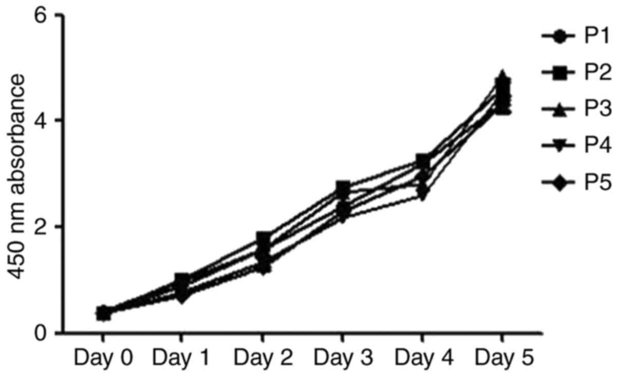
Fig. 1 represents the results. The
proliferation curves of mADSCs at different passages were similar
from day 0 to 5. Also, no difference was found in the 450-nm
absorbance on days 0 to 5 between mADSCs at different passages. The
results suggested that several cell passages had no significant
effect on the proliferation of mADSCs.
Spontaneous adipogenic differentiation
potential of mADSCs at different passages in vitro
Preadipocytes in the primary ADSCs tended to undergo
adipogenic differentiation spontaneously (1,2).
Also, the heterogeneity of ADSCs reduced with cell passages
(11). The spontaneous adipogenic
differentiation potential of mADSCs at different passages was
observed to find out whether it decreased with increasing cell
passages.
Spontaneous adipogenic differentiation potential of
mADSCs at different passages in vitro was examined by
staining with Oil Red O, and the relative expression levels of
Cebpa and Pparg were determined using real-time PCR
and western blot analysis. The mADSCs at different passages were
seeded at a density of 3×10⁴ cells/cm2 in a
culture vessel containing DMEM with 10% serum and antibiotics. The
timing of the experiment was recorded as day 0 one-day after
confluence. Then, the cells were washed and replenished using a
fresh medium every 48 h. Without treatment with adipogenic
differentiation medium and without further passaging, the cells
were collected on days 0, 3, 5 and 7 for RNA extraction and
collected on day 7 for protein extraction. The cells were stained
with Oil Red O on days 14 and 28 (Fig.
2A), and the absorbance was monitored at 510 nm. Without
induced adipogenic differentiation, the spontaneous adipogenic
potential of mADSCs at different passages was different in
vitro. The absorbance of extracted dye after staining with Oil
Red O on days 14 and 28 was statistically different (Fig. 2B). The 510-nm absorbance obtained
for mADSCs from passage 1 was higher than that for mADSCs from
passages 2 to 5. However, no difference in absorbance was found
between mADSCs from passages 2 to 5 on days 14 and 28. The relative
expression levels of Cebpa and Pparg from different
passages were undifferentiated on day 0, but the expression levels
of these both genes from passage 1 were significantly higher
compared with those of mADSCs from passages 2 to 5 on days 3, 5 and
7 (Fig. 2C). However, no
difference in the expression levels of C/EBPα and PPARγ proteins
was observed between mADSCs from passages 1 to 5 on day 7 (Fig. 2D).
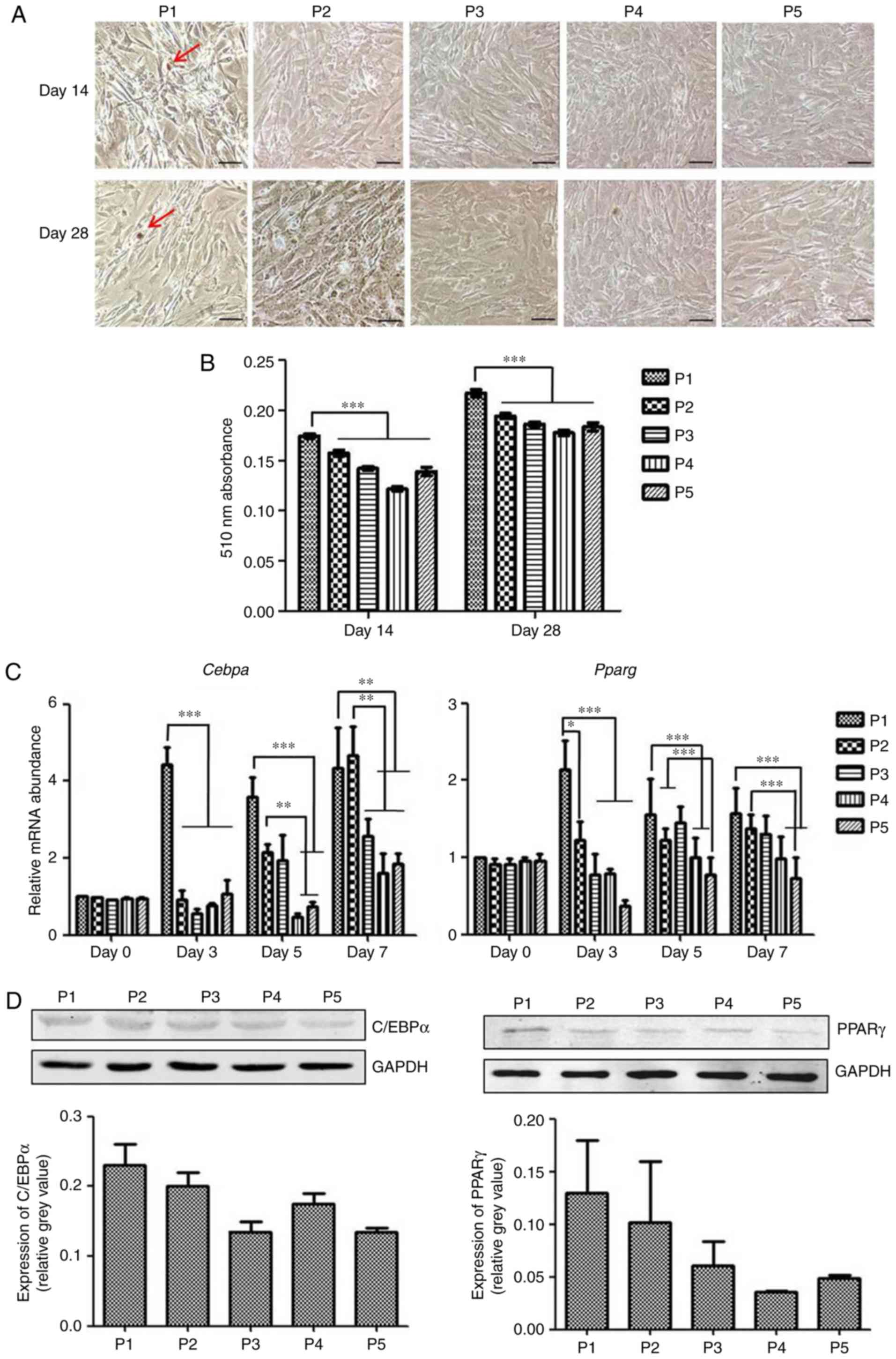 | Figure 2.Spontaneous adipogenic
differentiation potential of mADSCs at different passages in
vitro. (A) Oil Red O staining of mADSCs on days 14 and 28
without induced adipogenic differentiation. Scale bars, 100 µm;
magnification, ×100. The arrows indicate the lipids present in
adipocytes (red staining). (B) The absorbance of extracted dye from
mADSCs at different passages following staining with Oil Red O on
days 14 and 28. (C) Without adipogenic differentiation, the
relative expression levels of Cebpa and Pparγ from
mADSCs at different passages were detected on days 0, 3, 5 and 7.
Expression levels of these 2 genes from P1 were significantly
higher when compared with those from P2 to 5 on days 3, 5 and 7.
(D) Expression of C/EBPα and PPARγ proteins was detected on day 7
using western blot analysis. No difference was identified in the
protein expression levels of mADSCs at different passages.
Experiments were repeated at least 3 times with similar results.
Data are presented as the mean ± standard error of mean.
*P<0.05, **P<0.01 and ***P<0.001, as indicated. P,
passage; mADSCs, mouse adipose-derived stem cells; Cebpa and
C/EBPα, CCAAT/enhancer binding protein α; PPARγ and Pparg,
peroxisome proliferator-activated receptor γ. |
Therefore, mADSCs had the potential to undergo
spontaneous adipogenic differentiation, and the adipogenic
differentiation potential decreased quickly with several cell
passages. Then, the induced adipogenic differentiation potential of
mADSCs at different passages (cells cultured with adipogenic
differentiation medium) was compared to know whether this induced
differentiation was also affected by the number of cell
passages.
Induced adipogenic differentiation of
mADSCs at different passages in vitro
The mADSCs were cultured with adipogenic
differentiation medium to detect whether the adipogenesis of mADSCs
at different passages under induced adipogenic differentiation was
discrepant. The timing of the experiment was recorded as day 0 one
day after the confluence, then cells were switched to adipogenic
differentiation medium. The mRNA expression levels of Cebpa
and Pparg were analyzed on days 0, 3, 5 and 7. The cells
were collected for detecting the protein expression levels of
C/EBPα and PPARγ on day 7, and the extracted dye was monitored
after staining with Oil Red O on days 7 and 14. The real-time PCR
analysis showed that the mRNA expression levels of Pparg for
mADSCs from passages 3 to 5 were significantly higher than those
for mADSCs from passage 1 on days 3, 5 and 7. However, no
difference in the expression level of Cebpa was observed
between mADSCs at different passages on days 3, 5 and 7 (Fig. 3C). The results of Oil Red O
absorbance showed no difference between cells from passages 1 to 5
on days 7 and 14 under induced adipogenic differentiation (Fig. 3A and B). Also, western blot
analysis showed no difference in the protein expression levels of
C/EBPα and PPARγ between mADSCs at different passages on day 7
(Fig. 3D).
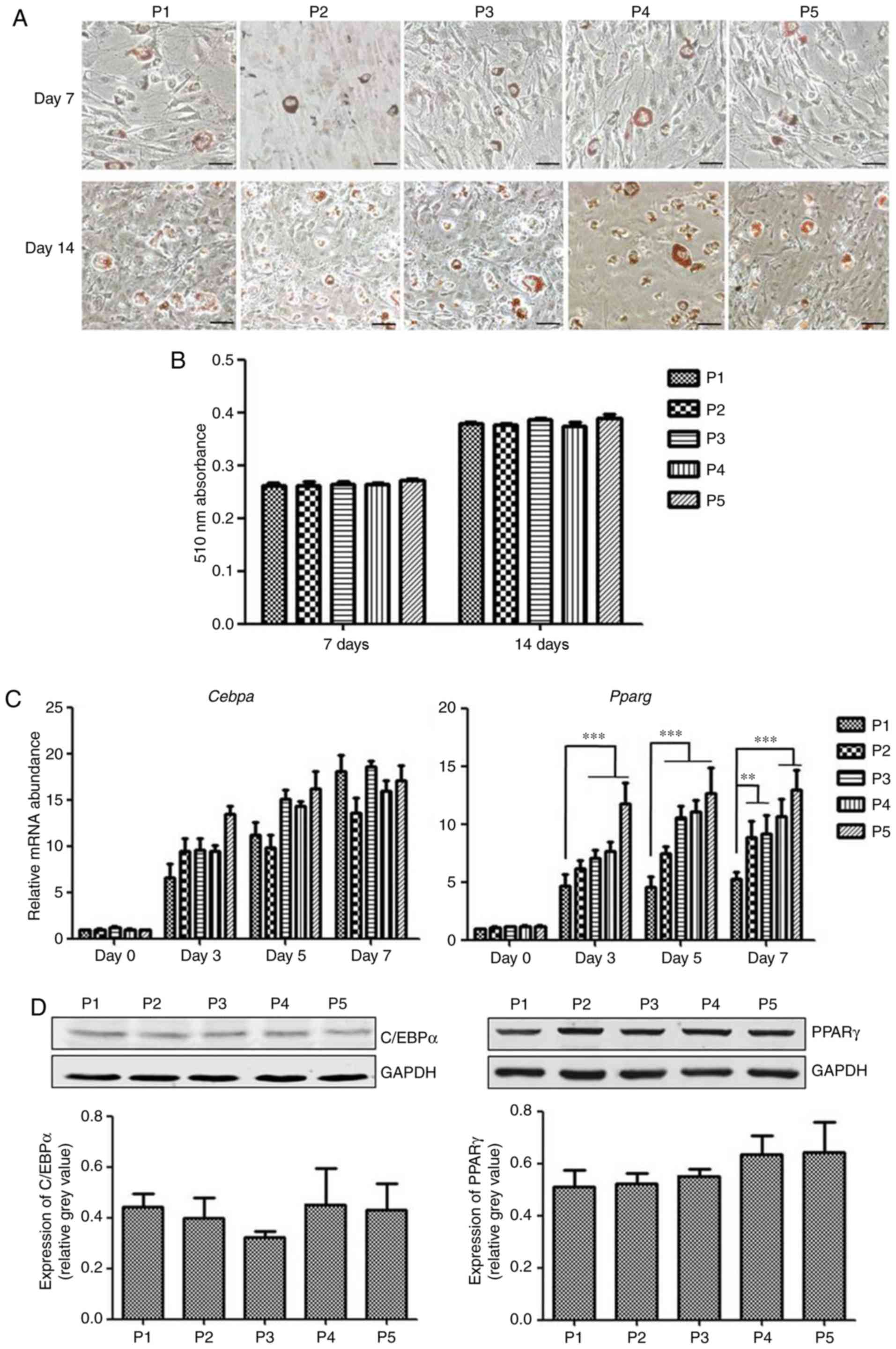 | Figure 3.Adipogenic differentiation of mADSCs
at different passages in vitro. (A) mADSCs cultured with
adipogenic differentiation medium on days 7 and 14. Lipid
production was shown using Oil Red O staining. Scale bars, 100 µm;
magnification, ×100. (B) Absorbance of extracted dye following
mADSCs were stained with Oil Red O; the absorbance of extracted dye
from mADSCs at different passages was undifferentiated on days 7
and 14. (C) Cultured with adipogenic differentiation medium, the
relative expression levels of Cebpa and Pparg from
mADSCs at different passages were detected on days 0, 3, 5 and 7.
(D) Western blot analysis revealed the expression levels of
C/EBPa and PPARγ proteins under induced adipogenic
differentiation on day 7. Experiments were repeated at least three
times with similar results. Data are presented as the mean ±
standard error of mean. **P<0.01 and ***P<0.001, as
indicated. P, passage; mADSCs, mouse adipose-derived stem cells;
Cebpa and C/EBPα, CCAAT/enhancer binding protein α; PPARγ
and Pparg, peroxisome proliferator-activated receptor γ. |
Hence, the induced adipogenic differentiation
potential was not significantly different between mADSCs from
passages 1 to 5 (except the expression levels of Pparγ).
Thus, the induced adipogenic differentiation was not affected
significantly by limited continuous cell passages.
Discussion
Generally, ADSCs are isolated in pools that contain
a mixture of cell types including mesenchymal stem cells and other
types of cells such as preadipocytes (2). Preadipocytes, which are the specific
precursor cells, have the potential for spontaneous adipogenic
differentiation. For more effective use of ADSCs, how the
adipogenic differentiation of ADSCs is influenced by preadipocytes
needs to be understood and the influence of spontaneous adipogenic
differentiation needs to be minimized when ADSCs are used in
regenerative medicine.
The content of preadipocytes is less in the primary
ADSCs ‘mix’ (23,25,26)
and preadipocytes share several cell markers with the primary
adipose derived stem cells, like Lin−, CD29+,
CD34+, Sca-1+, and CD45−
CD31− (23,25), making it difficult to screen for
preadipocytes alone from the primary ADSC ‘mix’ using
fluorescence-activated cell sorting. In the clinical practice of
regenerative medicine, generally, the ADSCs was used and observed
as a whole (3–6). For these reasons above, in this
study, the primary ADSC ‘mix’ was observed as a whole instead of
screening for preadipocytes. The potential of spontaneous
adipogenic differentiation of ADSC ‘mix’ was investigated in this
study.
Culturing with increasing cell passages was found to
reduce the heterogeneity of ADSCs (11). Studies indicated that continuous
cell passages reduced the number of cells with a CD34+
surface marker, which differentiated into adipocytes more
efficiently (25,27). However, whether spontaneous
adipogenic differentiation of ADSCs would decrease with serial cell
passages is not yet clear.
In this study, the spontaneous and induced
adipogenic differentiation potentials of mADSCs at different
passages were explored in vitro. ADSCs were found to
differentiate into adipocytes spontaneously, and the ability of
spontaneous differentiation decreased quickly with the increase in
the number of cell passages.
The ability of spontaneous adipogenic
differentiation can have clinical implications, which may be
beneficial or damaging (8,28). For chondrogenic or osteogenic
regeneration, the spontaneous adipogenic differentiation of ADSCs
should be minimized as possible. Adipogenic, chondrogenic, and
osteogenic differentiation was observed in passage 3 in many
studies (29–34). The results of the present study
indicated that the impact of spontaneous adipogenic differentiation
reduced sufficiently after passage 3, and the cells passaged more
than three times were probably suitable for chondrogenic and
osteogenic regeneration. For adipogenic regeneration, the early
passages of ADSCs may be more suitable.
The adipogenic differentiation potential was
correlated with the number of mesenchymal stem cells in the ADSC
‘mix’. No significant difference was found between the adipogenic
differentiation potential of cells from passages 1 to 5, suggesting
that the adipogenic differentiation of mesenchymal stem cells was
not affected by continuous cell passages. Liu et al
(17) reported that the adipogenic
potential of ADSCs decreased and the osteogenic differentiation
increased when the cells were passaged more than 12 times. In this
study, the adipogenic differentiation potential was not
significantly different between cells from passages 1 to 5 (except
the expression levels of Pparg) suggesting that the
adipogenic potential of ADSCs was not affected by limited cell
passages.
ADSCs from passages 3 and 4 were reported to be used
in regenerative medicine (13–15).
The results of this study also indicated no significant difference
in spontaneous and induced adipogenic differentiation potential
between cells of passages 3 and 4. It suggested similar
differentiation results in adipogenic regeneration when using cells
from passages 3 and 4. Whether the potential of chondrogenic and
osteogenic differentiation from cells of passages 3 and 4 is
different needs a follow-up study.
This study also analyzed the proliferation of mADSCs
at different passages. No difference in proliferation was observed
between cells from passages 1 to 5. Kim et al (35) reported that long-term culture and
cell passages of ADSCs could impair the activity of cells and
provoke cellular senescence, leading to low efficacy in
vivo. The results indicated that several cell passages had no
significant effect on the proliferation of mADSCs.
In conclusion, the present study showed that mADSCs
could undergo adipogenic differentiation spontaneously, and the
differentiation potential decreased with continuous cell passages.
The induced adipogenic differentiation was not affected
significantly by cell passages. The proliferation was not affected
by limited cell passages.
Acknowledgements
Not applicable.
Funding
No funding was received.
Availability of data and materials
The datasets used and analysed during the present
study are available from the corresponding author on reasonable
request.
Authors' contributions
NL isolated and cultured mouse adipose-derived stem
cells. GZ performed the oil red staining. NL and GZ statistically
analyzed and interpreted the data. DY designed the study, performed
the reverse transcription-quantitative polymerase chain reaction,
western blotting and statistical analysis, and was a major
contributor in writing the manuscript. All authors read and
approved the final manuscript.
Ethics approval and consent to
participate
All animal experiments were approved by the
Institutional Animal Care and Use Committee of Beijing Shijitan
Hospital of Capital Medical University (Beijing, China) (approval
no. 2017-KL16).
Consent for publication
Not applicable.
Competing interests
The authors declare that they have no competing
interests.
References
|
1
|
Rodeheffer MS, Birsoy K and Friedman JM:
Identification of white adipocyte progenitor cells in vivo. Cell.
135:240–249. 2008. View Article : Google Scholar : PubMed/NCBI
|
|
2
|
Yoshimura K, Shigeura T, Matsumoto D, Sato
T, Takaki Y, Aiba-Kojima E, Sato K, Inoue K, Nagase T, Koshima I,
et al: Characterization of freshly isolated and cultured cells
derived from the fatty and fluid portions of liposuction aspirates.
J Cell Physiol. 208:64–76. 2006. View Article : Google Scholar : PubMed/NCBI
|
|
3
|
Bunnell BA, Flaat M, Gagliardi C, Patel B
and Ripoll C: Adipose-derived stem cells: Isolation, expansion and
differentiation. Methods. 45:115–120. 2008. View Article : Google Scholar : PubMed/NCBI
|
|
4
|
Gimble JM, Guilak F and Bunnell BA:
Clinical and preclinical translation of cell-based therapies using
adipose tissue-derived cells. Stem Cell Res Ther. 1:192010.
View Article : Google Scholar : PubMed/NCBI
|
|
5
|
Gir P, Oni G, Brown SA, Mojallal A and
Rohrich RJ: Human adipose stem cells: Current clinical
applications. Plast Reconstr Surg. 129:1277–1290. 2012. View Article : Google Scholar : PubMed/NCBI
|
|
6
|
Harasymiak-Krzyżanowska I, Niedojadło A,
Karwat J, Kotula L, Gil-Kulik P, Sawiuk M and Kocki J: Adipose
tissue-derived stem cells show considerable promise for
regenerative medicine applications. Cell Mol Biol Lett. 18:479–493.
2013. View Article : Google Scholar : PubMed/NCBI
|
|
7
|
Tobita M, Orbay H and Mizuno H:
Adipose-derived stem cells: Current findings and future
perspectives. Discov Med. 11:160–170. 2011.PubMed/NCBI
|
|
8
|
Romagnoli C and Brandi ML: Adipose
mesenchymal stem cells in the field of bone tissue engineering.
World J Stem Cells. 6:144–152. 2014. View Article : Google Scholar : PubMed/NCBI
|
|
9
|
Roxburgh J, Metcalfe AD and Martin YH: The
effect of medium selection on adipose-derived stem cell expansion
and differentiation: Implications for application in regenerative
medicine. Cytotechnology. 68:957–967. 2016. View Article : Google Scholar : PubMed/NCBI
|
|
10
|
Baer PC and Geiger H: Adipose-derived
mesenchymal stromal/stem cells: Tissue localization,
characterization, and heterogeneity. Stem Cells Int.
2012:8126932012. View Article : Google Scholar : PubMed/NCBI
|
|
11
|
Locke M, Windsor J and Dunbar PR: Human
adipose-derived stem cells: Isolation, characterization and
applications in surgery. ANZ J Surg. 79:235–244. 2009. View Article : Google Scholar : PubMed/NCBI
|
|
12
|
Fu Y, Deng J, Jiang Q, Wang Y, Zhang Y,
Yao Y, Cheng F, Chen X, Xu F, Huang M, et al: Rapid generation of
functional hepatocyte-like cells from human adipose-derived stem
cells. Stem Cell Res Ther. 7:1052016. View Article : Google Scholar : PubMed/NCBI
|
|
13
|
Orbay H, Devi K, Williams PA, Dehghani T,
Silva EA and Sahar DE: Comparison of endothelial differentiation
capacities of human and rat adipose-derived stem cells. Plast
Reconstr Surg. 138:1231–1241. 2016. View Article : Google Scholar : PubMed/NCBI
|
|
14
|
Dufrane D and Lafosse A: A simple method
to determine the purity of adipose-derived stem cell-based cell
therapies. Stem Cells Transl Med. 5:1575–1579. 2016. View Article : Google Scholar : PubMed/NCBI
|
|
15
|
Lin YC, Harn HJ, Lin PC, Chuang MH, Chen
CH, Lin SZ and Chiou TW: Commercial production of autologous stem
cells and their therapeutic potential for liver cirrhosis. Cell
Transplant. 26:449–460. 2017. View Article : Google Scholar : PubMed/NCBI
|
|
16
|
Krähenbühl SM, Grognuz A, Michetti M,
Raffoul W and Applegate LA: Enhancement of human adipose-derived
stem cell expansion and stability for clinical use. Int J Stem Cell
Res Ther. 2:72015.
|
|
17
|
Liu Y, Zhang Z, Zhang C, Deng W, Lv Q,
Chen X, Huang T and Pan L: Adipose-derived stem cells undergo
spontaneous osteogenic differentiation in vitro when passaged
serially or seeded at low density. Biotech Histochem. 91:369–376.
2016. View Article : Google Scholar : PubMed/NCBI
|
|
18
|
Lowe CE, O'Rahilly S and Rochford JJ:
Adipogenesis at a glance. J Cell Sci. 124:2681–2686. 2011.
View Article : Google Scholar : PubMed/NCBI
|
|
19
|
Zhang LJ, Guerrero-Juarez CF, Hata T,
Bapat SP, Ramos R, Plikus MV and Gallo RL: Innate immunity. Dermal
adipocytes protect against invasive Staphylococcus aureus skin
infection. Science. 347:67–71. 2015. View Article : Google Scholar : PubMed/NCBI
|
|
20
|
Muto J, Morioka Y, Yamasaki K, Kim M,
Garcia A, Carlin AF, Varki A and Gallo RL: Hyaluronan digestion
controls DC migration from the skin. J Clin Invest. 124:1309–1319.
2014. View
Article : Google Scholar : PubMed/NCBI
|
|
21
|
Zuk PA, Zhu M, Ashjian P, De Ugarte DA,
Huang JI, Mizuno H, Alfonso ZC, Fraser JK, Benhaim P and Hedrick
MH: Human adipose tissue is a source of multipotent stem cells. Mol
Biol Cell. 13:4279–4295. 2002. View Article : Google Scholar : PubMed/NCBI
|
|
22
|
Ramirez-Zacarías JL, Castro-Muñozledo F
and Kuri-Harcuch W: Quantitation of adipose conversion and
triglycerides by staining intracytoplasmic lipids with Oil Red O.
Histochemistry. 97:493–497. 1992. View Article : Google Scholar : PubMed/NCBI
|
|
23
|
Bajek A, Gurtowska N, Olkowska J,
Kazmierski L, Maj M and Drewa T: Adipose-derived stem cells as a
tool in cell-based therapies. Arch Immunol Ther Exp (Warsz).
64:443–454. 2016. View Article : Google Scholar : PubMed/NCBI
|
|
24
|
de Witte SF, Lambert EE, Merino A, Strini
T, Douben HJ, O'Flynn L, Elliman SJ, de Klein AJ, Newsome PN, Baan
CC, et al: Aging of bone marrow- and umbilical cord-derived
mesenchymal stromal cells during expansion. Cytotherapy.
19:798–807. 2017. View Article : Google Scholar : PubMed/NCBI
|
|
25
|
Bajek A, Gurtowska N, Olkowska J, Maj M,
Kaźmierski Ł, Bodnar M, Marszałek A, Dębski R and Drewa T: Does the
harvesting technique affect the properties of adipose-derived stem
cells?-the comparative biological characterization. J Cell Biochem.
118:1097–1107. 2017. View Article : Google Scholar : PubMed/NCBI
|
|
26
|
González-Cruz RD and Darling EM:
Adipose-derived stem cell fate is predicted by cellular mechanical
properties. Adipocyte. 2:87–91. 2013. View Article : Google Scholar : PubMed/NCBI
|
|
27
|
Wang QF, Huang Y, He GC, Wang HS, Chen ZH,
Cai XH, Xie YH and Liu Q: Osteoblast differentiation of rabbit
adipose-derived stem cells by polyethylenimine-mediated BMP-2 gene
transfection in vitro. Genet Mol Res. 16:2017. View Article : Google Scholar
|
|
28
|
Aoyagi Y, Kuroda M, Asada S, Bujo H,
Tanaka S, Konno S, Tanio M, Ishii I, Aso M and Saito Y: Fibrin glue
increases the cell survival and the transduced gene product
secretion of the ceiling culture-derived adipocytes transplanted in
mice. Exp Mol Med. 43:161–167. 2011. View Article : Google Scholar : PubMed/NCBI
|
|
29
|
Güven S, Mehrkens A, Saxer F, Schaefer DJ,
Martinetti R, Martin I and Scherberich A: Engineering of large
osteogenic grafts with rapid engraftment capacity using mesenchymal
and endothelial progenitors from human adipose tissue.
Biomaterials. 32:5801–5809. 2011. View Article : Google Scholar : PubMed/NCBI
|
|
30
|
Jang Y, Jung H, Nam Y, Rim YA, Kim J,
Jeong SH and Ju JH: Centrifugal gravity-induced BMP4 induces
chondrogenic differentiation of adipose-derived stem cells via SOX9
upregulation. Stem Cell Res Ther. 7:1842016. View Article : Google Scholar : PubMed/NCBI
|
|
31
|
Papadimitropoulos A, Scherberich A, Güven
S, Theilgaard N, Crooijmans HJ, Santini F, Scheffler K, Zallone A
and Martin I: A 3D in vitro bone organ model using human progenitor
cells. Eur Cell Mater. 21:445–458. 2011. View Article : Google Scholar : PubMed/NCBI
|
|
32
|
Leong DT, Abraham MC, Rath SN, Lim TC,
Chew FT and Hutmacher DW: Investigating the effects of preinduction
on human adipose-derived precursor cells in an athymic rat model.
Differentiation. 74:519–529. 2006. View Article : Google Scholar : PubMed/NCBI
|
|
33
|
Maglione M, Spano S, Ruaro ME, Salvador E,
Zanconati F, Tromba G and Turco G: In vivo evaluation of
chitosan-glycerol gel scaffolds seeded with stem cells for
full-thickness mandibular bone regeneration. J Oral Sci.
59:225–232. 2017. View Article : Google Scholar : PubMed/NCBI
|
|
34
|
Ye X, Liao C and Liu G: Age-related
changes in the regenerative potential of adipose-derived stem cells
isolated from the prominent fat pads in human lower. eyelids.
11:e01665902016.
|
|
35
|
Kim S, Piao J, Son Y and Hong HS:
Substance P enhances proliferation and paracrine potential of
adipose-derived stem cells in vitro. Biochem Biophys Res Commun.
485:131–137. 2017. View Article : Google Scholar : PubMed/NCBI
|