Introduction
The prevalence of obesity, prediabetes and diabetes
has increased in the last three decades (1–5).
Diabetes and obesity are characterized by low grade chronic
inflammation status and insulin resistance in adipose tissue
(6). Several researchers
identified infiltration of macrophages in adipose tissue of
diabetic or obese individuals by immunohistochemistry, polymerase
chain reaction (PCR) or flow cytometry with F4/80 or CD68
antibodies (7–9).
Interleukin (IL)29 exhibits multiple immune
regulatory activities, including antiviral, antiproliferation and
antitumor properties. Previous studies have suggested that IL29 is
involved in the regulation of Th (T helper) 1/Th2 responses and
function of B cells, plasmacytoma dendritic cells and macrophages
(10–14). IL29 enhanced lipopolysaccharide
(LPS)-induced inflammatory cytokine production from toll like
receptor 4 signaling pathway in RAW264.7 cells (15).
Tissue inhibitor of metalloproteinase
(TIMP)-associated disorders have been implicated in
pathophysiological processes of obesity and diabetes in humans.
Concentrations of matrix metalloproteinases (MMPs) and TIMPs are
increased in the plasma of diabetic and obese humans (16). A previous study demonstrated that
high glucose and interferon-γ increased MMP1 expression in U937
mononuclear cells (17). However,
the TIMP1 expression pattern following stimulation with IL29 in
Raw264.7 cells and primary macrophages remains to be elucidated.
Therefore, the present study investigated the effects of IL29 and
high glucose on TIMP1 in Raw264.7 cells and primary macrophages.
The results of the present study may aid in elucidating the
underlying mechanisms of obesity and diabetes mellitus.
Materials and methods
Human study
Adipose tissues were collected from patients with
obesity (8 males, 40–50 years old, BMI>28) and age and sex
matched lean controls (8 males, 40–50 years old, BMI<24) during
abdominal surgery from the First Affiliated Hospital of Jinzhou
Medical University (Jinzhou, China) from December 2016 to January
2017. All volunteers signed written informed consent forms prior to
the study. All experiments were approved by the Medical Ethics
Committee of the First Affiliated Hospital of Jinzhou Medical
University.
Raw264.7 cell culture
Raw264.7 cells were purchased from American Type
Culture Collection (Manassas, VA, USA) and cultured in glucose free
1640 medium (Thermo Fisher Scientific, Inc., Waltham. MA, USA) with
5 mM glucose, supplemented with 10% fetal bovine serum (FBS;
Tiangen Biotech Co., Ltd., Beijing, China) and 1%
penicillin-streptomycin (P/S). The medium was replaced at least
once every two days. Cells were harvested for passaging when plates
were 90% confluent.
Primary macrophage extraction and
culture
TLR2−/− or TLR4−/− mice
backcrossed on C57BL/6J from the Jackson Laboratory (Ben Harbor,
ME, USA or Sacramento, CA, USA) were housed with C57BL/6J mice from
Beijing HFK Bioscience Co., Ltd. (Beijing, China) to get TLR2 or
4+/+ and TLR2 or 4−/− sex and age matched
littermate mice used for studies in vitro. A total number of
315 mice were used. Primary macrophages were extracted from
abdominal cavities of TLR2+/+ or TLR 4+/+ and
TLR2−/− or TLR 4−/− mice. Briefly, 10 ml
bacteria free PBS was injected to the abdominal cavities multiple
times in each fresh sacrificed mouse in order to generate
macrophages, and then was pumped out following massaging for 5 min.
Cells in the PBS extracted from abdominal cavities were centrifuged
at 700 × g for 5 min at 20–25°C and cultured in the aforementioned
1640 medium. The medium was replaced following 12 h of culture to
remove unadherent cells from macrophages. All animal studies were
approved by the Ethics Committee for Experimental Research, Jinzhou
Medical University (Jinzhou, China).
IL29 and high glucose-activation
assays, and antibody-mediated inhibition assays
Cells (Raw264.7 cells and primary macrophages) were
cultured in the 1640 medium as previously described with 5 mM
glucose, 10% FBS and 1% P/S for two days, and then cultured in FBS
free medium for IL29 and high glucose-activated assays. Activation
was performed by treatment with IL29 at a concentration of 0, 10,
30 or 100 ng/ml or 25 mM glucose for 24 h (low dose group=5 mM and
high dose group=30 mM). Antibodies against TLR2 or 4 (dilution,
1:100; cat no. 10472R; Beijing Biosynthesis Biotechnology Co.,
Ltd., Beijing, China) were added to the medium at 37°C for 24 h to
inhibit TLR2 or TLR4. Subsequently, cells were harvested with a
cell scraper followed by centrifugation at 700 × g for 5 min at
20–25°C for further research.
Western blotting
Western blotting was performed according to a
previously described protocol (18). Total proteins from adipose tissue
were extracted using radioimmunoprecipitation assay buffer
(Beyotime Institute of Biotechnology, Haimen, China) (with 1%
phenylmethane sulfonyl fluoride or phenylmethylsulfonyl fluoride).
Subsequently, a bicinchoninic acid assay was performed to measure
protein concentration in middle extracting solution. Proteins were
separated by SDS-PAGE on 10% gels, transferred to polyvinylidene
fluoride membranes and blocked with skimmed milk for 2 h at room
temperature. Rabbit-anti-mouse (or human) primary antibodies to
TIMP1 (1:500; cat no. wl02342; Wanleibo Co., Ltd., Shanghai,
China), myeloid differentiation primary response protein MyD88
(MyD88; 1:300; cat no. bs-1047R; Beijing Biosynthesis Biotechnology
Co., Ltd.), IL29 (1:1,500; cat no. ab38569; Abcam, Cambridge, MA,
USA) and β-actin (1:3,000; cat no. AB21800; Absci, Vancouver, WA,
USA) were used to bind target proteins at 4°C overnight. Following
incubation with goat-anti-rabbit secondary antibody (1:5,000; cat
no. ABL3012-2; Absci, USA) for 2 h at 20–25°C, an Enhanced
Chemiluminescence kit (Wanleibo Co., Ltd.) was used to detect
protein expression.
Immunofluorescence
Raw264.7 cells were collected by centrifugation (700
× g for 5 min at 20–25°C) and seeded on dish climbing glasses
(https://item.taobao.com/item.htm?id=13540518880) in
24-well plate for 24 h. IL29 and high glucose-activated assays were
performed as described previously, for 24 h. Raw264.7 cells were
rinsed 3 times with PBS and fixed with 4% paraformaldehyde for 30
min at 20–25°C. Non-specific binding was blocked in PBS containing
5% normal goat serum (cat no. SL03B; Beijing Solarbio Science &
Technology Co., Ltd., Beijing, China) at room temperature for 1 h.
Cells were initially incubated with primary antibodies [rabbit
anti-mouse TLR2 (cat no. 10472R; 1:300) or TLR4 (cat no. 1021R;
1:300) at 4°C on a shaker overnight]. Subsequently, climbing
glasses with Raw264.7 cells were washed 3 times in PBS at room
temperature prior to incubation with goat anti-rabbit fluorescein
isothiocyanate (green; Wanleibo Co., Ltd., Shanghai, China; 1:200;
cat no. WLA031a) or Alexa Fluor 594 (red; Thermo Fisher Scientific,
Inc,. 1:300; cat no. A-11032) secondary antibodies in the dark at
room temperature for 1 h. Following washing with PBS 3 times, TLR2
and 4 expression levels were observed under a fluorescence
microscope.
ELISA
TIMP1 in conditioned media was quantified using
sandwich ELISA kits (cat no. MTM100) according to the
manufacturers' protocol (R&D Systems, Inc., Minneapolis, MN,
USA).
Reverse transcription-quantitative
(RT-q)PCR assay
Total RNA was extracted from the Raw264.7 cell line
using TRIzol® reagent (Thermo Fisher Scientific, Inc.)
and dissolved in RNase free water with RNase inhibitor added. Prior
to cDNA synthesis, RNA concentration was measured by Nanodrop 2000
(Thermo Fisher Scientific, Inc.) and modified to 100 µg/µl with
diethyl pyrocarbonate (DEPC) water (Life Technologies; Thermo
Fisher Scientific, Inc., cat no. AM9915G). All experiments were
performed according to the manufacturers' protocol of PrimeScriptTM
RT reagent kit with gDNA Eraser (Takara Bio, Inc., Otsu, Japan).
DNA was removed in a 10 µl reaction with RNA sample (1 µl), 5X gDNA
Eraser Buffer (2 µl), gDNA Eraser (1 µl) and RNase Free
ddH2O (6 µl) for 2 min at 42°C. The cDNA was synthesized
in a 20 µl reaction with Primer Script RT Enzyme Mix (1 µl), RT
Primer Mix (1 µl), 5X Primer Script Buffer (4 µl), RNase Free
dH2O (4 µl), and the 10 µl reaction mixture from the
previous step. The RT was performed in 20 µl reactions at 25°C for
10 min followed by 37°C for 15 min and final denaturation at 85°C
for 5 min. The cDNA was stored at −80°C until further use. The
expression of IL-6 mRNA and tumor necrosis factor-α (TNF-α) mRNA
was analyzed in 20 µl reactions with 2X GreenStar Master Mix (10
µl), forward primer (1 µl, 10 pmol/µl), reverse primer (1 µl, 10
pmol/µl), DEPC water (6 µl) and template DNA (2 µl) from reverse
transcription. Primers were designed and synthesized by Sangon
Biotech (Sangon Biotech Co., Ltd, Shanghai, China; Table I). qPCR experiments were performed
according to the manufacturers' protocol of AccuPower®
2X GreenStar qPCR Master Mix kit with gDNA Eraser (Bioneer
Corporation, Daejeon, Korea). Reactions were performed in 20 µl
volume and the following thermocycling conditions were used for
PCR: Initial denaturation for 30 sec at 95°C; 45 cycles of 95°C for
5 sec and 60°C for 34 sec. Relative quantification of gene
expression was performed using comparative 2−ΔΔCq
method. GAPDH was used as a reference gene.
 | Table I.Primers for IL6, TNFα and GAPDH. |
Table I.
Primers for IL6, TNFα and GAPDH.
| Gene | Sequence (5′-3′) |
|---|
| IL6 |
|
|
Forward |
ATGAAGTTCCTCTCTGCAAGAGACT |
|
Reverse |
CACTAGGTTTGCCGAGTAGATCTC |
| TNFα |
|
|
Forward |
TGTCTCAGCCTCTTCTCATT |
|
Reverse |
AGATGATCTGAGTGTGAGGG |
| GAPDH |
|
|
Forward |
TTGTCAAGCTCATTTCCTGGTATG |
|
Reverse |
GGATAGGGCCTCTCTTGCTCA |
Statistical analysis
Data are presented as the mean ± standard error of
the mean, and analyzed by one-way analysis of variance between
multiple groups, followed by the least significant difference
post-hoc test. Interaction effects were analyzed by analysis of
variance of factorial design. SPSS software (version 20.0; IBM
SPSS, Armonk, NY, USA) was used for data analysis. P<0.05 was
considered to indicate a statistically significant difference.
Results
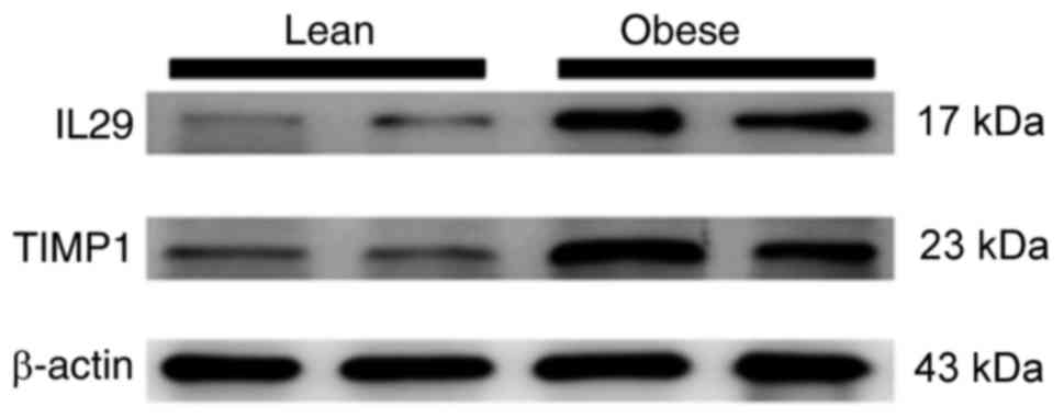
IL29 and TIMP1 levels are increased in
adipose tissue of patients with obesity
In this study, elevated levels of IL29 and TIMP1 in
adipose tissue of obese individuals compared with lean individuals
was observed (Fig. 1).
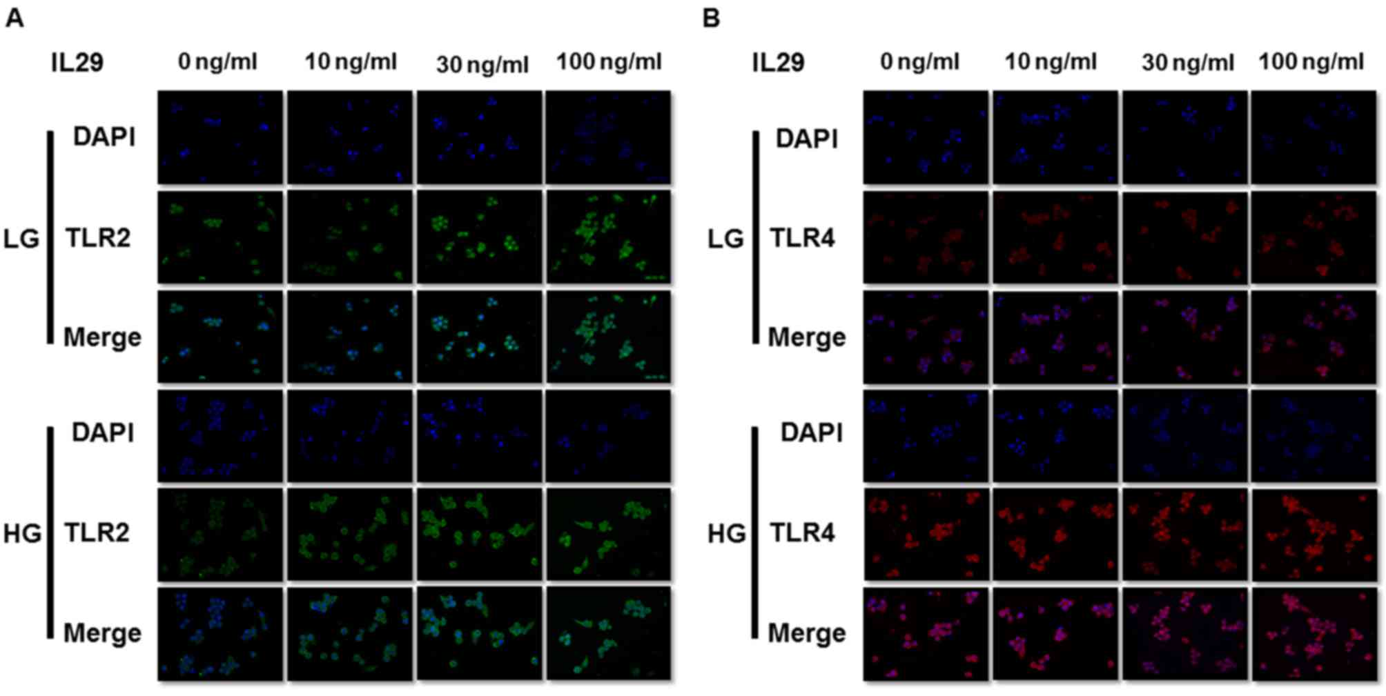
IL29 and high glucose activate TIMP1
release in Raw264.7 cells
In the present study, IL29 and high glucose
stimulated TLR2 and TLR4 expression respectively (Fig. 2A and B), as well as TIMP1 in a
concentration-independent manner (Fig.
3). Upregulated MyD88 protein expression level was detected by
western blotting in high glucose group compared with low glucose
group (Fig. 3A). Moreover, MyD88
protein expression was upregulated in a IL29
concentration-dependent manner. mRNA expression levels of
downstream inflammation factors TNF-α and IL-6 were also
up-regulated (Fig. 3C and D).
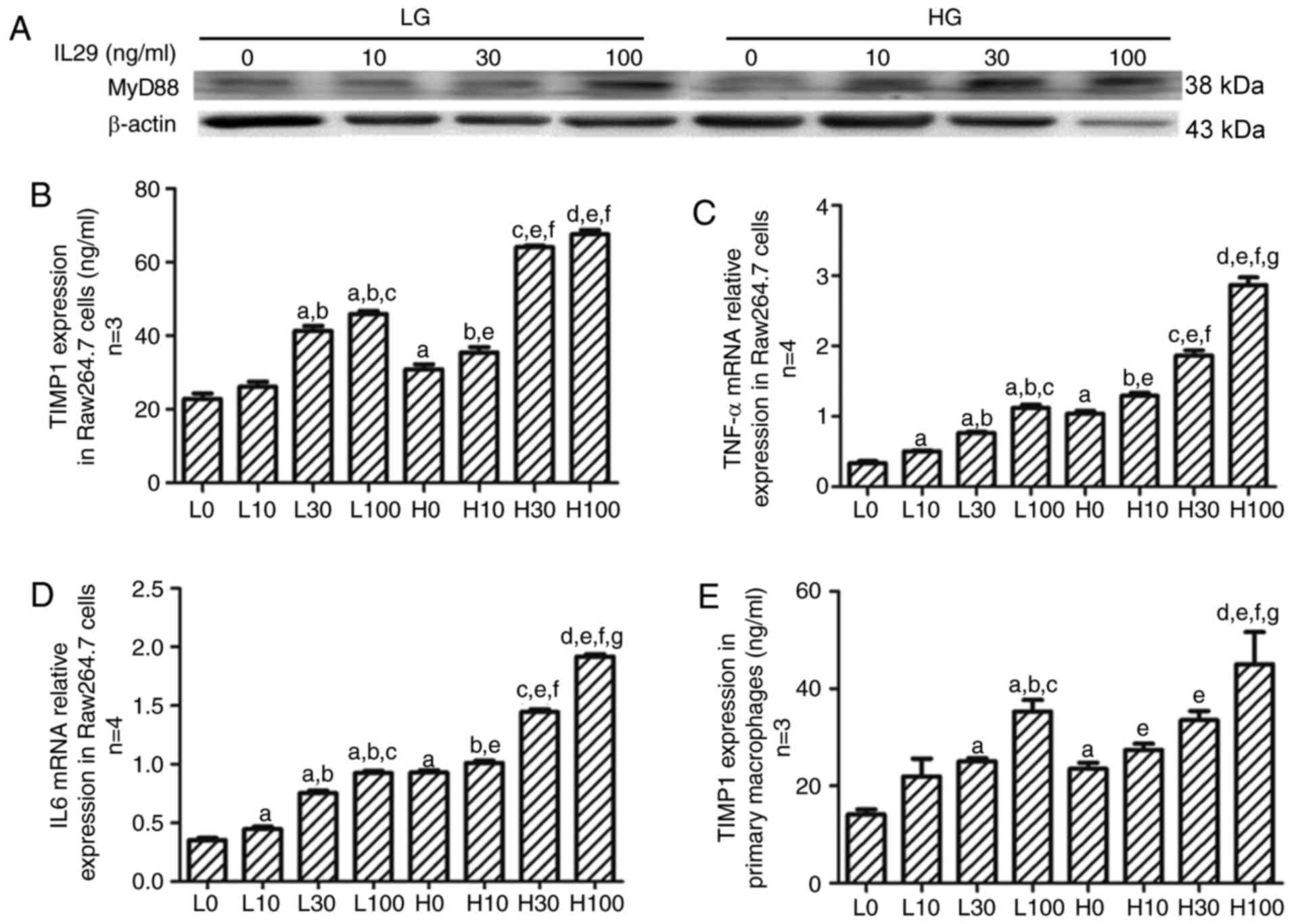 | Figure 3.IL29 and high glucose stimulate TIMP1,
MyD88, TNF-α and IL6 expression in Raw264.7 cells as well as TIMP1
levels in primary macrophages. (A) Western blotting detected
elevated levels of MyD88 protein in Raw264.7 cells. (B) ELISA assay
was performed to study the effects of IL29 and high glucose on
TIMP1 expression in Raw264.7 cells. Expression of (C) TNF-α and (D)
IL6 mRNA induced by IL29 and high glucose stimulation in Raw264.7
cells. (E) ELISA assay was performed to study the effects of IL29
and high glucose on TIMP1 expression in primary macrophages.
aP<0.05 vs. L0, bP<0.05 vs. L10,
cP<0.05 vs. L30, dP<0.05 vs. L100,
eP<0.05 vs. H0, fP<0.05 vs. H10,
gP<0.05 vs. H30. L0, low glucose and 0 ng/ml IL29
group; L10, low glucose and 10 ng/ml IL29 group; L30, low glucose
and 30 ng/ml IL29 group; L100, low glucose and 100 ng/ml IL29
group; H0, high glucose and 0 ng/ml IL29 group; H10, high glucose
and 10 ng/ml IL29 group; H30, high glucose and 30 ng/ml IL29 group;
H100, high glucose and 100 ng/ml IL29 group; IL, interleukin;
TIMP1, metalloproteinase inhibitor 1; TNF-α, tumor necrosis
factor-α; MyD8, myeloid differentiation primary response protein
MyD88. |
IL29 and high glucose activate TIMP1
release in primary macrophages
TIMP1 expression pattern in primary macrophages was
similar to that in Raw264.7 cells. IL29 and high glucose, activated
TIMP1 expression in primary macrophages in a concentration
independent manner (Fig. 3E).
Indeed, it was found that TIMP1 expression was significantly
increased in the 100 ng/ml group compared with the 0, 10 and 30
ng/ml groups.
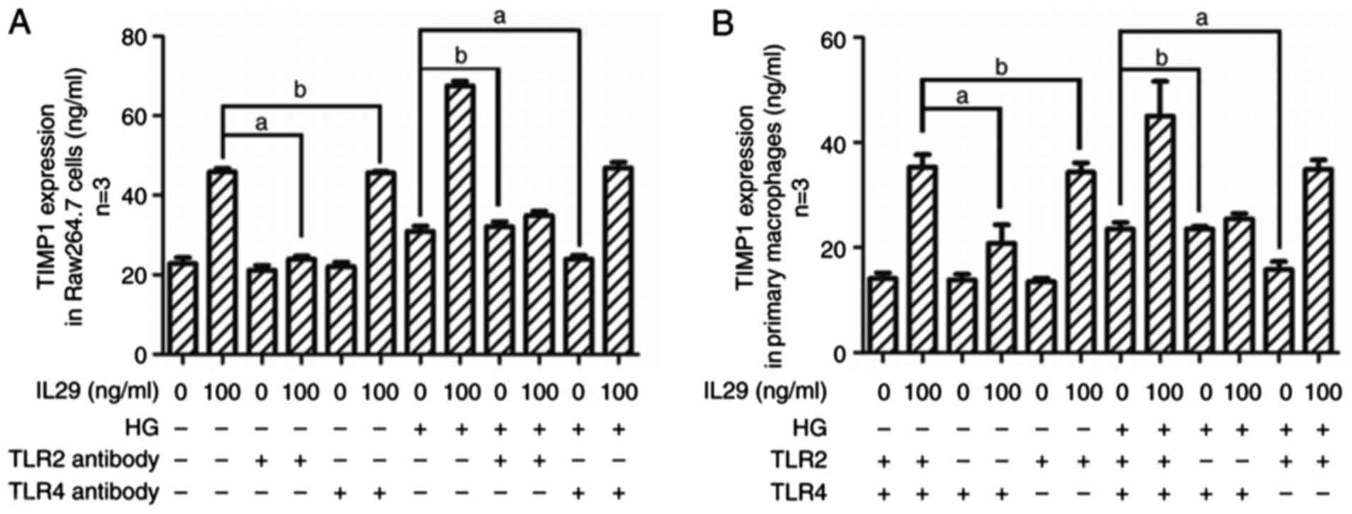
Antibody-mediated inhibition of TLR2
and 4 inhibits TIMP1 expression
Antibody-mediated inhibition of TLR2 signaling
suppressed IL29-stimulated TIMP1 expression but did not affect high
glucose activated TIMP1 release in the medium. In addition, high
glucose-stimulated TIMP1 levels were inhibited by TLR4 antibody.
However, TLR4 antibody could not suppress upregulated TIMP1 caused
by IL29 (Fig. 4A).
TLR2 knockout suppresses
IL29-stimulated TIMP1 expression and TLR4 knockout inhibits high
glucose-activated TIMP1 level
Similar to TLR2 and 4 antibody-mediated inhibition
studies, IL29 did not increase TIMP1 expression in primary
macrophages extracted from TLR2−/− mice compared with
macrophages extracted from TLR2+/+ mice. In addition,
increased TIMP1 expression level in high glucose medium was
inhibited by TLR4 gene knockout in primary macrophages (Fig. 4B).
Discussion
Diabetes mellitus and obesity patients (1–5) are
characterized by disorders of MMPs and TIMPs. Extracellular matrix
(ECM) substrate of MMPs deposition is associated with adipose
tissue fibrosis and local and systemic insulin sensitivity
(19,20). TIMPs could inhibit the activity of
MMPs. The expression level of MMPs exhibit various patterns in
obesity. It had been reported that increased expression levels of
MMP 2,3,11,12,13,14 and 19, and decreased expression levels of MMP
7,9,16 and 24 are present in high-fat-diet-induced obese mice or
genetic ob/ob mice (16). TIMP1, a
kind of inhibitor of ECM degradation shared the same tendency of
ECM in obesity (19,20). IL29 exhibits multiple immune
regulatory activities and enhances production of LPS-induced
inflammatory cytokines in Raw264.7 cells, accounting for the TLR4
signaling pathway (15). However,
whether IL29 induces inflammatory cytokine expression without LPS,
at least, with low grade LPS in obesity remains unknown. Increased
IL29 levels have previously been detected in adipose tissue of
humans with obesity. This is consistent with previous research. In
previous work, researchers found low grade chronic inflammation in
adipose tissue of obese individuals (6).
The remolding of obese adipose tissue is associated
with ECM accumulation in intercellular substance (21). SVF (stromal vascular fraction)
cells contribute to ECM synthesis and degradation. Macrophages, a
type of SVF cell, are inflammatory regulators in adipose tissue
that may induce TIMP1 expression. In the present study, IL29 was
used to stimulate TIMP1 in Raw264.7 cells and primary macrophages
from mice. IL29 and high glucose increased TIMP1 level in a
concentration-dependent manner in the medium of Raw264.7 cells. In
addition, similar results were obtained for assays using primary
macrophages from C57BL/6J mice treated with IL29 and high glucose.
To investigate the mechanism underlying the observed alterations in
TIMP1 expression, the present study determined expression of TLR2
and 4 by immunostaining. Furthermore, expression levels of MyD88,
TNFα and IL6 were investigated in the medium of Raw264.7 cells.
Treatment with IL29 increased TLR2 expression, while high glucose
upregulated TLR4 expression. MyD88 and downstream TNFα and IL6
levels were stimulated by IL29 and high glucose in a synergistic
manner.
The present study investigated the roles of TLR2 and
4 in modulation of TIMP1 expression in Raw264.7 cells and primary
macrophages by TLR2 or 4 antibody-mediated inhibition or gene
knockout. IL29-stimulated TIMP1 expression was inhibited by
antibody-mediated inhibition of TLR2 or TLR2 gene knockout.
However, TLR2 inhibition or TLR2 gene knockout did not affect high
glucose-mediated TIMP1 expression. Conversely, TLR4 inhibition or
knockout suppressed TIMP1 levels increased by high glucose
treatment, however not by IL29-mediated stimulation.
In conclusion, the results of the present study
indicate that TLR2 is involved in IL29-stimulated TIMP1 expression
in Raw264.7 cells and primary macrophages. High glucose activated
TIMP1 expression may by mediated by TLR4 signaling.
Acknowledgements
The present study was supported by the Scientific
Foundation for Doctoral Research (grant no. 20131067) and the
Natural Scientific Foundation (grant no. 201602308) from Liaoning
Science and Technology Administration Bureau. Professors Zheng and
Zhai in the Key Laboratory of Brain and Spinal Cord Injury in
Liaoning province in the First Affiliated Hospital of Jinzhou
Medical University made contributions to this study. Professor
Zheng provided TLR2 and TLR4 knockout mice for this study.
Professor Zhai provided the Raw264.7 cell line for this study.
References
|
1
|
Chan JC, Zhang Y and Ning G: Diabetes in
China: A societal solution for a personal challenge. Lancet
Diabetes Endocrinol. 2:969–979. 2014. View Article : Google Scholar : PubMed/NCBI
|
|
2
|
Flegal KM, Carroll MD, Ogden CL and Curtin
LR: Prevalence and trends in obesity among US adults, 1999–2008.
JAMA. 303:235–241. 2010. View Article : Google Scholar : PubMed/NCBI
|
|
3
|
Flegal KM, Kruszon-Moran D, Carroll MD,
Fryar CD and Ogden CL: Trends in obesity among adults in the United
States, 2005 to 2014. JAMA. 315:2284–2291. 2016. View Article : Google Scholar : PubMed/NCBI
|
|
4
|
Ogden CL, Carroll MD, Lawman HG, Fryar CD,
Kruszon-Moran D, Kit BK and Flegal KM: Trends in obesity prevalence
among Children and adolescents in the United States, 1988–1994
through 2013–2014. JAMA. 315:2292–2299. 2016. View Article : Google Scholar : PubMed/NCBI
|
|
5
|
Xu Y, Wang L, He J, Bi Y, Li M, Wang T,
Wang L, Jiang Y, Dai M, Lu J, et al: Prevalence and control of
diabetes in Chinese adults. JAMA. 310:948–959. 2013. View Article : Google Scholar : PubMed/NCBI
|
|
6
|
Kawasaki T and Kawai T: Toll-like receptor
signaling pathways. Front Immunol. 5:4612014. View Article : Google Scholar : PubMed/NCBI
|
|
7
|
Korkmaz H, Bozdag Z, Akarsu E, Tarakcioglu
M, Ulusal H and Gökalp MA: Macrophage infiltration into
subcutaneous adipose tissue is associated with local levels of
11BHSD1. Exp Clin Endocrinol Diabetes. 124:474–480. 2016.
View Article : Google Scholar : PubMed/NCBI
|
|
8
|
Nam YR, Won SB, Chung YS, Kwak CS and Kwon
YH: Inhibitory effects of Doenjang, Korean traditional fermented
soybean paste, on oxidative stress and inflammation in adipose
tissue of mice fed a high-fat diet. Nutr Res Pract. 9:235–241.
2015. View Article : Google Scholar : PubMed/NCBI
|
|
9
|
Xie L, Zhang K, Rasmussen D, Wang J, Wu D,
Roemmich JN, Bundy A, Johnson WT and Claycombe K: Effects of
prenatal low protein and postnatal high fat diets on visceral
adipose tissue macrophage phenotypes and IL-6 expression in Sprague
Dawley rat offspring. PLoS One. 12:e01695812017. View Article : Google Scholar : PubMed/NCBI
|
|
10
|
de Groen RA, Boltjes A, Hou J, Liu BS,
McPhee F, Friborg J, Janssen HL and Boonstra A: IFN-λ-mediated
IL-12 production in macrophages induces IFN-γ production in human
NK cells. Eur J Immunol. 45:250–259. 2015. View Article : Google Scholar : PubMed/NCBI
|
|
11
|
Hou W, Wang X, Ye L, Zhou L, Yang ZQ,
Riedel E and Ho WZ: Lambda interferon inhibits human
immunodeficiency virus type 1 infection of macrophages. J Virol.
83:3834–3842. 2009. View Article : Google Scholar : PubMed/NCBI
|
|
12
|
Jordan WJ, Eskdale J, Srinivas S, Pekarek
V, Kelner D, Rodia M and Gallagher G: Human interferon lambda-1
(IFN-lambda1/IL-29) modulates the Th1/Th2 response. Genes Immun.
8:254–261. 2007. View Article : Google Scholar : PubMed/NCBI
|
|
13
|
Megjugorac NJ, Gallagher GE and Gallagher
G: Modulation of human plasmacytoid DC function by IFN-lambda1
(IL-29). J Leukoc Biol. 86:1359–1363. 2009. View Article : Google Scholar : PubMed/NCBI
|
|
14
|
Novak AJ, Grote DM, Ziesmer SC, Rajkumar
V, Doyle SE and Ansell SM: A role for IFN-lambda1 in multiple
myeloma B cell growth. Leukemia. 22:2240–2246. 2008. View Article : Google Scholar : PubMed/NCBI
|
|
15
|
Xu D, Yan S, Wang H, Gu B, Sun K, Yang X,
Sun B and Wang X: IL-29 enhances LPS/TLR4-mediated inflammation in
rheumatoid arthritis. Cell Physiol Biochem. 37:27–34. 2015.
View Article : Google Scholar : PubMed/NCBI
|
|
16
|
Lin, Chun TH and Kang L: Adipose
extracellular matrix remodelling in obesity and insulin resistance.
Biochem Pharmacol. 119:8–16. 2016. View Article : Google Scholar : PubMed/NCBI
|
|
17
|
Lu Z, Li Y, Samuvel DJ, Jin J, Zhang X,
Lopes-Virella MF and Huang Y: MD-2 is involved in the stimulation
of matrix metalloproteinase-1 expression by interferon-gamma and
high glucose in mononuclear cells-a potential role of MD-2 in
Toll-like receptor 4-independent signalling. Immunology.
140:301–313. 2013.PubMed/NCBI
|
|
18
|
Song B, Ding L, Zhang H, Chu Y, Chang Z,
Yu Y, Guo D, Zhang S and Liu X: Ginsenoside Rb1 increases insulin
sensitivity through suppressing 11β-hydroxysteroid dehydrogenase
type I. Am J Transl Res. 9:1049–1057. 2017.PubMed/NCBI
|
|
19
|
Zhang H, Wang R and Song B: Deposition of
collagen I in adipose tissue of obese mice was inhibited by toll
like receptor 2 knockout. Chin J Endocrinol Metab. 33:408–412.
2017.
|
|
20
|
Hopps E and Caimi G: Matrix
metalloproteinases in metabolic syndrome. Eur J Intern Med.
23:99–104. 2012. View Article : Google Scholar : PubMed/NCBI
|
|
21
|
Luo T, Nocon A, Fry J, Sherban A, Rui X,
Jiang B, Xu XJ, Han J, Yan Y, Yang Q, et al: AMPK activation by
metformin suppresses abnormal extracellular matrix remodeling in
adipose tissue and ameliorates insulin resistance in obesity.
Diabetes. 65:2295–2310. 2016. View Article : Google Scholar : PubMed/NCBI
|