Introduction
Human periodontal ligament, a band of fibrous
connective tissue, has shock absorption ability and prevents tooth
and alveolar bone injury during chewing (1). As the most abundant cells in human
periodontal ligament, human periodontal ligament fibroblasts
(HPDLCs) are involved in pathological changes of the human
periodontal ligament and periodontal tissue regeneration (2). Besides that, HPDLCs have been
demonstrated to have the capacity to generate extracellular matrix
and regulate osteoclastic differentiation within periodontal tissue
(3). For these reasons, HPDLCs was
considered to play a vital role in the damage repair of periodontal
diseases.
Icariin
(2-(4′-methoxyphenyl)-3-rhamnosido-5-hydroxyl-7-glucosido-8-(3′-methyl-2-butylenyl)-4-chromanone,
the major active ingredient of epimedii, possesses a broad spectrum
of pharmacological applications, such as the protection of bone,
cartilage, neuro and cardiovascular system (4). In addition, it has been suggested and
supported by studies that icarrin could induce rat adipose-derived
stem cells differentiation into osteoblasts (5) and regulate extracellular matrix (ECM)
synthesis (6). However, little is
known that whether icariin could affect ECM synthesis and
osteoblastic differentiation in HPDLCs.
Matrix metalloproteinases (MMPs) are a family of
proteolytic enzymes that degrade components of ECM (7). According to published reports,
numerous pathways such as Toll-like receptors (TLRs) (8), NF-κB (9) play important roles in MMPs
expressions. However, activation of NF-κB and TLRs inhibit
osteogenic differentiation in various cells, including
pre-osteoblasts (10), human bone
marrow mesenchymal stem cells (11). Therefore, TLR/NF-κB pathway maybe a
major target for the prevention and treatment of damaged
HPDLCs-induced periodontal diseases.
In the present study, HPDLCs were isolated and
cultured in the absence or presence of icariin at different
concentrations to explore the molecular mechanisms of icarrin in
HPDLCs. These results strongly demonstrated that icariin promote
HPDLCs to differentiate into osteoblasts and stimulate ECM
synthesis via suppressing TLR-4 and phosphorylation of NF-κB.
Materials and methods
Primary culture of HPDLCs
HPDLCs were isolated from the molar of a female
patient (25 years old) at Shanghai Stomatological Hospital.
Briefly, periodontal ligament tissue dissected from patients were
chopped into pieces and digested with 0.25% Trypsin/EDTA (Gibco;
Thermo Fisher Scientific, Inc., Waltham, MA, USA) at 37°C for 1 h.
Then, cells were centrifugated at 1,000 × g for 10 min and
subsequently cultured in Dulbecco's modified Eagle's medium (DMEM)
(Gibco; Thermo Fisher Scientific, Inc.) containing 10% fetal bovine
serum (FBS) at 37°C with 5% CO2. A complete clinical
examination was performed in all participants, clinical data were
recorded and written consent was obtained from each subject. The
experimental protocols were approved by the Ethics Committee of
Shanghai Stomatological Hospital (Shanghai, China).
CCK-8 assay
Icariin was obtained from the Chinese National
Institute for Control of Pharmaceutical and Biological Products
(Beijing, China). Cell viability was measured using Cell Counting
Kit-8 (CCK-8) (Dojindo Molecular Technologies, Inc., Kumamoto,
Japan). Briefly, HPDLCs were planted into a 96-well plate at the
concentration of 5×10³/well. After adherence, cells were
treated with icariin at different concentrations for different
times. Then, CCK-8 solution was added into cells, and cells were
incubated for another 4 h. The average value of optic density was
detected using a Microplate Spectrophotometer (Thermo Fisher
Scientific, Inc.) at a wavelength of 450 nm.
Detection of cell apoptosis
HPDLCs were cultured in the in the absence and
presence of various concentrations of icariin for 24 h. Then, cells
packed by centrifugation at 1,000 × g. After washing with PBS for 3
times, cells were stained with Hoechst 33258 at 37°C for 5 min in
the dark. Images (magnificaion, ×400) were captured under a
confocal laser scanning microscope (Carl Zeiss AG, Oberkochen,
Germany).
Transwell assay
2×10⁴ cells in each group were seeded
into serum-free medium in the insert coated with Matrigel (BD
Biosciences, San Jose, CA, USA), and the lower chamber was filled
with DMEM containing 10% FBS. After incubation for 24 h, the
invaded cells were stained with crystal violet. Cells were counted
in at least 6 randomly selected fields under a light microscope
(Olympus, Tokyo, Japan).
Western blot analysis
Proteins were isolated from HPDLCs using a Total
Protein Extraction kit (Applygen Technologies, Inc., Beijing,
China). Then, proteins were separated by 10% sodium dodecyl
sulfate-polyacrylamide gel electrophoresis (SDS-PAGE) and
transferred onto polyvinylidene difluoride (PVDF) membranes (EMD
Millipore, Billerica, MA, USA). After blocking with 5% BSA,
membranes were incubated with primary antibodies specific for
matrix matalloproteinase-1 (MMP-1), bone morphogenetic protein 2
(BMP2), collagen I (Col I), osteoprotegerin (OPG), toll-like
receptor 4 (TLR-4), nuclear factor-κB (NF-κB), GAPDH (Abcam,
Cambridge, MA, USA) at 4°C overnight, followed by incubation with
horseradish peroxidase-conjugated IgG at room temperature for 1.5
h. Blots were observed using enhanced chemiluminescence.
ELISA assay
HPDLCs were planted into a 96-well plate at the
concentration of 2×10⁵ cells/ml. After different
treatment, the cells were incubated for another 24 h. Then, culture
supernatants were collected and ALP level was assessed using ELISA
kits (R&D Systems, Inc., Minneapolis, MN, USA). The experiment
performed strictly according to the manufacturer's
instructions.
Detection of p65 nuclear
translocation
HPDLCs were cultured in chamber slides, followed by
different treatment. Then, cells were fixed in 4% paraformaldehyde,
permeabilized with 0.1% Trion and blocked with 5% BSA in PBS for 1
h at room temperature. Then, cells were incubated with NF-κB p65
(Abcam) at 4°C overnight, followed by incubation with second
antibody for 30 min. After washing, cells were counterstained with
DAPI. Cells were observed under a confocal laser scanning
microscope (Carl Zeiss Group, Germany).
Flow cytometry
Cell apoptosis was measured using Annexin V
Apoptosis Detection kit (BD Biosciences). Briefly, HPDLCs were
treated with Icariin at different concentrations for 24 h. Then,
cells were stained with Annexin V-FITC and PI for 15 min. Cell
apoptosis was detected using FACS Calibur flow cytometer (BD
Biosciences).
Statistical analysis
All statistical analysis was carried out using SPSS
17.0 (SPSS, Inc., Chicago, IL, USA). Data are presented as the mean
± standard deviation. Analysis was performed using one-way analysis
of variance followed by a Bonferroni post hoc test. P<0.05 was
considered to indicate a statistically significant difference.
Results
The effect of icariin on HPDLCs
viability
The structure of icariin was shown in Fig. 1A. To select the proper
concentration of icariin for subsequent experiments, HPDLCs were
treated with icariin at different concentration (0.1, 0.25, 0.5, 1,
2.5, 5, 10, 20, 50, 100, 200, 300 and 400 µM) for 24 h. As
illustrated in Fig. 1B, cell
viability was remarkably suppressed in HPDLCs cells treated with
icariin at the concentration of 50 µM above. To exclude cell
toxicity, the concentrations of 10, 20 and 50 µM were chosen for
the next experiments. CCK-8 assay results indicated that cell
viability was increased in a dose-dependent manner when treated by
icariin at day 2, 3 and 4 (Fig.
1C).
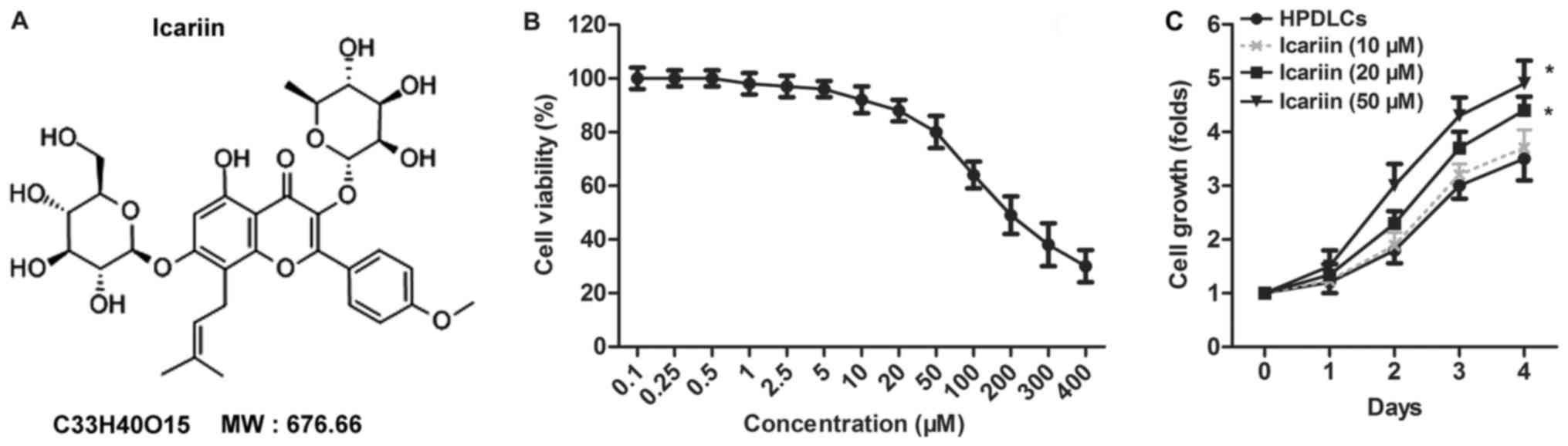 | Figure 1.Effect of icariin on HPDLC
proliferation. (A) The structure of icariin. (B) HPDLC viability
was detected by CCK-8 assay. Cells were treated with icariin at
different concentrations (0.1, 0.25, 0.5, 1, 2.5, 5, 10, 20, 50,
100, 200, 300 and 400 µM) for 24 h. (C) Cell viability was measured
using CCK-8 in HPDLC cells treated with icariin at different
concentrations (0, 10, 20 and 50 µM) and different times (1, 2, 3
and 4 days). The experiments were repeated at least 3 times with
similar results, and data are presented as the mean ± standard
deviation. *P<0.05 vs. HPDLC. CCK, Cell Counting Kit; HPDLC,
human periodontal ligament fibroblast. |
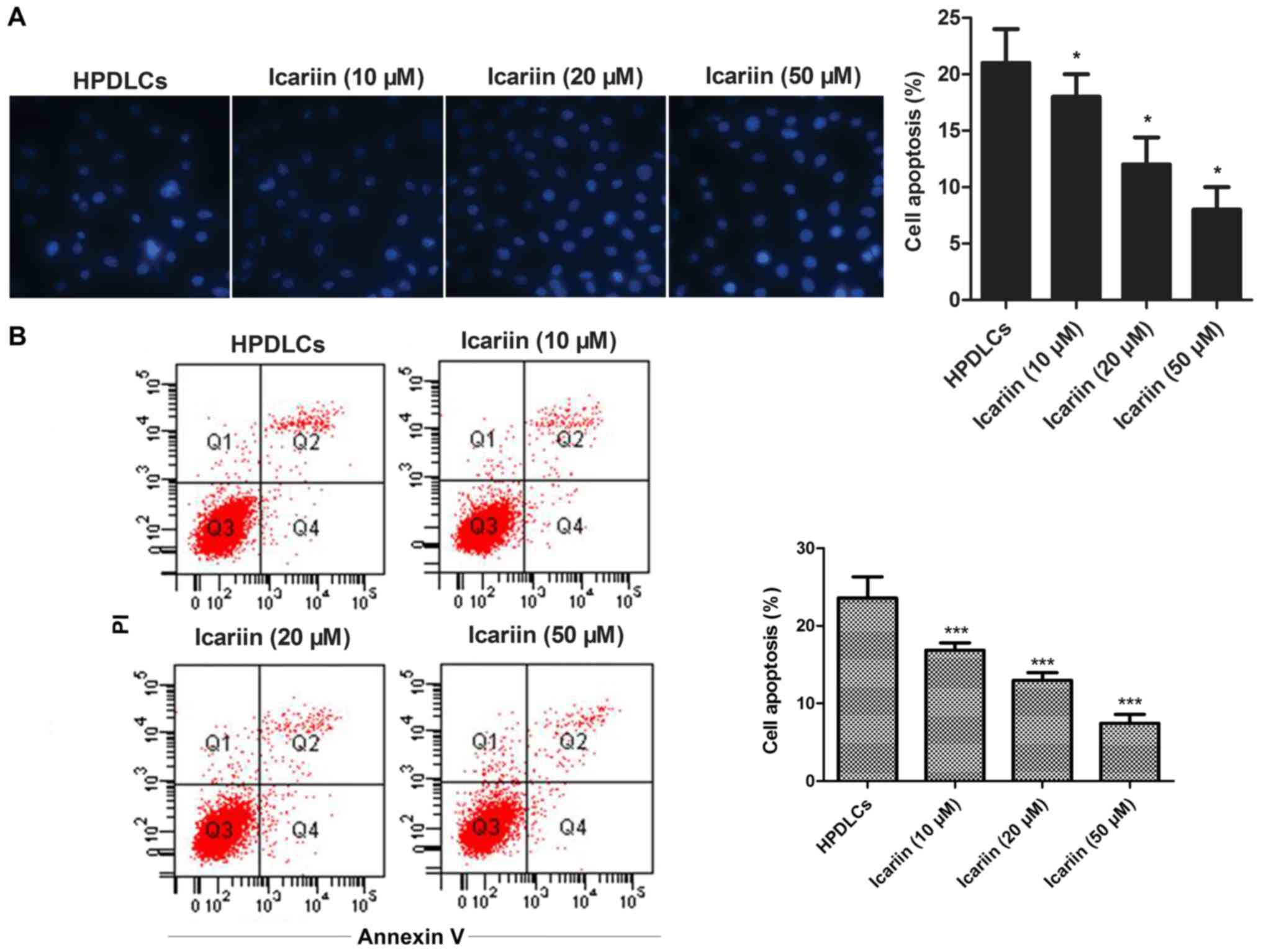
Icariin suppresses the apoptosis of
HPDLCs
To investigate whether icariin was related to cell
apoptosis, the morphological changes of icariin-treated HPDLCs were
measured by Hoechst 33258 staining and flow cytometry. Results
suggested that the icariin treatment decreased the cell apoptosis
in a dose-dependent manner (Fig.
2).
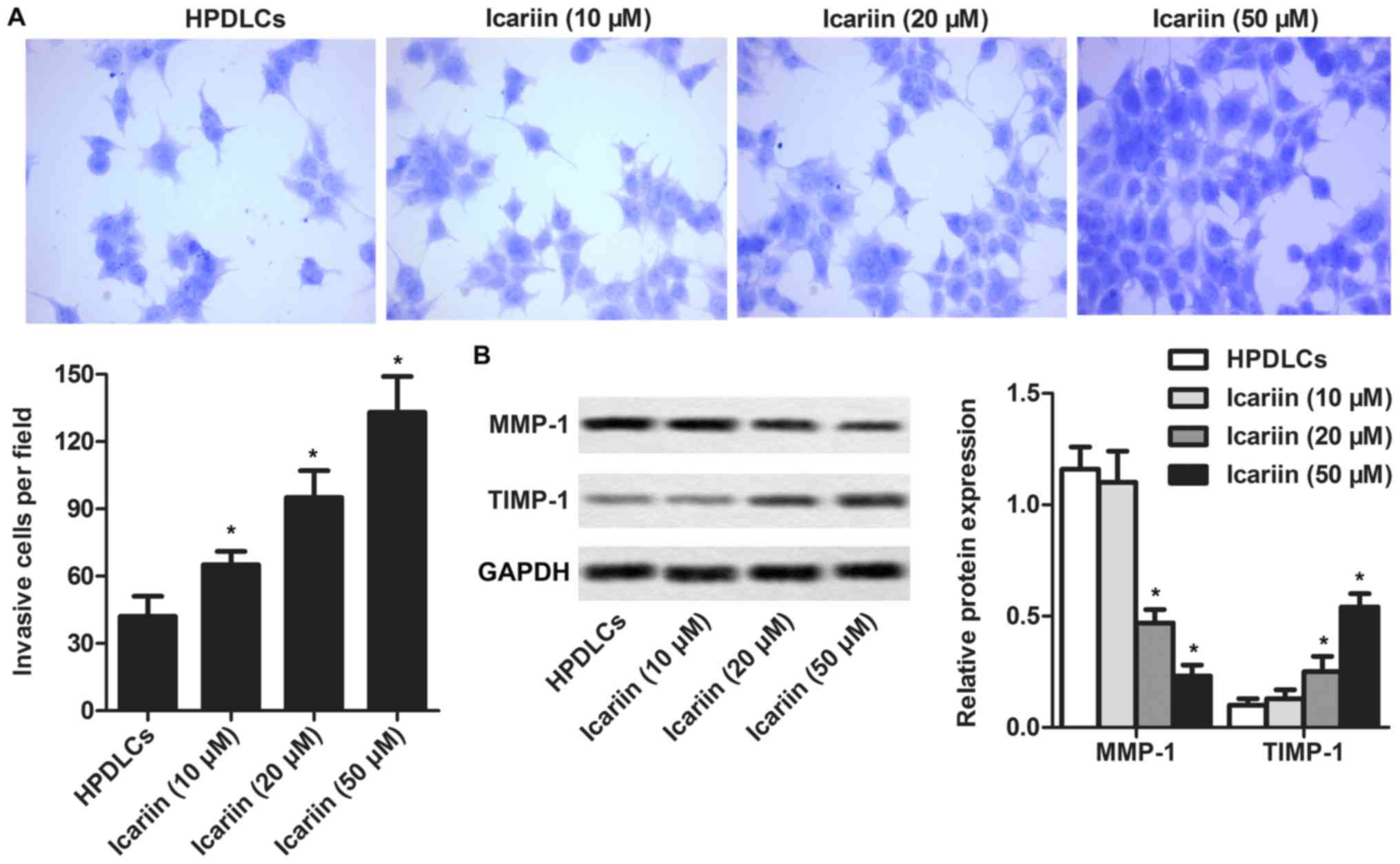
Icariin induces HPDLCs motility and
fibrosis
To explore the effect of icariin on the motility of
HPDLCs, cell motility was measured using Transwell. As illustrated
in Fig. 3A, cell motility
increased in a dose-dependent manner when treated with icariin.
Besides that, MMP-1 expression was reduced and TIMP-1 expression
was elevated with the increasing concentration of icariin (Fig. 3B), indicating that icariin play a
vital role in inducing HPDLCs fibrosis.
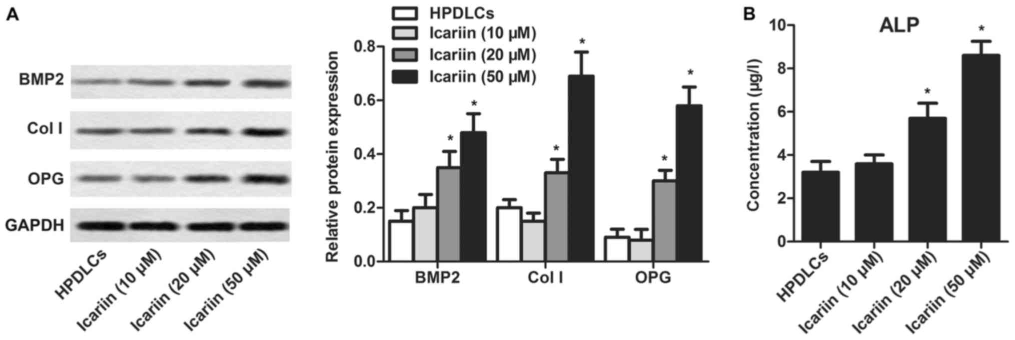
Icariin enhances HPDLCs osteogenic
differentiation ability
To determine whether icariin could affect the
osteogenic differentiation of HPDLCs, the level of related proteins
were measured using western blotting. Results suggested that, BMP2,
Col I and OPG expressions were slightly elevated in icariin (20 µM)
group and remarkably increased in icariin (50 µM) group compared
with control group (Fig. 4A). In
addition, ELISA assay indicated that icariin treatment enhanced ALP
level in a dose-dependent manner in the supernatant of HPDLCs
(Fig. 4B). These results indicated
that HPDLCs osteogenic differentiation ability could be enhanced by
icariin treatment.
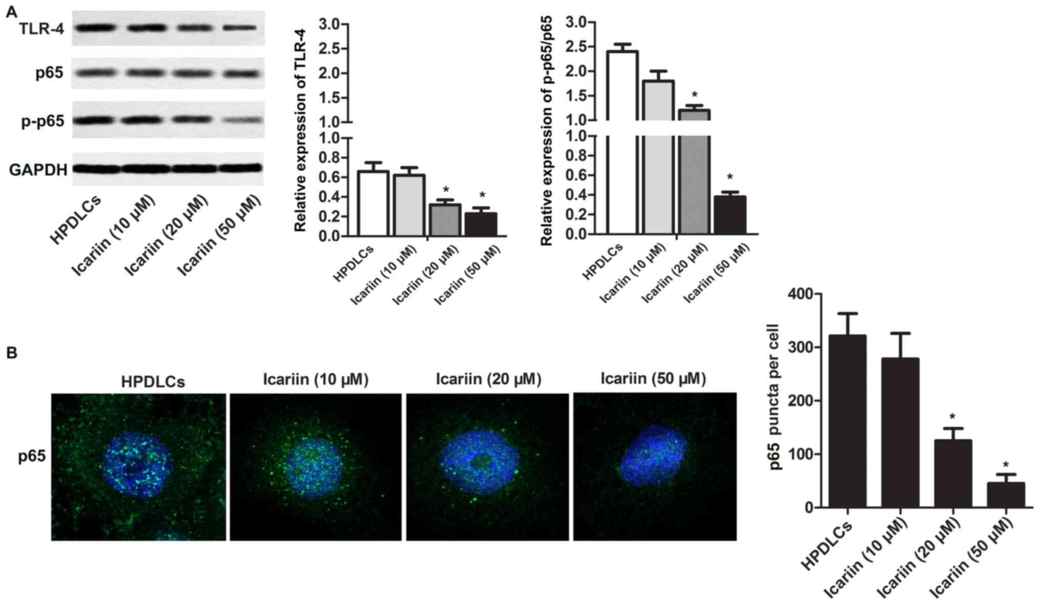
Icariin inactivates TLR-4/NF-κB
pathway
Recently, various researches have suggested that the
activation of TLR-4/NF-κB pathway could inhibit osteogenic
differentiation, cell viability and motility. Thus, TLR-4/NF-κB
pathway was examined. It is observed that icarrin decreased TLR-4
expression and p65 phosphorylation in a dose-dependent manner
(Fig. 5A). Moreover, p65 nuclear
translocation was remarkably suppressed by icariin treatment
(Fig. 5B). These results indicated
that TLR-4/NF-κB pathway could be remarkably suppressed by icariin
treatment.
Discussion
HPDLCs is one of the most important cells in
periodontal ligament. Due to the stem cell-like property, HPDLCs
have potent proliferation, differentiation and migration abilities
(12). According to published
reports, HPDLCs could differentiate into osteoblastic cells and
collagen-forming cells (13).
Accumulated studies have indicated that HPDLCs play a vital role in
maintaining the integrity and homeostasis of the periodontal
ligament during alveolar bone remodeling (14). Thus, improving the proliferation,
differentiation and migration capacity is one of the beneficial
strategies to suppress periodontal ligament degradation.
Both proliferation and migration of HPDLCs are
essential for repair and regeneration. The results from current
studies have demonstrated that icariin could promote the
proliferation and migration of various cells. For example, icariin
was found to tremendously enhance the osteoblast proliferation and
Col I level using membrane chromatography coupled with liquid
chromatography and time-of-flight mass spectrometry (15). Icariin treatment increased the
proliferation of human neural stem cells and the formation of
neurospheres (16). Moreover,
human umbilical cord mesenchymal stem cells treated with icariin
(100 µg/ml) exhibit remarkable cell migration to kidney tissue in
mice with acute kidney (17).
Endothelial cell migration could be promoted by icariin treatment
(18). Similarly, in our research,
the proliferation and migration of HPDLCs were tremendously
elevated when the concentration of icariin was 20 and 50 µM.
Previous studies have demonstrated that ECM
degradation is closely related to MMPs and TIMPs, for TIMPs help to
regenerate ECM via binding to MMPs to decrease their activity
(19). Icariin was reported to be
involved in the expression of MMPs and TIMPs. According to Wang
et al (20), icariin
suppressed the MMP-1, MMP-3 and MMP-13 expression via MAPK pathways
in IL-1β-induced SW1353 chondrosarcoma cells. Icariin treatment
inhibits MMP-9 expression and carbonic anhydrase II in the tibia of
glucocorticoid-induced hypocalcemia and hypercalciuria mice
(21). Lipopolysaccharide-induced
the increase in the level of MMP-1, MMP-3, MMP-13, cyclooxygenase-2
and iNOS could be remarkably suppressed by the pretreatment of
icariin in neonatal mice chondrocytes (22). Among the MMPs, MMP-1 is the major
enzyme to participate in degrading collagen types I and III, which
are the most abundant components of the periodontal tissue matrix
(23). In our research, icariin
treatment decreased MMP-1 expression and elevated TIMP-1 expression
in a dose-dependent manner, indicating ECM degradation could be
restrained by icariin in HPDLCs. According to published stidies,
MMP-1 expression is always associated with enhanced cell mobility,
which is not consistent to what they found. Thus, we speculated
that icariin may be involved in some other mechanism that has a
moderation effect on MMP-1 expression and cell mobility, which need
further exploration.
Apoptosis, a common cellular behavior, has
irreplaceable functions in multicellular organisms. Accumulated
studies have indicated that HPDLCs apoptosis is closely associated
with the development of periodontitis (24). Icariin was reported to possess the
anti-apoptosis ability. According to published researches,
H2O2-induced apoptosis was inhibited by
icariin via the PI3K/Akt Pathway in rat nucleus pulposus
intervertebral disc cells (25).
Besides that, oral administration of icariin remarkably suppressed
cardiomyocyte apoptosis and attenuated left heart ventricle
remodeling and abnormal mitochondria (26). Similarly, our research indicated
that the apoptosis of HPDLCs was reduced by icariin in a
dose-dependent manner.
BMP2, Col I, OPG and ALP have potent capacity of
promoting osteoblast differentiation and inducing osteogenesis
(27,28). Arising studies have demonstrated
that icarrin play a vital role in regulating these genes. Wang
et al used ALP activity assay to observe that icariin
stimulate BMP2 osteogenesis in a concentration-dependent manner
(29). Besides, the expression of
osteogenic genes (Col I, Runx2, osteopotin and DLX5) were
significantly elevated by icariin in rat bone marrow stromal cells
(30). Moreover, Mok et al
(31) demonstrated that icariin
inhibited the loss of bone mass and strength in the distal pat of
the femur and elevated the mRNA level of OPG in the ovariectomized
mouse. A similar result was drawn in our research, the expression
levels of BMP2, Col I, OPG and ALP were tremendously enhanced by
icariin at a dose-dependent manner.
It has been suggested and supported by studies that
the inactivation of TLR-4 and NF-κB was involved in ECM synthesis
and osteogenic differentiation (11,32).
The results obtained from the current studies have demonstrated
that icarrin was closely related to TLR-4/NF-κB pathway. According
to Zhang et al, icariin treatment could remarkably suppress
NF-κB nuclear translocation and the activation of Nlrp3
inflammasome in IgAN rats (33).
In addition, icariin can elevate the osteogenic differentiation
activity, decrease the NF-κB gene and protein expression, increase
the OPG expression, enhance of ALP gene expression level in
MC3T3-E1 cells (34). A similar
result was drawn in our research, the expression of TLR-4 and P-65
was dramatically blocked by icarrin treatment. Moreover, icarrin
further suppressed NF-κB p65 nuclear translocation in HPDLCs.
In conclusion, the current study has illustrated
that icarrin treatment decreased the apoptosis and increased the
viability and migration of HPDLCs. However, HPDLCs have stem
cell-like characteristics and the osteogenic differentiation and
ECM synthesis abilities could be remarkably enhanced by icariin
treatment via inactivating of TLR-4/NF-κB pathway. Our study is
valuable for unraveling the underlying mechanism of icarrin as a
candidate drug for periodontal diseases.
Acknowledgements
The authors would like to thank Shanghai
Stomatological Hospital (Shanghai, China) for providing advice and
technical support during the present study.
Funding
No funding was received.
Availability of data and materials
The datasets used and/or analyzed during the current
study are available from the corresponding author on reasonable
request.
Authors' contributions
HJL analyzed and interpreted the main data regarding
the cell function study and immunofluorescence. XYL was responsible
for study design and the drafting of the manuscript. DBJ conducted
the statistical analysis. All authors read and approved the final
manuscript.
Ethics approval and consent to
participate
The experimental protocols were approved by the
Institute Research Medical Ethics Committee of Shanghai
Stomatological Hospital (Shanghai, China); written informed consent
was obtained from all participants.
Patient consent for publication
Written informed consent was obtained from all
participants.
Competing interests
The authors declare that they have no competing
interests.
Glossary
Abbreviations
Abbreviations:
|
ECM
|
extracellular matrix
|
|
MMP-1
|
matrix matalloproteinase-1
|
|
TIMP-1
|
tissue inhibitor of
metalloproteinase-1
|
|
BMP2
|
bone morphogenetic protein 2
|
|
Col I
|
collagen I
|
|
OPG
|
osteoprotegerin
|
|
TLR-4
|
Toll-like receptor 4
|
|
NF-κB
|
nuclear factor-κB
|
References
|
1
|
Naveh GR, Chattah Lev-Tov N, Zaslansky P,
Shahar R and Weiner S: Tooth-PDL-bone cmplex: Response to
compressive loads encountered during mastication-a review. Arch
Oral Biol. 57:1575–1584. 2012. View Article : Google Scholar : PubMed/NCBI
|
|
2
|
Roberts WE, Mozsary PG and Klingler E:
Nuclear size as a cell-kinetic marker for osteoblast
differentiation. Am J Anat. 165:373–384. 1982. View Article : Google Scholar : PubMed/NCBI
|
|
3
|
Konstantonis D, Papadopoulou A, Makou M,
Eliades T, Basdra EK and Kletsas D: Senescent human periodontal
ligament fibroblasts after replicative exhaustion or ionizing
radiation have a decreased capacity towards osteoblastic
differentiation. Biogerontology. 14:741–751. 2013. View Article : Google Scholar : PubMed/NCBI
|
|
4
|
Li C, Li Q, Mei Q and Lu T:
Pharmacological effects and pharmacokinetic properties of icariin,
the major bioactive component in Herba Epimedii. Life Sci.
126:57–68. 2015. View Article : Google Scholar : PubMed/NCBI
|
|
5
|
Ye Y, Jing X, Li N, Wu Y, Li B and Xu T:
Icariin promotes proliferation and osteogenic differentiation of
rat adipose-derived stem cells by activating the RhoA-TAZ signaling
pathway. Biomed Pharmacother. 88:384–394. 2017. View Article : Google Scholar : PubMed/NCBI
|
|
6
|
Li M, Zhang C, Zhong Y and Zhao J: A novel
approach to utilize icariin as icariin-derived ecm on small
intestinal submucosa scaffold for bone repair. Ann Biomed Eng.
45:2673–2682. 2017. View Article : Google Scholar : PubMed/NCBI
|
|
7
|
Parks WC, Wilson CL and López-Boado YS:
Matrix metalloproteinases as modulators of inflammation and innate
immunity. Nat Rev Immunol. 4:617–629. 2004. View Article : Google Scholar : PubMed/NCBI
|
|
8
|
Medzhitov R, Shevach EM, Trinchieri G,
Mellor AL, Munn DH, Gordon S, Libby P, Hansson GK, Shortman K, Dong
C, et al: Highlights of 10 years of immunology in nature reviews
immunology. Nat Rev Immunol. 11:693–702. 2011. View Article : Google Scholar : PubMed/NCBI
|
|
9
|
Shih YL, Chou HM, Chou HC, Lu HF, Chu YL,
Shang HS and Chung JG: Casticin impairs cell migration and invasion
of mouse melanoma B16F10 cells via PI3K/AKT and NF-κB signaling
pathways. Environ Toxicol. 32:2097–2112. 2017. View Article : Google Scholar : PubMed/NCBI
|
|
10
|
Wang LM, Zhao N, Zhang J, Sun QF, Yang CZ
and Yang PS: Tumor necrosis factor-alpha inhibits osteogenic
differentiation of pre-osteoblasts by downregulation of EphB4
signaling via activated nuclear factor-κB signaling pathway. J
Periodontal Res. 53:66–72. 2018. View Article : Google Scholar : PubMed/NCBI
|
|
11
|
Wang YJ, Zhang HQ, Han HL, Zou YY, Gao QL
and Yang GT: Taxifolin enhances osteogenic differentiation of human
bone marrow mesenchymal stem cells partially via NF-κB pathway.
Biochem Biophys Res Commun. 490:36–43. 2017. View Article : Google Scholar : PubMed/NCBI
|
|
12
|
Lim SS, Kook SH and Lee JC: COMP-Ang1
enhances DNA synthesis and cell cycle progression in human
periodontal ligament cells via Tie2-mediated phosphorylation of
PI3K/Akt and MAPKs. Mol Cell Biochem. 416:157–168. 2016. View Article : Google Scholar : PubMed/NCBI
|
|
13
|
Kook SH, Jeon YM, Park SS and Lee JC:
Periodontal fibroblasts modulate proliferation and osteogenic
differentiation of embryonic stem cells through production of
fibroblast growth factors. J Periodontol. 85:645–654. 2014.
View Article : Google Scholar : PubMed/NCBI
|
|
14
|
Lekic P and McCulloch CA: Periodontal
ligament cell population: The central role of fibroblasts in
creating a unique tissue. Anat Rec. 245:327–341. 1996. View Article : Google Scholar : PubMed/NCBI
|
|
15
|
Wang N, Zhang Q, Xin H, Shou D and Qin L:
Osteoblast cell membrane chromatography coupled with liquid
chromatography and time-of-flight mass spectrometry for screening
specific active components from traditional Chinese medicines. J
Sep Sci. 40:4311–4319. 2017. View Article : Google Scholar : PubMed/NCBI
|
|
16
|
Yang P, Guan YQ, Li YL, Zhang L, Zhang L
and Li L: Icariin promotes cell proliferation and regulates gene
expression in human neural stem cells in vitro. Mol Med Rep.
14:1316–1322. 2016. View Article : Google Scholar : PubMed/NCBI
|
|
17
|
Cui H, Liu Z, Wang L, Bian Y, Li W, Zhou
H, Chu X and Zhao Q: Icariin-treated human umbilical cord
mesenchymal stem cells decrease chronic liver injury in mice.
Cytotechnology. 69:19–29. 2017. View Article : Google Scholar : PubMed/NCBI
|
|
18
|
Chung BH, Kim JD, Kim CK, Kim JH, Won MH,
Lee HS, Dong MS, Ha KS, Kwon YG and Kim YM: Icariin stimulates
angiogenesis by activating the MEK/ERK- and PI3K/Akt/eNOS-dependent
signal pathways in human endothelial cells. Biochem Biophys Res
Commun. 376:404–408. 2008. View Article : Google Scholar : PubMed/NCBI
|
|
19
|
Belibasakis GN and Guggenheim B: Induction
of prostaglandin E(2) and interleukin-6 in gingival fibroblasts by
oral biofilms. FEMS Immunol Med Microbiol. 63:381–386. 2011.
View Article : Google Scholar : PubMed/NCBI
|
|
20
|
Wang Z, Ding L, Zhang S, Jiang T, Yang Y
and Li R: Effects of icariin on the regulation of the
OPG-RANKL-RANK system are mediated through the MAPK pathways in
IL-1β-stimulated human SW1353 chondrosarcoma cells. Int J Mol Med.
34:1720–1726. 2014. View Article : Google Scholar : PubMed/NCBI
|
|
21
|
Zhang J, Song J and Shao J: Icariin
attenuates glucocorticoid-induced bone deteriorations, hypocalcemia
and hypercalciuria in mice. Int J Clin Exp Med. 8:7306–7314.
2015.PubMed/NCBI
|
|
22
|
Liu MH, Sun JS, Tsai SW, Sheu SY and Chen
MH: Icariin protects murine chondrocytes from
lipopolysaccharide-induced inflammatory responses and extracellular
matrix degradation. Nutr Res. 30:57–65. 2010. View Article : Google Scholar : PubMed/NCBI
|
|
23
|
Hannas AR, Pereira JC, Granjeiro JM and
Tjäderhane L: The role of matrix metalloproteinases in the oral
environment. Acta Odontol Scand. 65:1–13. 2007. View Article : Google Scholar : PubMed/NCBI
|
|
24
|
Liu J, Jiang Y, Mao J, Gu B, Liu H and
Fang B: High levels of glucose induces a dose-dependent apoptosis
in human periodontal ligament fibroblasts by activating caspase-3
signaling pathway. Appl Biochem Biotechnol. 170:1458–1471. 2013.
View Article : Google Scholar : PubMed/NCBI
|
|
25
|
Deng X, Chen S, Zheng D, Shao Z, Liang H
and Hu H: Icariin prevents H2O2-induced
apoptosis via the PI3K/Akt pathway in rat nucleus pulposus
intervertebral disc cells. Evid Based Complement Alternat Med.
2017:26942612017. View Article : Google Scholar : PubMed/NCBI
|
|
26
|
Qian ZQ, Wang YW, Li YL, Li YQ, Ling-Zhu
and Yang DL: Icariin prevents hypertension-induced cardiomyocyte
apoptosis through the mitochondrial apoptotic pathway. Biomed
Pharmacother. 88:823–831. 2017. View Article : Google Scholar : PubMed/NCBI
|
|
27
|
Di Benedetto A, Posa F, Carbone C, Cantore
S, Brunetti G, Centonze M, Grano M, Lo Muzio L, Cavalcanti-Adam EA
and Mori G: NURR1 downregulation favors osteoblastic
differentiation of MSCs. Stem Cells Int. 2017:76170482017.
View Article : Google Scholar : PubMed/NCBI
|
|
28
|
Boumah CE, Selvamurugan N and Partridge
NC: Transcription in the osteoblast: Regulatory mechanisms utilized
by parathyroid hormone and transforming growth factor-beta. Prog
Nucleic Acid Res Mol Biol. 80:287–321. 2005. View Article : Google Scholar : PubMed/NCBI
|
|
29
|
Wang Q, Cao L, Liu Y, Zheng A, Wu J, Jiang
X and Ji P: Evaluation of synergistic osteogenesis between icariin
and BMP2 through a micro/meso hierarchical porous delivery system.
Int J Nanomedicine. 12:7721–7735. 2017. View Article : Google Scholar : PubMed/NCBI
|
|
30
|
Wei Q, Zhang J, Hong G, Chen Z, Deng W, He
W and Chen MH: Icariin promotes osteogenic differentiation of rat
bone marrow stromal cells by activating the ERα-Wnt/β-catenin
signaling pathway. Biomed Pharmacother. 84:931–939. 2016.
View Article : Google Scholar : PubMed/NCBI
|
|
31
|
Mok SK, Chen WF, Lai WP, Leung PC, Wang
XL, Yao XS and Wong MS: Icariin protects against bone loss induced
by oestrogen deficiency and activates oestrogen receptor-dependent
osteoblastic functions in UMR 106 cells. Br J Pharmacol.
159:939–949. 2010. View Article : Google Scholar : PubMed/NCBI
|
|
32
|
Zhang M, Hu X, Li S, Lu C, Li J, Zong Y,
Qi W and Yang H: Hepatoprotective effects of ethyl pyruvate against
CCl4-induced hepatic fibrosis via inhibition of TLR4/NF-κB
signaling and up-regulation of MMPs/TIMPs ratio. Clin Res Hepatol
Gastroenterol. 42:72–81. 2018. View Article : Google Scholar : PubMed/NCBI
|
|
33
|
Zhang L, Wang XZ, Li YS, Zhang L and Hao
LR: Icariin ameliorates IgA nephropathy by inhibition of nuclear
factor kappa b/Nlrp3 pathway. FEBS Open Bio. 7:54–63. 2016.
View Article : Google Scholar : PubMed/NCBI
|
|
34
|
Zhang S, Feng P, Mo G, Li D, Li Y, Mo L,
Yang Z and Liang D: Icariin influences adipogenic differentiation
of stem cells affected by osteoblast-osteoclast co-culture and
clinical research adipogenic. Biomed Pharmacother. 88:436–442.
2017. View Article : Google Scholar : PubMed/NCBI
|