Introduction
Traditional Chinese medicine (TCM) has been gaining
more attention due to its satisfactory clinical results and the
reduced side effects when utilized for cancer treatment. TCM is an
important part of complementary and alternative medicines as it has
standardized diagnostic and therapeutic systems and is implemented
worldwide (1). There is increasing
evidence that various herbs and compounds derived from natural
products with antitumor effects can induce apoptosis in various
tumor cells (2–4). Toosendanin is a triterpenoid
extracted from the root bark of Melia toosendan. Toosendanin
was found to inhibit tumor cell proliferation and promote tumor
cell apoptosis (5–7). In addition, a previous study showed
that toosendanin can block osteosarcoma tumorigenesis (8). However, there has been no research on
the effects of toosendanin on Ewing's sarcoma (ES).
ES is a rare invasive tumor in the primitive
neuroectodermal tumor family (9),
which is common in children and adolescents (10,11).
ES is extremely malignant, with a short disease course, rapid
recurrence and high transfer rate. Although neoadjuvant
chemotherapy and limb salvage surgery are widely used clinically,
the 5-year survival rate for ES patients with early metastasis or
recurrence is still less than 50% and the 10-year survival rate is
less than 30% (12,13). In addition, most neoadjuvant
chemotherapy drugs have the risk of unstable effectiveness and
serious side effects such as cardiotoxicity and nephrotoxicity.
Therefore, there is an urgent need for safer and more effective
anticancer drugs in clinical practice.
Studies have shown that baicalein extracted from the
TCM Astragalus membranaceus can induce apoptosis through the
mitochondrial apoptosis and death receptor pathways in ES cells
(14). Therefore, we hypothesize
that toosendanin may also have antitumor and/or pro-apoptotic
activity in ES. Subsequently, we studied the effects of toosendanin
on cell activity and apoptosis of the human ES cell line SK-ES-1
and further elucidated the relevant molecular mechanisms.
Materials and methods
Cell culture
The human ES cell lines SK-ES-1 and RD-ES were
obtained from the American Type Culture Collection (ATCC; Manassas,
VA, USA). The cells were cultured in RPMI-1640 medium supplemented
with 10% (v/v) fetal bovine serum (FBS), 100 U/ml penicillin and
100 µg/ml streptomycin. The cells were incubated at 37°C in a
humidified atmosphere containing 5% CO2. Cells were
passaged when they reached 80% confluency. Exponential-phase cells
were used in the experiments, and the passage number was
<20.
Reagents and antibodies
Purified toosendanin (source: root bark and bark of
Melia toosendan; molecular formula (MF):
C30H38O11; molecular weight (MW):
574.62; purity ≥98%) was purchased from Sigma-Aldrich (Merck KGaA,
Darmstadt, Germany). RPMI-1640 medium, phosphate-buffered saline
(PBS), dimethyl sulfoxide (DMSO), bovine serum albumin (BSA), Cell
Counting Kit-8 (CCK-8) and horseradish peroxidase-conjugated goat
anti-rabbit secondary antibodies (dilution 1:2,500; cat. no. HS101)
were purchased from TransGen Biotech, Inc. (Beijing, China). FBS
was obtained from HyClone Laboratories (GE Healthcare, Chicago, IL,
USA), while the Annexin V-FITC/Propidium Iodide (PI) Apoptosis
Assay kit was obtained from Becton Dickinson (BD Biosciences, San
Jose, CA, USA). The Hoechst 33258 staining kit was purchased from
Nanjing KeyGen Biotech Co., Ltd. (Nanjing, China). Antibodies
against B-cell lymphoma 2 (Bcl-2; dilution 1:2,000; cat. no.
ab182858), Bcl-2-associated X protein (Bax; dilution 1:1,000; cat.
no. ab32503), cytochrome c (dilution 1:5,000; cat. no.
ab133504), caspase-3 (dilution 1:5,000; cat. no. ab32351),
caspase-8 (dilution 1:1,000; cat. no. ab108333), caspase-9
(dilution 1:1,000; cat. no. ab32539), poly(ADP-ribose) polymerase
(PARP; dilution 1:1,000; cat. no. ab32138) and GAPDH (dilution
1:2,500; cat. no. ab9485) were purchased from Abcam (Cambridge,
UK).
Determination of cell viability by the
CCK-8 method
SK-ES-1 and RD-ES cells were cultured in 96-well
plates (5×10³ cells/well). Cells were treated with
different concentrations (0, 1, 2, 5, 10, 20, 40, 50 and 60 µM) of
toosendanin for 24, 48 and 72 h and control cells were treated with
<0.1% (v/v) DMSO. After the indicated incubation times, 10 µl of
CCK-8 was added to the plates and incubated for an additional 1–4 h
at 37°C. Thereafter, the absorbance was measured at 450 nm using an
ELISA plate reader (ELx800; BioTek Instruments, Inc., Winooski, VT,
USA).
Hoechst 33258 nuclear staining. Cells
(5×10⁴ cells/well) were incubated with 0, 25 or 50 µM
toosendanin in 24-well plates for 24 h at 37°C. The cells were then
fixed with 4% paraformaldehyde for 30 min. Thereafter, the cells
were washed three times with pre-cooled PBS and stained with 10
mg/l Hoechst 33258 solution for 10 min at 25°C in the dark.
Subsequently, the stained nuclei were observed under a fluorescence
microscope (Olympus Corp., Tokyo, Japan) at 350 nm excitation and
460 nm emission wavelengths (magnification, ×200).
Annexin V-FITC/PI apoptosis assay
SK-ES-1 cells were cultured for 24 h with 0, 25, or
50 µM toosendanin, washed twice with ice-cold PBS, and resuspended
at a concentration of 1×10⁶ cells/ml in 1X binding
buffer. The cell suspension (100 µl) was incubated with 1 µl
Annexin V-FITC and 2 µl propidium iodine (PI) solution for 15 min
at 25°C in the dark. After addition of 150 µl 1X binding buffer,
the samples were analyzed using a FACSVerse flow cytometer (BD
Biosciences, San Jose, CA, USA). Apoptosis rates were analyzed
using FlowJo v7.6 software (Tree Star, Inc., Ashland, OR, USA).
Western blot analysis
SK-ES-1 cells were cultured in a 6-well plate at a
density of 2×10⁵ cells/well. After treatment with 0, 25
or 50 µM toosendanin for 24 h, cells were harvested and lysed in
RIPA buffer containing a protease inhibitor cocktail
(Sigma-Aldrich; Merck KGaA). The lysate was centrifuged at 12,000 ×
g for 10 min at 4°C. The supernatant was then collected, and the
protein concentration was determined by the BCA method. The same
protein amounts (10 µg in each lane) were loaded and separated by
10% SDS-PAGE, followed by transfer onto polyvinylidene difluoride
(PVDF) membranes. The membranes were blocked with 5% (w/v) fat-free
milk in Tris-buffered saline containing 0.05% Tween-20 (TBS-T) and
then incubated with primary antibodies at 4°C overnight. The next
day, the PVDF membranes were washed three times in TBS-T and
incubated with HRP-conjugated secondary antibodies for 2 h at room
temperature. Immunoreactive proteins were detected by an ECL kit
(Thermo Fisher Scientific, Inc.) and then developed on an X-ray
film (Kodak). The proteins were quantified via densitometry using
ImageJ software (version 1.51j81; National Institutes of
Health).
Statistical analysis
Data are expressed as mean ± standard deviation (SD)
and analyzed by GraphPad Prism 7.0 software (GraphPad Software,
Inc., Chicago, IL, USA). One-way analysis of variance (ANOVA) was
conducted with the Newman-Keuls method to determine the
significance of the differences between the experimental
conditions. All experiments were repeated at least three times.
Differences in means were considered statistically significant at
*P<0.05, **P<0.01 and ***P<0.001 (as indicated in the
figure legends).
Results
Toosendanin inhibits cell growth of
SK-ES-1 and RD-ES cells
To study the effect of toosendanin on ES cell
activity, SK-ES-1 and RD-ES cells were exposed to different
concentrations of toosendanin for 24 h. CCK-8 results demonstrated
that toosendanin inhibited the viability of SK-ES-1 and RD-ES cells
in a dose-dependent manner and that SK-ES-1 cells were more
sensitive to toosendanin (Fig.
1A). The half maximal inhibitory concentration
(IC50) of SK-ES-1 cells treated with toosendanin was
32.95 µM at 24 h. Furthermore, it was observed that toosendanin
inhibited the viability of SK-ES-1 in a time- and dose-dependent
manner (Fig. 1B). Subsequently,
SK-ES-1 cells were treated with toosendanin at concentrations of 0,
25 and 50 µM for 24 h in the following assays.
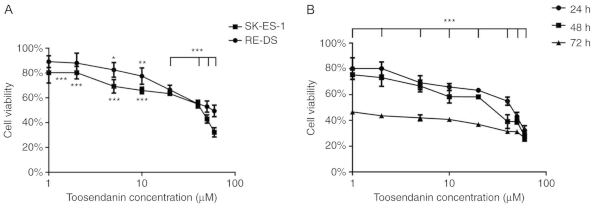 | Figure 1.(A) SK-ES-1 cells and RD-ES cells were
treated with different concentrations of toosendanin (0, 1, 2, 5,
10, 20, 40, 50 and 60 µM) for 24 h and a CCK-8 assay was performed
to detect cell viability after treatment. Data are expressed as the
mean ± SD of three independent experiments. *P<0.05, **P<0.01
and ***P<0.001 vs. the control. (B) SK-ES-1 cells were treated
with different concentrations of toosendanin (0, 1, 2, 5, 10, 20,
40, 50 and 60 µM) for 24, 36, and 48 h and then a CCK-8 assay was
performed to detect cell viability after treatment. Data are
expressed as the mean ± SD of three independent experiments.
***P<0.001 vs. the control. |
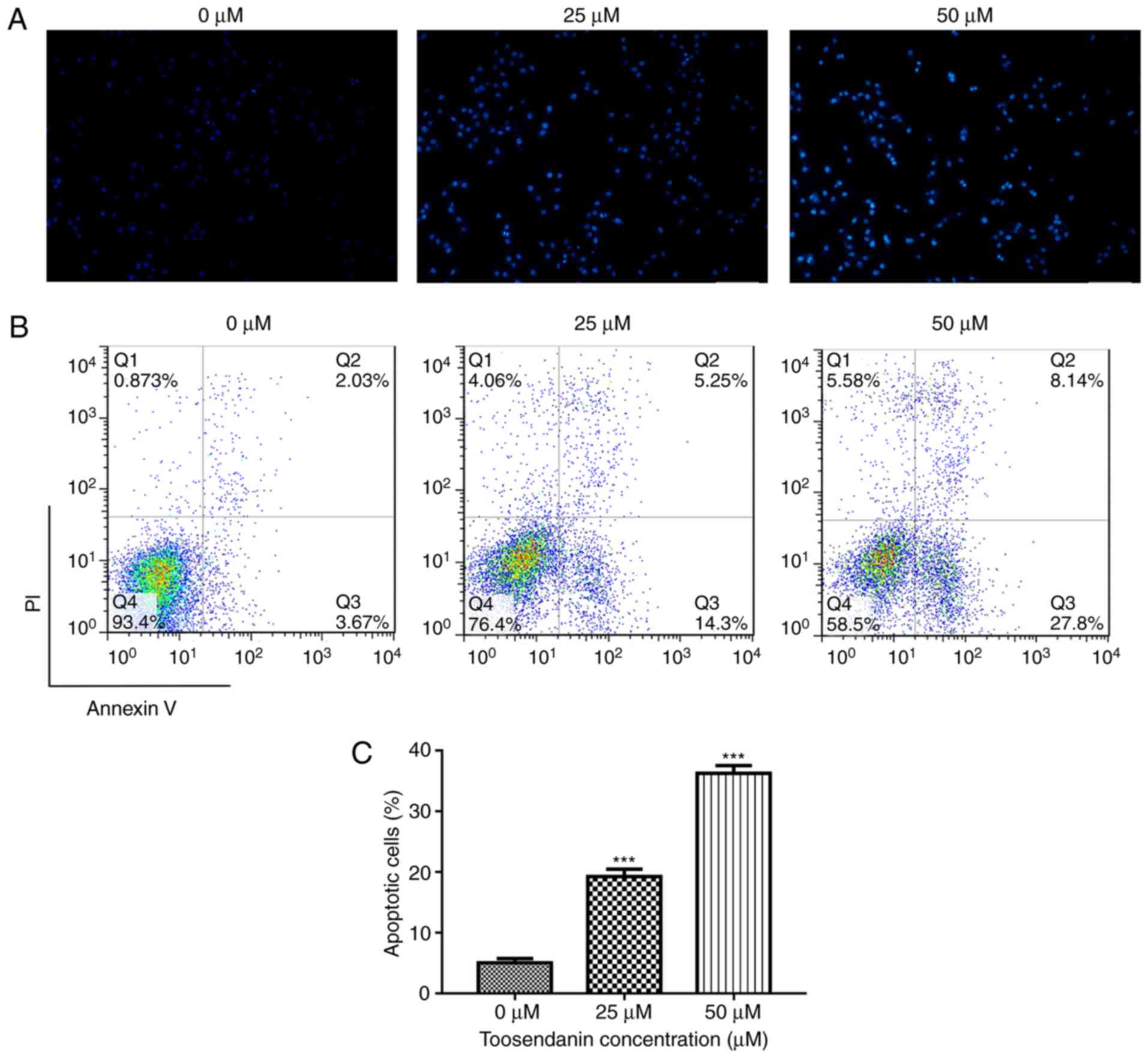
Toosendanin results in morphological
changes in SK-ES-1 cells
SK-ES-1 cells treated with toosendanin and stained
with Hoechst 33258 showed concentrated and broken nuclei in a
dose-dependent manner, which are typical morphological features of
apoptotic cells (Fig. 2A).
Toosendanin induces apoptosis
Apoptosis was measured by flow cytometry with
Annexin V-FITC/PI double labeling. The apoptosis rate of the
control group (sum of early and late apoptosis) was 5.03±0.71%.
After 24 h of treatment with 25 or 50 µM toosendanin, the apoptotic
rate increased to 19.32±1.26 and 36.28±1.28%, respectively
(Fig. 2B). Thus, toosendanin
induced apoptosis in a dose-dependent manner (Fig. 2C).
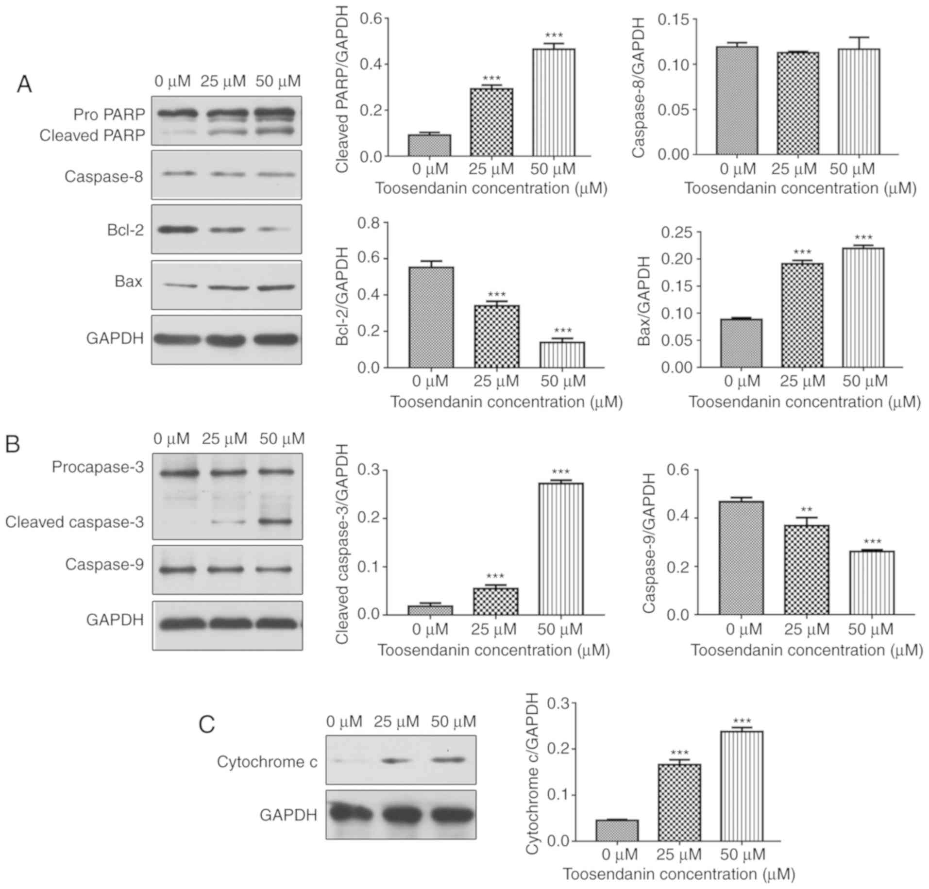
Toosendanin modulates the expression
levels of caspase and Bcl-2 family proteins
Western blot analysis was used to analyze expression
levels of apoptosis-related protein Bcl-2, pro-apoptotic Bax,
cytochrome c, caspase-3, −8 and −9 and PARP to identify the
mechanism of toosendanin-induced SK-ES-1 apoptosis. The results
showed that toosendanin increased Bax protein levels (Fig. 3A) and cytochrome c release
(Fig. 3C) as well as decreased
Bcl-2 protein levels (Fig. 3A).
This indicates that toosendanin activates the mitochondrial
apoptotic pathway in SK-ES-1 cells by modulating the expression of
Bcl-2 family proteins. At the same time, we evaluated the protein
expression levels of caspase-3, −8 and −9. Cleavage of caspase-3
and the key cellular substrate PARP was observed as well as
downregulation of procaspase-3 and procaspase-9 expression levels;
caspase-3 and caspase-9 were cleaved and downregulated in a
dose-dependent manner (Fig. 3A and
B). In contrast, expression levels of caspase-8 did not change
(Fig. 3A). These results indicate
that toosendanin-induced apoptosis involves the caspase cascade and
is triggered by the mitochondrial apoptotic pathway.
Discussion
As malignant tumors are the result of unchecked cell
proliferation, inhibiting tumor cell proliferation and promoting
tumor cell apoptosis are effective means to prevent tumor growth
and eliminate tumors (15).
Apoptosis, also known as programmed cell death, is the orderly
death of cells controlled by genes that maintain internal
environment stability. One of the characteristics of tumor cells is
their ability to resist apoptosis (16) and thus, induction of tumor cell
apoptosis is the mechanism of action of many antitumor drugs
(17). With increasing attention
on toosendanin, its antitumor effect has become a ‘hot topic’ in
research. Tada et al (18)
found that toosendanin exhibits strong cytotoxicity against human
cancer cell lines (KB cells) and that the toxicity mechanism may be
related to the C-14/C-15 epoxy structure of toosendanin. Zhang
et al (19) reported that
toosendanin may induce apoptosis in lymphoma U937 cells by
arresting cells in G0/G1 and S phases.
Furthermore, He et al (20)
demonstrated that toosendanin possesses strong anticancer effects
in vivo and in vitro via inducing
mitochondria-dependent apoptosis in hepatocellular carcinoma cells.
Meanwhile, Ju et al (21)
discovered that the pro-apoptotic effects of toosendanin on human
promyelocytic leukemia HL-60 cells were mediated through the c-Jun
N-terminal kinase (JNK) signaling pathway. The above studies have
shown that toosendanin is a potential antitumor drug with
remarkable effects; however, the mechanism of action in regards to
Ewing's sarcoma (ES) cells has not been elucidated. To this end, we
employed human SK-ES-1 cells to explore the mechanism of action of
toosendanin in regards to ES.
The present study found that toosendanin has an
inhibitory effect on the proliferation of the SK-ES-1 cell line in
a time- and dose-dependent manner. These findings are consistent
with the literature reported on other types of tumors, which also
have identified toosendanin as an anticancer agent. To the best of
our knowledge, this is the first study to explore the effects of
toosendanin on ES in vitro.
To further explore the mechanism of apoptosis
induction in SK-ES-1 cells by toosendanin, the changes in
expression levels of caspase-3, −8 and −9, and other
apoptosis-related genes were determined by western blotting. There
are two main pathways of apoptosis, the mitochondrial pathway and
the death receptor pathway. The mitochondrial pathway involves the
ratio of pro-apoptotic protein Bax to anti-apoptotic protein Bcl-2,
which affects the release of many apoptotic proteins in the
mitochondrial membrane space, such as cytochrome c.
Cytochrome c activates caspase-9 and reactivates caspase-3.
The Fas/tumor necrosis factor (TNF) receptor located in the cell
membrane of the death receptor pathway is activated by apoptosis,
which activates caspase-8 that goes on to activate caspase-3.
Subsequently, activated caspase-3 causes cleavage or degradation of
key cellular substrates including PARP, resulting in cell
morphological changes, DNA double-strand breaks, and other
characteristics of apoptotic cells (22,23).
In the present study, toosendanin-induced apoptosis was accompanied
by a change in the ratio of Bax/Bcl-2, which activated caspase-9
and caspase-3. In addition, PARP cleavage was also observed. These
findings indicate that SK-ES-1 cell apoptosis induced by
toosendanin is triggered by the mitochondrial pathway.
In conclusion, toosendanin was found to inhibit ES
cell viability and apoptosis through the mitochondrial apoptosis
pathway. Our findings support the utilization of toosendanin as a
potential therapeutic agent for the treatment of ES. However, the
present study has certain limitations. First, we only employed
in vitro experiments and thus further research is needed to
elucidate the in vivo effects of toosendanin on SK-ES-1
×enograft tumors in nude mice. Second, we cannot rule out whether
toosendanin participates in other signaling pathways in the
induction of ES cell apoptosis. Furthermore, safety must be
guaranteed before clinical application of ‘natural medicine’.
Previous studies have reported that high doses of toosendanin (80
mg/kg) result in serious liver injury in mice (24), while the effect of toosendanin on
normal or healthy cells in vitro remains unclear. Our in
vitro experiments demonstrated that the IC50 value
of toosendanin in SK-ES-1 cells was 32.95 µM, and the cytotoxicity
of this concentration must be evaluated prior to use. Thus, future
research must comprehensively evaluate the effects of toosendanin
on normal cells through in vivo and in vitro
experiments to understand the therapeutic range of toosendanin.
Acknowledgements
Not applicable.
Funding
The present study was supported by the Jiangxi
Provincial Department of Science and Technology (grant no.
20171ACG70006).
Availability of data and materials
The datasets used and/or analyzed during the present
study are available from the corresponding author on reasonable
request.
Authors' contributions
TG, AX, BZ and MD conceived and designed the study;
TG, AX and XL performed the experiments; BZ and HZ analyzed the
data; TG, BZ and MD wrote the paper; XL and JZ prepared the
figures; TG, AX, XL, HZ, BZ and MD reviewed and edited the
manuscript. All authors read and approved the manuscript and agree
to be accountable for all aspects of the research in ensuring that
the accuracy or integrity of any part of the work are appropriately
investigated and resolved.
Ethics approval and consent to
participate
Not applicable.
Patient consent for publication
Not applicable.
Competing interests
The authors declare that they have no competing
interests.
Glossary
Abbreviations
Abbreviations:
|
TCM
|
traditional Chinese medicine
|
|
ES
|
Ewing's sarcoma
|
|
Bcl-2
|
B-cell lymphoma
|
|
Bax
|
Bcl-2-associated X protein
|
|
PARP
|
poly(ADP-ribose) polymerase
|
|
FBS
|
fetal bovine serum
|
References
|
1
|
Ahmed S, Anuntiyo J, Malemud CJ and Haqqi
TM: Biological basis for the use of botanicals in osteoarthritis
and rheumatoid arthritis: A review. Evid Based Complement Alternat
Med. 2:301–308. 2005. View Article : Google Scholar : PubMed/NCBI
|
|
2
|
Zhang K, Wang X, Wang C, Zheng H, Li T,
Xiao S, Wang M, Fei C, Zhang L and Xue F: Investigation of
quinocetone-induced mitochondrial damage and apoptosis in HepG2
cells and compared with its metabolites. Environ Toxicol Pharmacol.
39:555–567. 2015. View Article : Google Scholar : PubMed/NCBI
|
|
3
|
Zhang J, Song J, Wu D, Wang J and Dong W:
Hesperetin induces the apoptosis of hepatocellular carcinoma cells
via mitochondrial pathway mediated by the increased intracellular
reactive oxygen species, ATP and calcium. Med Oncol. 32:1012015.
View Article : Google Scholar : PubMed/NCBI
|
|
4
|
Pieme CA, Santosh GK, Tekwu EM, Askun T,
Aydeniz H, Ngogang JY, Bhushan S and Saxena AK: Fruits and barks
extracts of Zanthozyllum heitzii a spice from Cameroon induce
mitochondrial dependent apoptosis and G0/G1 phase arrest in human
leukemia HL-60 cells. Biol Res. 47:542014. View Article : Google Scholar : PubMed/NCBI
|
|
5
|
Zhou Q, Wu X, Wen C, Wang H, Wang H, Liu H
and Peng J: Toosendanin induces caspase-dependent apoptosis through
the p38 MAPK pathway in human gastric cancer cells. Biochem Biophys
Res Commun. 505:261–266. 2018. View Article : Google Scholar : PubMed/NCBI
|
|
6
|
Shi YL and Li MF: Biological effects of
toosendanin, a triterpenoid extracted from Chinese traditional
medicine. Prog Neurobiol. 82:1–10. 2007. View Article : Google Scholar : PubMed/NCBI
|
|
7
|
Pei Z, Fu W and Wang G: A natural product
toosendanin inhibits epithelial-mesenchymal transition and tumor
growth in pancreatic cancer via deactivating Akt/mTOR signaling.
Biochem Biophys Res Commun. 493:455–460. 2017. View Article : Google Scholar : PubMed/NCBI
|
|
8
|
Zhang T, Li J, Yin F, Lin B, Wang Z, Xu J,
Wang H, Zuo D, Wang G, Hua Y and Cai Z: Toosendanin demonstrates
promising antitumor efficacy in osteosarcoma by targeting STAT3.
Oncogene. 36:6627–6639. 2017. View Article : Google Scholar : PubMed/NCBI
|
|
9
|
Windsor R, Strauss S, Seddon B and Whelan
J: Experimental therapies in Ewing's sarcoma. Expert Opin Investig
Drugs. 18:143–159. 2009. View Article : Google Scholar : PubMed/NCBI
|
|
10
|
Teicher BA, Bagley RG, Rouleau C, Kruger
A, Ren Y and Kurtzberg L: Characteristics of human Ewing/PNET
sarcoma models. Ann Saudi Med. 31:174–182. 2011. View Article : Google Scholar : PubMed/NCBI
|
|
11
|
Zhang Z, Huang L, Yu Z, Chen X, Yang D,
Zhan P, Dai M, Huang S, Han Z and Cao K: Let-7a functions as a
tumor suppressor in Ewing's sarcoma cell lines partly by targeting
cyclin-dependent kinase 6. DNA Cell Biol. 33:136–147. 2014.
View Article : Google Scholar : PubMed/NCBI
|
|
12
|
Ross KA, Smyth NA, Murawski CD and Kennedy
JG: The biology of ewing sarcoma. ISRN Oncol.
2013:7597252013.PubMed/NCBI
|
|
13
|
Scotlandi K: Targeted therapies in Ewing's
sarcoma. Adv Exp Med Biol. 587:13–22. 2006. View Article : Google Scholar : PubMed/NCBI
|
|
14
|
Ye C, Yu X, Zeng J, Dai M and Zhang B:
Effects of baicalein on proliferation, apoptosis, migration and
invasion of Ewing's sarcoma cells. Int J Oncol. 51:1785–1792. 2017.
View Article : Google Scholar : PubMed/NCBI
|
|
15
|
Cohen GM: Caspases: The executioners of
apoptosis. Biochem J. 326:1–16. 1997. View Article : Google Scholar : PubMed/NCBI
|
|
16
|
Singhal S, Vachani A, Antin-Ozerkis D,
Kaiser LR and Albelda SM: Prognostic implications of cell cycle,
apoptosis, and angiogenesis biomarkers in non-small cell lung
cancer: A review. Clin Cancer Res. 11:3974–3986. 2005. View Article : Google Scholar : PubMed/NCBI
|
|
17
|
Viktorsson K, Lewensohn R and Zhivotovsky
B: Apoptotic pathways and therapy resistance in human malignancies.
Adv Cancer Res. 94:1432005. View Article : Google Scholar : PubMed/NCBI
|
|
18
|
Tada K, Takido M and Kitanaka S: Limonoids
from fruit of Melia toosendan and their cytotoxic activity.
Phytochemistry. 51:787–791. 1999. View Article : Google Scholar : PubMed/NCBI
|
|
19
|
Zhang B, Wang ZF, Tang MZ and Shi YL:
Growth inhibition and apoptosis-induced effect on human cancer
cells of toosendanin, a triterpenoid derivative from chinese
traditional medicine. Invest New Drugs. 23:547–553. 2005.
View Article : Google Scholar : PubMed/NCBI
|
|
20
|
He Y, Wang J, Liu X, Zhang L, Yi G, Li C,
He X, Wang P and Jiang H: Toosendanin inhibits hepatocellular
carcinoma cells by inducing mitochondria-dependent apoptosis.
Planta Med. 76:1447–1453. 2010. View Article : Google Scholar : PubMed/NCBI
|
|
21
|
Ju J, Qi Z, Cai X, Cao P, Liu N, Wang S
and Chen Y: Toosendanin induces apoptosis through suppression of
JNK signaling pathway in HL-60 cells. Toxicol In Vitro. 27:232–238.
2013. View Article : Google Scholar : PubMed/NCBI
|
|
22
|
Wang X: The expanding role of mitochondria
in apoptosis. Genes Dev. 15:2922–2933. 2001.PubMed/NCBI
|
|
23
|
Wang C and Youle RJ: The role of
mitochondria in apoptosis*. Annu Rev Genet. 43:95–118. 2009.
View Article : Google Scholar : PubMed/NCBI
|
|
24
|
Lu X, Ji C, Tong W, Lian X, Wu Y, Fan X
and Gao Y: Integrated analysis of microRNA and mRNA expression
profiles highlights the complex and dynamic behavior of
toosendanin-induced liver injury in mice. Sci Rep. 6:342252016.
View Article : Google Scholar : PubMed/NCBI
|