Introduction
Pancreatic cancer is a highly malignant neoplasm of
the digestive system that accounts for >200,000 deaths/year
globally (1). The incidence of
pancreatic cancer is low compared with that of lung, breast,
colorectal and gastric cancers; however, it is associated with a
very high mortality rate. It has been reported that the incidence
of pancreatic cancer is very similar to the associated mortality
rate; the reported 5-year survival rate of patients with pancreatic
cancer is <6% (2). The
mortality rate of patients with pancreatic cancer ranks fourth
among common cancers, and is predicted to rise to second within a
decade (3). A number of factors
have been identified as contributing to the etiopathogenesis of
pancreatic cancer, including heredity, smoking, high-fat diet,
chronic pancreatitis and consumption of nitrous acid compounds
(4). Due to the latency of
pancreatic cancer, the majority of patients are diagnosed at an
advanced stage, when tumor tissue has already infiltrated the
surrounding tissues and has formed distant metastases, decreasing
the usefulness of surgical interventions (5). As a result of drug resistance, the
efficacy of postoperative adjuvant therapy has also been very
unsatisfactory (6). Carbohydrate
antigen 19-9 (CA19-9) is the most frequently used marker for the
clinical diagnosis of cancer; the reported sensitivity and
specificity of CA19-9 for the diagnosis of pancreatic cancer is
69-93 and 46–98%, respectively (7). Therefore, early diagnosis and
treatment are important to improve the prognosis and survival of
patients with pancreatic cancer.
At present, high-throughput sequencing is employed
in a variety of contexts, such as the discovery of gene mutations
and chromosomal translocations that are closely associated with the
occurrence and development of tumors (8–10).
High-throughput sequencing may be useful for the diagnosis of
cancer and development of targeted therapies. These analyses may
provide novel insights to guide subsequent research.
Materials and methods
Microarray data
The gene expression profile of GSE62165 (11) was downloaded from the GEO database
(12). The data were created using
the GPL13667 Affymetrix® Human Genome U219 array
(Affymetrix; Thermo Fisher Scientific, Inc.). GSE62165 contained
data on 118 pancreatic ductal adenocarcinoma (PDAC) samples and 13
control samples. Data were standardized using the robust
multi-array average (RMA) algorithm using limma package (version
3.38.3) (13). In addition, a
separate dataset, GSE28735 (14,15),
was used to verify the results. The expression profiles included 45
matched pairs of pancreatic tumor and adjacent non-tumor tissues
from 45 patients with PDAC. The Cancer Genome Atlas (TCGA;
http://cancergenome.nih.gov/) contains
genomic sequencing data involving 33 species of cancer.
Identification of differentially
expressed genes (DEGs)
The limma package (version 3.38.3) (13) was used to identify DEGs between
pancreatic cancer tissue and normal pancreatic tissue samples in R
software (version 3.5; http://www.R-project.org). |log2 Fold Change
(FC)|>3.0 and adjusted P-value <0.05 were considered to be
the threshold for differential gene identification.
Gene Ontology (GO) and Kyoto
Encyclopedia of genes and genomes (KEGG) pathway analysis of
DEGs
GO (http://www.geneontology.org/) and the KEGG (https://www.kegg.jp/) (16–19)
were used to analyze the function of DEGs using the cluster
Profiler R package (20).
P<0.05 was considered to indicate a statistically significant
difference in functional enrichment analysis.
Core genes screening from the
protein-protein interaction (PPI) network
A PPI network for the DEGs was generated using the
STRING database (https://string-db.org/). Then, Cytoscape (version
3.6.1) (21) was employed, and a
plug-in termed cytohubba (22) was
integrated into the software. The plug-in provides 12 types of
topological analysis methods [Maximal Clique Centrality, Maximum
Neighborhood Component (MNC), Density of MNC, Degree, Edge
Percolated Component, Bottleneck, EcCentricity, Closeness,
Radiality, Betweenness, Stress and Clustering Coefficient). Using
12 analysis methods, we identified the top 18 genes as core
genes.
Expression levels and survival
analysis of core genes in pancreatic cancer
UALCAN (http://ualcan.path.uab.edu/index.html) (23) was employed to perform survival
analysis based on the information of TCGA database. Survival
analysis was performed via the Kaplan-Meier method using 18
identified core genes, based on their core gene expression levels
in pancreatic adenocarcinoma (PAAD). P<0.05 was considered to be
statistically significant. The P-value was calculated using
log-rank test. The ‘scaled_estimate’ column provided the potential
transcripts produced by each gene. The ‘scaled_estimate’ was
multiplied by 106 to obtain a transcripts per million
(TPM) expression value (24). Gene
expression levels in tumor tissues exhibited notable
inter-individual variability. High expression indicated that the
TPM value was above the upper quartile value. Low expression
indicated that the TPM value was equal or below the upper quartile
value.
Verification of results
The findings from the bioinformatics analyses were
validated using the dataset GSE28735 from the GEO database. The
expression profiles included 45 matched pairs of pancreatic tumor
and adjacent non-tumor tissues from 45 patients with PDAC. The
online analysis tool GEO2R (https://www.ncbi.nlm.nih.gov/geo/geo2r/) was used to
determine the expression of DEGs. We further verified the
expression of COL17A1.
Results
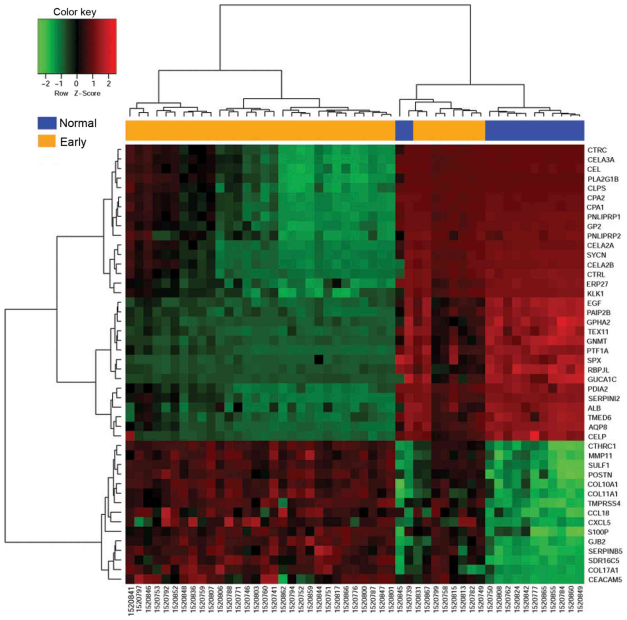
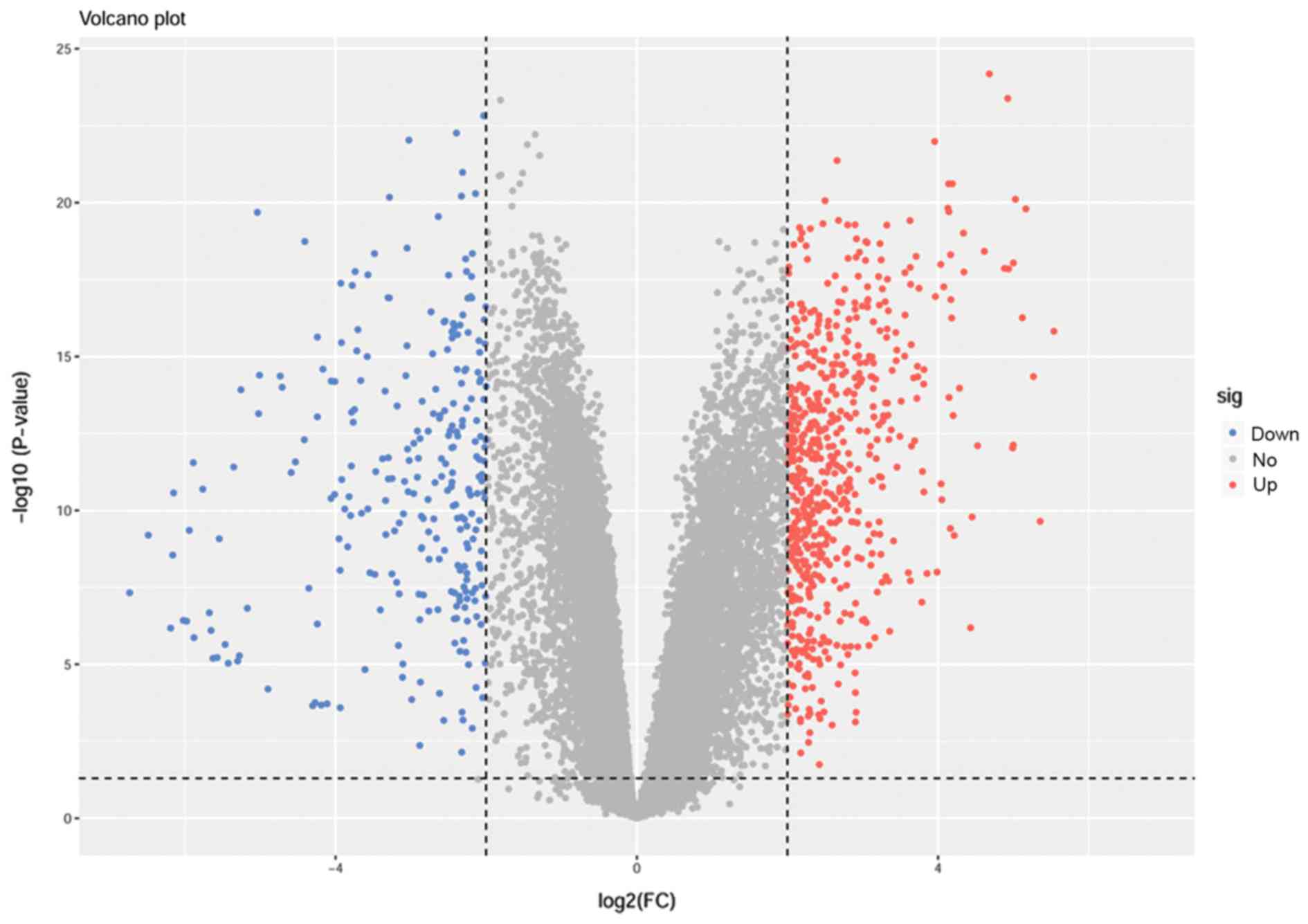
Analysis of DEGs
The selected chipset GSE62165 included 118 PDAC
samples and 13 control samples. Differences in gene expression
profiles were analyzed using 38 early-stage tumors and 13 normal
tissues. A total of 240 DEGs (adjusted P-value <0.05;
|log2FC|≥3.0) were identified using R version 3.5
software, including 137 upregulated genes and 103 downregulated
genes (Table I). The heat map of
genes with upregulated expression is presented in Fig. 1. A volcanic map of all genes is
presented in Fig. 2.
 | Table I.Top 20 differentially expressed genes
in early-stage pancreatic cancer tissues based on Log2FC. |
Table I.
Top 20 differentially expressed genes
in early-stage pancreatic cancer tissues based on Log2FC.
| A, Upregulated
genes |
|---|
|
|---|
| Gene symbol | Log2FC | Adjusted
P-value |
|---|
| COL1A1 | 5.9240335 |
3.17×10⁻¹⁶ |
| KRT17 | 5.5334643 |
4.10×10⁻¹² |
| CEACAM5 | 5.3990511 |
1.66×10−09 |
| S100P | 5.2665291 |
1.66×10⁻¹³ |
| COL10A1 | 5.2258147 |
1.17×10⁻¹⁷ |
| SERPINB5 | 5.1642588 |
5.50×10⁻¹⁵ |
| GJB2 | 5.0747799 |
7.98×10⁻¹⁸ |
| COL17A1 | 5.0501325 |
1.80×10⁻¹¹ |
| CXCL5 | 5.0384043 |
1.28×10⁻¹¹ |
| TMPRSS4 | 5.0203823 |
2.37×10⁻¹⁶ |
| SDR16C5 | 4.9961337 |
3.16×10⁻¹⁶ |
| CTHRC1 | 4.9626426 |
7.77×10⁻²⁰ |
| COL11A1 | 4.9350078 |
6.41×10⁻¹⁷ |
| SLC6A14 | 4.8841916 |
2.47×10⁻¹⁵ |
| MMP11 | 4.8824426 |
3.14×10⁻¹⁶ |
| SULF1 | 4.721966 |
2.96×10⁻¹⁷ |
| FN1 | 4.6424864 |
2.99×10⁻¹⁶ |
| POSTN | 4.6415794 |
1.33×10⁻¹⁶ |
| CCL18 | 4.5489901 |
1.41×10⁻¹¹ |
| MUC4 | 4.5022059 |
1.25×10−09 |
|
| B, Downregulated
genes |
|
| Gene
symbol | Log2FC | Adjusted
P-value |
|
| SYCN | −6.6535946 |
2.74×10−07 |
| SERPINI2 | −6.2894352 |
8.73×10−09 |
| AQP8 | −6.2356139 |
2.11×10⁻¹⁰ |
| AMY1A | −6.1790263 |
4.38×10⁻¹⁰ |
| ALB | −6.1165814 |
2.16×10−08 |
| CELA2A | −6.0845313 |
2.83×10−06 |
| PNLIPRP1 | −6.0676353 |
6.52×10⁻¹⁰ |
| CTRL | −5.9224624 |
1.73×10−06 |
| PDIA2 | −5.9185276 |
4.65×10−09 |
| CPA1 | −5.804844 |
5.21×10−06 |
| TMED6 | −5.7967792 |
2.37×10⁻¹⁰ |
| CELP | −5.7183603 |
1.15×10⁻¹⁰ |
| AQP12A | −5.6598636 |
3.88×10⁻¹⁴ |
| CUZD1 | −5.5766969 |
1.68×10−06 |
| CELA2B | −5.5649112 |
2.23×10−05 |
| CPA2 | −5.55513 |
3.43×10−06 |
| CELA3A | −5.5508171 |
1.84×10−05 |
| GP2 | −5.5087922 |
1.20×10−06 |
| ERP27 | −5.4765153 |
7.39×10−09 |
| CPA1 | −5.4417426 |
1.81×10−05 |
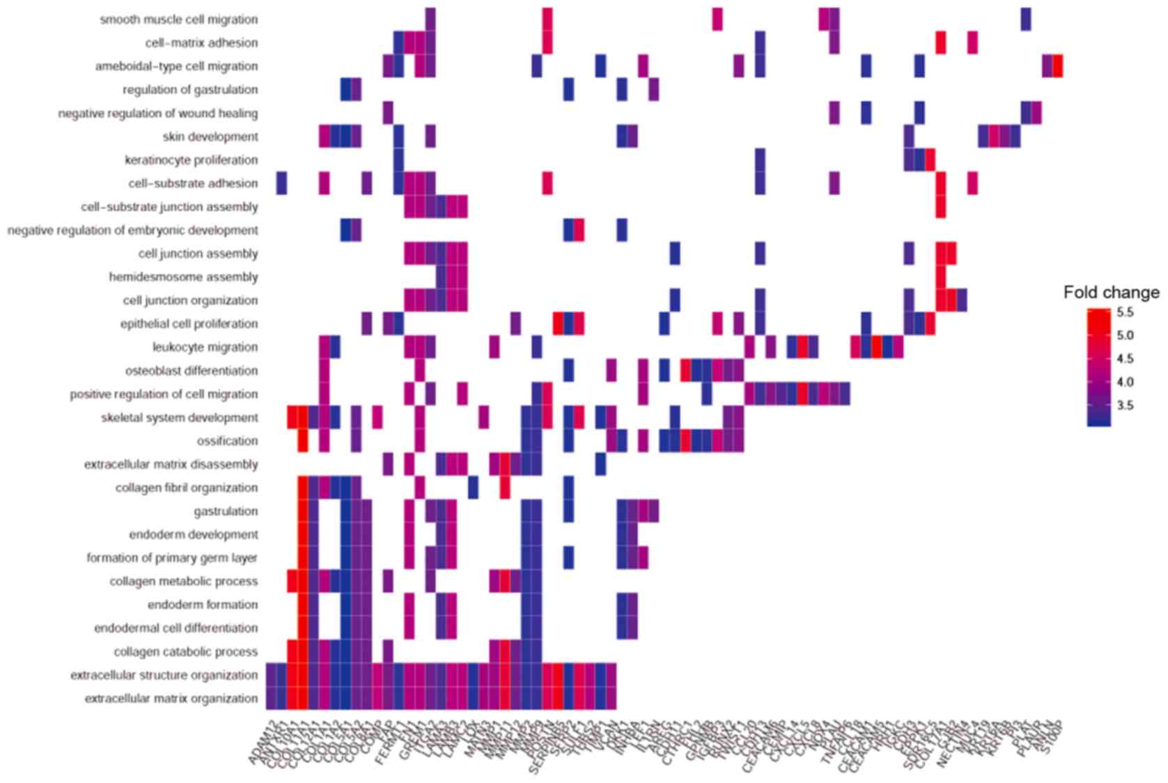
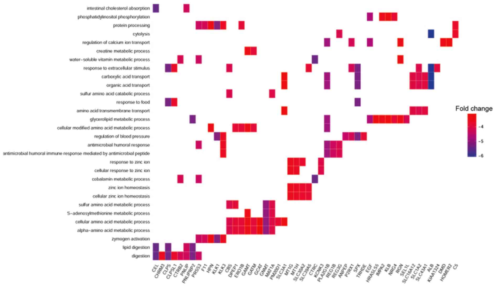
Enrichment analysis of DEGs
To investigated the distribution of DEGs, GO and
KEGG analysis of upregulated and downregulated genes was conducted.
GO analysis revealed that the ‘biological processes’ (BPs) of
upregulated genes mainly included extracellular matrix
organization, extracellular structure organization and collagen
catabolic process. ‘Molecular functions’ (MFs) of upregulated DEGs
primarily included extracellular matrix structural constituents,
glycosaminoglycan binding and cytokine activity. For the ‘cell
components’ (CCs) identified by GO analysis, proteinaceous
extracellular matrix, extracellular matrix component and
endoplasmic reticulum lumen were the most prominent (Table II). For downregulated DEGs, the
main enriched BPs were digestion, lipid digestion and sulfur amino
acid metabolic process, whereas the primary MFs were exopeptidase
activity, serine-type endopeptidase activity and serine-type
peptidase activity (Table II).
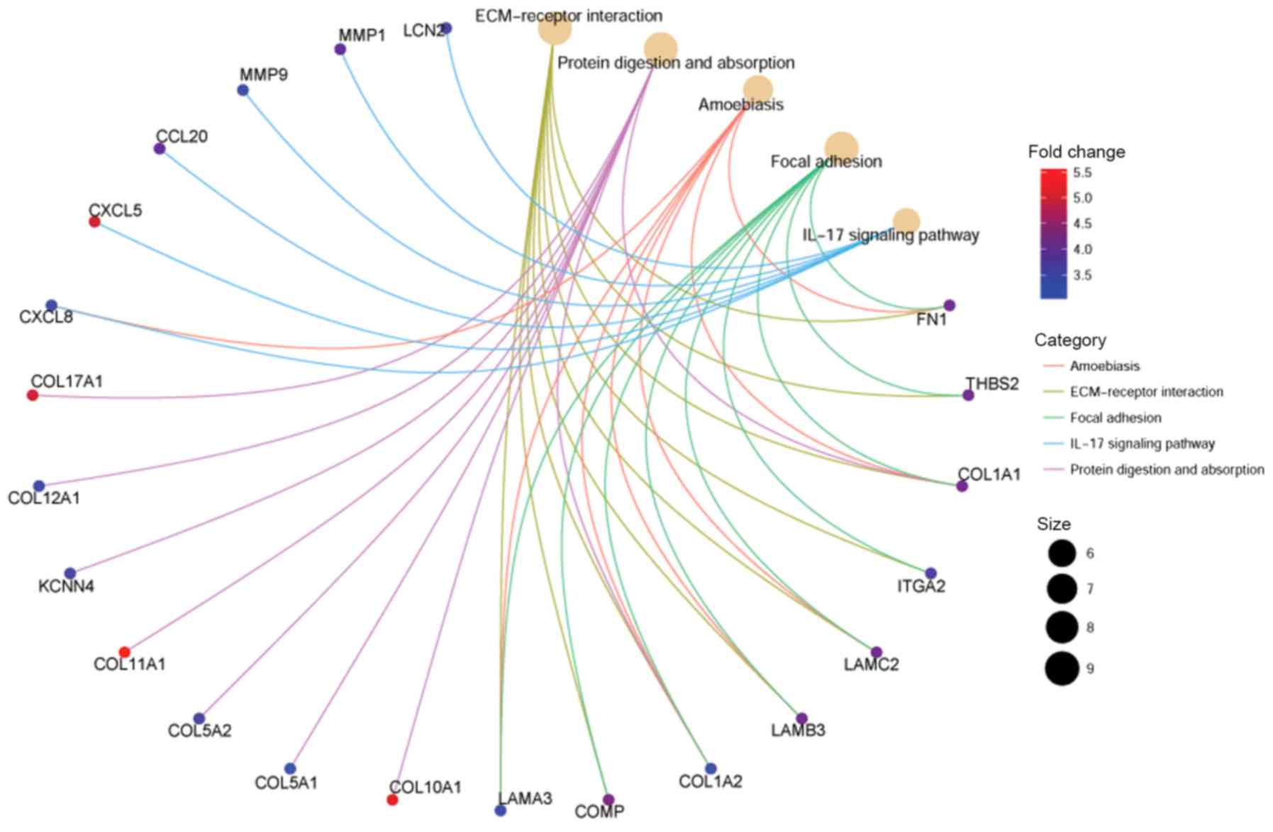
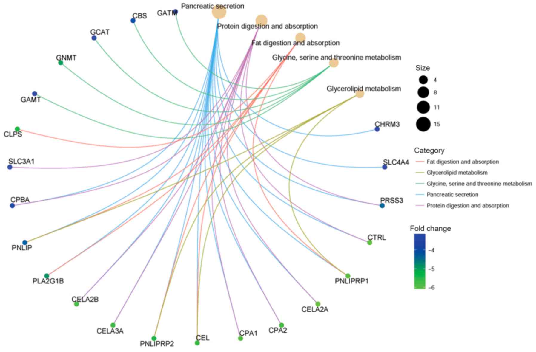
Figs. 3 and 4 present the associations between genes
and enrichment results, indicating the genes that were highly
changed between the two conditions.
| Table II.GO analysis of differentially
expressed genes in pancreatic cancer. |
Table II.
GO analysis of differentially
expressed genes in pancreatic cancer.
| A, Upregulated
genes |
|---|
|
|---|
| Category | ID | Description | Count | P-value |
|---|
| GOBP | GO:0030198 | Extracellular
matrix organization | 22 |
9.83×10⁻²¹ |
| GOBP | GO:0043062 | Extracellular
structure organization | 22 |
1.05×10⁻²⁰ |
| GOBP | GO:0030574 | Collagen catabolic
process | 10 |
2.16×10⁻¹³ |
| GOBP | GO:0044243 | Multicellular
organismal catabolic process | 10 |
6.89×10⁻¹³ |
| GOBP | GO:0032963 | Collagen metabolic
process | 11 |
4.16×10⁻¹² |
| GOMF | GO:0005201 | Extracellular
matrix structural constituent | 8 |
1.09×10−09 |
| GOMF | GO:0005539 | Glycosaminoglycan
binding | 7 |
2.85×10−05 |
| GOMF | GO:0005125 | Cytokine
activity | 7 |
3.50×10−05 |
| GOMF | GO:0008009 | Chemokine
activity | 4 |
4.62×10−05 |
| GOMF | GO:1901681 | Sulfur compound
binding | 7 |
4.91×10−05 |
| GOCC | GO:0005578 | Proteinaceous
extracellular matrix | 20 |
1.05×10⁻¹⁷ |
| GOCC | GO:0044420 | Extracellular
matrix component | 10 |
4.54×10⁻¹¹ |
| GOCC | GO:0005788 | Endoplasmic
reticulum lumen | 13 |
1.70×10⁻¹⁰ |
| GOCC | GO:0005581 | Collagen
trimer | 8 |
2.20×10−09 |
| GOCC | GO:0098644 | Complex of collagen
trimers | 5 |
1.32×10−08 |
|
| B, Downregulated
genes |
|
|
Category | ID |
Description | Count | P-value |
|
| GOBP | GO:0007586 | Digestion | 10 |
7.20×10⁻¹⁰ |
| GOBP | GO:0044241 | Lipid
digestion | 5 |
1.00×10−08 |
| GOBP | GO:0000096 | Sulfur amino acid
metabolic process | 4 |
1.27×10−05 |
| GOBP | GO:0009235 | Cobalamin metabolic
process | 3 |
6.62×10−05 |
| GOBP | GO:1901605 | α-Amino acid
metabolic process | 6 |
1.76×10⁻⁴ |
| GOMF | GO:0008238 | Exopeptidase
activity | 8 |
7.37×10−09 |
| GOMF | GO:0004252 | Serine-type
endopeptidase activity | 10 |
2.66×10−08 |
| GOMF | GO:0008236 | Serine-type
peptidase activity | 10 |
7.39×10−08 |
| GOMF | GO:0008235 | Metalloexopeptidase
activity | 6 |
8.31×10−08 |
| GOMF | GO:0017171 | Serine hydrolase
activity | 10 |
8.76×10−08 |
Table III
presents KEGG pathway analysis of the DEGs, revealing that the
upregulated genes were mainly located in extracellular matrix
(ECM)-receptor interaction, protein digestion and absorption, and
focal adhesion pathways. Conversely, downregulated genes were
primarily located in pancreatic secretion, protein digestion and
absorption, and fat digestion and absorption pathways. Figs. 5 and 6 present the distribution of the major
KEGG pathways generated using clusterProfiler. It was observed that
ECM-receptor interactions (Fig. 5)
and pancreatic secretion (Fig. 6)
were the pathways most enriched with up- and downregulated DEGs,
respectively.
| Table III.KEGG pathway analysis of
differentially expressed genes in pancreatic cancer. |
Table III.
KEGG pathway analysis of
differentially expressed genes in pancreatic cancer.
| A, Upregulated
genes |
|---|
|
|---|
| ID | Description | Count | P-value |
|---|
| hsa04512 | Extracellular
matrix-receptor interaction | 7 |
2.23×10−07 |
| hsa04974 | Protein digestion
and absorption | 7 |
4.25×10−07 |
| hsa04510 | Focal adhesion | 7 |
8.15×10−05 |
| hsa04657 | Interleukin-17
signaling pathway | 5 |
1.32×10⁻⁴ |
| hsa05146 | Amebiasis | 5 |
1.53×10⁻⁴ |
|
| B, Downregulated
genes |
|
| ID |
Description | Count | P-value |
|
| hsa04972 | Pancreatic
secretion | 13 |
3.63×10⁻¹⁶ |
| hsa04974 | Protein digestion
and absorption | 8 |
1.03×10−08 |
| hsa04975 | Fat digestion and
absorption | 6 |
4.10×10−08 |
| hsa00561 | Glycerolipid
metabolism | 4 |
2.27×10⁻⁴ |
| hsa00260 | Glycine, serine and
threonine metabolism | 3 |
1.01×10⁻³ |
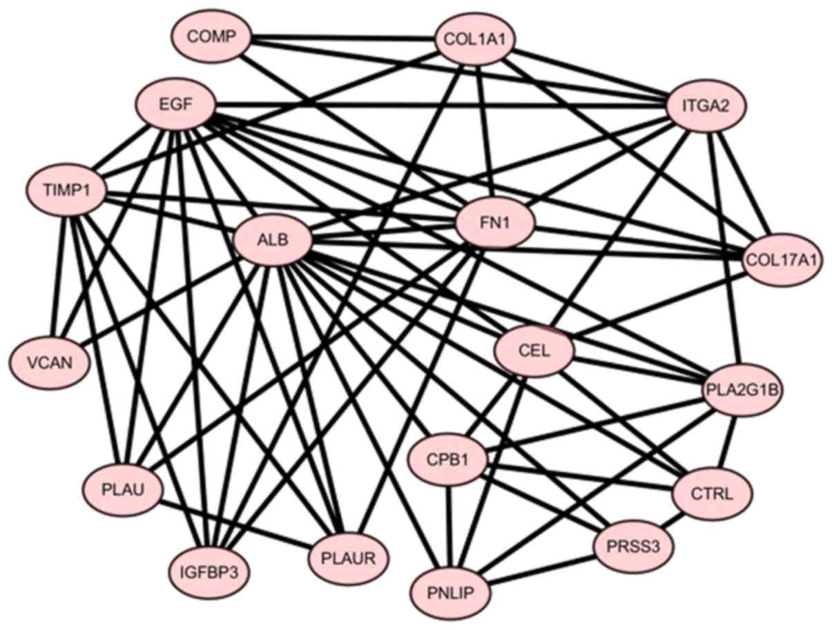
Screening of core genes in the
PPI
Based on the information in the STRING database and
using 12 types of calculation methods in Cytoscape, the following
18 core genes were identified: EGF, ALB, COL17A1, FN1, TIMP1,
PLAU, PLA2G1B, IGFBP3, PLAUR, VCAN, COL1A1, PNLIP, CTRL, PRSS3,
COMP, CPB1, ITGA2 and CEL. These core genes were
associated with each other and may exhibit synergistic effects in
the development of pancreatic cancer (Fig. 7). According to the previous
enrichment analysis, the core genes, were mainly located in
pancreatic secretion, protein digestion and absorption, and focal
adhesion pathways.
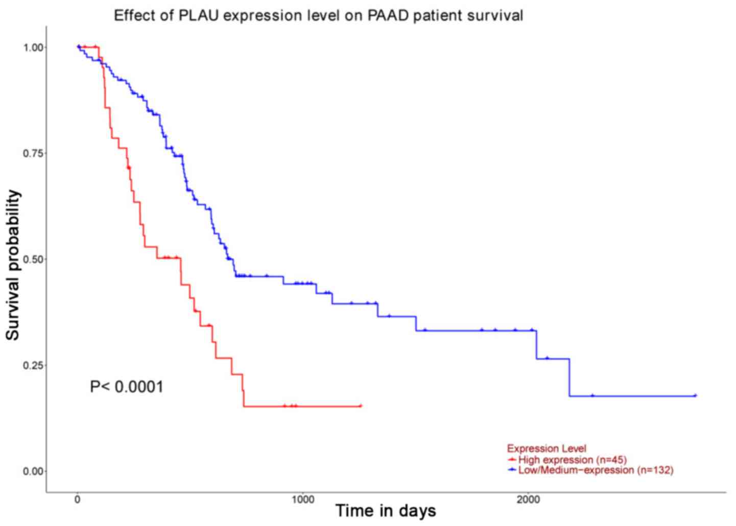
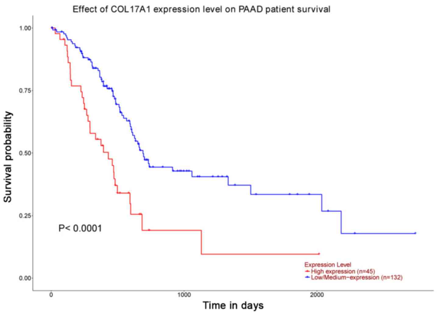
Gene expression level and survival
analysis
Notably, COL17A1 and PLAU genes were the only genes
associated with survival. Following the identification of core
genes, survival analysis for PAAD was performed using UALCAN.
PLAU [which encodes the serine protease urokinase-type
plasminogen activator (uPA); Fig.
8] and COL17A1 [which encodes collagen type XVII α1
chain (COL17A1); Fig. 9] were
demonstrated to be significantly associated with survival
(P<0.05). Subsequently, the expression levels of genes in
primary pancreatic cancer were compared; only one gene was
identified to be significantly differentially expressed,
COL17A1, whereas PLAU was not significantly differentially
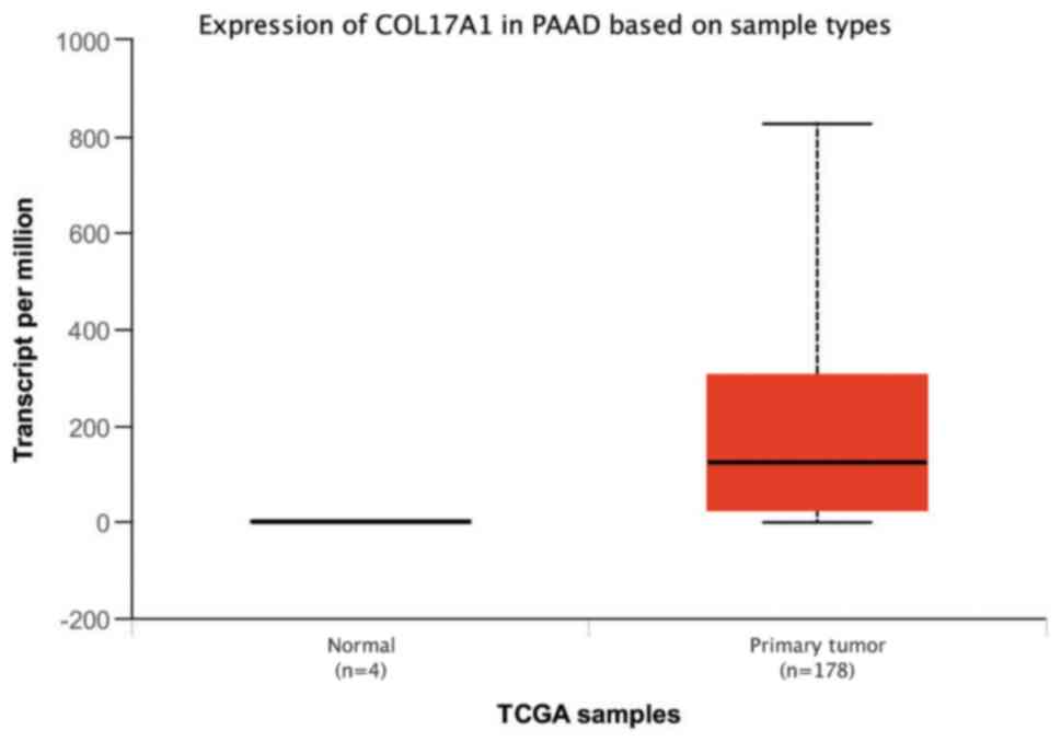
expressed. The expression levels of COL17A1 were analyzed in
TCGA database, and the results were consistent with those of the
aforementioned differential gene analysis; COL17A1 was
significantly upregulated in PAAD tumor tissues compared with
normal tissues (P=1.62×10⁻¹²; Fig. 10).
Verification of COL17A1
Differences in gene expression between 45 pancreatic
cancer patients and 45 normal pancreatic tissues were analyzed. In
particular, the expression level of COL17A1 was investigated. The
results of the analysis to verify the importance of COL17A1
are presented in Table IV; it was
observed that COL17A1 was significantly upregulated in
pancreatic tumor tissue in the two GEO databases.
| Table IV.Differential expression of COL17A1 in
pancreatic cancer tissues in two databases. |
Table IV.
Differential expression of COL17A1 in
pancreatic cancer tissues in two databases.
| Database | Gene | Log2FC | Adjusted
P-value |
|---|
| GSE62165 | COL17A1 | 5.0501325 |
1.8×10⁻¹¹ |
| GSE28735 | COL17A1 | 1.893626 |
6.56×10⁻¹³ |
Discussion
The incidence of pancreatic cancer and the
associated mortality rates have exhibited an increasing trend in
previous years (3). Studies have
reported that patients with pancreatic cancer survive for only 4
months on average without treatment; even in patients who undergo
treatment, the survival is not significantly extended (25). Therefore, accurate early diagnosis
of pancreatic cancer and the development of effective targeted
therapy is of major importance.
A previous study identified core genes in pancreatic
cancer that were reported to be of diagnostic relevance (26). In the present study, the chipset
GSE62165 from the GEO was analyzed, containing data of 118 PDAC and
13 normal pancreatic tissues (11). Differences in gene expression
levels were only compared between normal tissues and early-stage
tumor tissue. A total of 240 DEGs (137 upregulated and 103
downregulated) were identified using R, and GO (27) and KEGG pathway analyses of DEGs
revealed the locations and functions of DEGs. Upregulated genes
were mainly located in the ECM and collagen trimers, and were
involved in ECM organization and ECM-receptor interactions, focal
adhesion, and protein digestion and absorption. Conversely,
downregulated genes were mainly enriched in digestion and
exopeptidase activity pathways. A PPI network was built, and 18
core genes were identified; the prognostic value of these genes for
patients with pancreatic cancer was analyzed using UACLAN.
PLAU and COL17A1 were significantly associated with
poorer survival; it was then revealed using data from TCGA that
COL17A1 was significantly upregulated in pancreatic cancer
tissues compared with control tissues, consistent with the results
of the differential gene analysis. It was predicted that these two
genes may be associated with the proliferation, invasion and
metastasis of pancreatic cancer.
PLAU encodes a serine protease, uPA (28). Following GO and KEGG analyses, the
functional enrichment of PLAU was investigated. PLAU is
mainly involved in the regulation of cell motility, cellular
component movement and locomotion (29). It is primarily expressed in the
endoplasmic reticulum lumen and invadopodium (30). PLAU plays a key role in regulating
cell migration and adhesion during tissue regeneration and
intracellular signaling (31).
Increased expression of COL17A1 leads to tumor cell invasion and
metastasis of tumor cells to surrounding tissues (32). PLAU is involved in predicting the
survival rate of patients with gastric cancer (33). It may serve an important role in
the invasion and metastasis of pancreatic cancer cells (34); however, the specific pathways
involved are yet to be determined. It is hypothesized that
PLAU may serve an important role in the diagnosis and
treatment of pancreatic cancer in the future.
COL17A1 is mainly located in the extracellular
matrix and collagen trimmers (35). Extracellular matrix molecules,
including proteoglycan and fibrin, have been reported to affect the
growth, migration and differentiation of cells (36). A study showed that COL17A1 can
inhibit the migration and invasion of breast cancer cells, acting
as a p53 transcriptional target gene (37). A previous study has reported that
the extracellular matrix is closely associated with the metastasis
of breast cancer (38). High
levels of collagen in breast and colorectal cancers have been
associated with tumor invasion (39,40).
A previous study that employed the
minimum-redundancy-maximum-relevance method also identified
COL17A1 as a core gene of pancreatic cancer (26); however, in the present study, the
upregulated expression of COL17A1 in pancreatic cancer was
verified in multiple datasets, and its effects on patient survival
were determined. Survival analysis using UACLAN based on data from
TCGA revealed that the expression levels of CLO17A1 were
closely associated with the survival of patients with pancreatic
cancer, and that CLO17A1 was highly expressed in primary
pancreatic tumor tissues. The present findings suggested that the
expression of COL17A1 is associated with the occurrence and
development of pancreatic cancer. Therefore, this bioinformatics
analysis may provide novel insight for future studies investigating
the pathogenesis of pancreatic cancer.
However, the present study presented certain
limitations. In examining the expression level of COL17A1, only
four normal samples were investigated, and further studies
examining a high number of control samples are required to confirm
the present results.
Acknowledgements
Not applicable.
Funding
The present work was supported by The ‘Six Talents
Summit’ Project in Jiangsu Province, miR-203 targets Survivin to
upregulate the expression of Caspase-3 and promote the apoptosis of
pancreatic cancer cells (grant no. WAW-008).
Availability of data and materials
The datasets used and/or analyzed in the present
study are available in the GEO (http://www.ncbi.nlm.nih.gov/geo) and UALCAN
(http://ualcan.path.uab.edu)
repositories.
Authors' contributions
JZ and LX conceived the study. JW, ZL, KW, KZ and DX
analyzed the data and drafted the manuscript. All authors reviewed
and approved the final manuscript.
Ethics approval and consent to
participate
Not applicable.
Patient consent for publication
Not applicable.
Competing interests
The authors declare that they have no competing
interests.
References
|
1
|
Kamisawa T, Wood LD, Itoi T and Takaori K:
Pancreatic cancer. Lancet. 388:73–85. 2016. View Article : Google Scholar : PubMed/NCBI
|
|
2
|
Michaud D: Epidemiology of pancreatic
cancer. Minerva Chir. 59:99–111. 2004.PubMed/NCBI
|
|
3
|
Siegel R, Ma J, Zou Z and Jemal A: Cancer
statistics, 2014. CA Cancer J Clin. 64:9–29. 2014. View Article : Google Scholar : PubMed/NCBI
|
|
4
|
Risch HA: Etiology of pancreatic cancer,
with a hypothesis concerning the role of N-nitroso compounds and
excess gastric acidity. J Natl Cancer Inst. 95:948–960. 2003.
View Article : Google Scholar : PubMed/NCBI
|
|
5
|
Kern SE, Shi C and Hruban RH: The
complexity of pancreatic ductal cancers and multidimensional
strategies for therapeutic targeting. J Pathol. 223:295–306. 2011.
View Article : Google Scholar : PubMed/NCBI
|
|
6
|
Grasso C, Jansen G and Giovannetti E: Drug
resistance in pancreatic cancer: Impact of altered energy
metabolism. Crit Rev Oncol Hematol. 114:139–152. 2017. View Article : Google Scholar : PubMed/NCBI
|
|
7
|
Eskelinen M and Haglund U: Developments in
serologic detection of human pancreatic adenocarcinoma. Scand J
Gastroenterol. 34:833–844. 1999. View Article : Google Scholar : PubMed/NCBI
|
|
8
|
Bass AJ, Lawrence MS, Brace LE, Ramos AH,
Drier Y, Cibulskis K, Sougnez C, Voet D, Saksena G, Sivachenko A,
et al: Genomic sequencing of colorectal adenocarcinomas identifies
a recurrent VTI1A-TCF7L2 fusion. Nat Genet. 43:964–968. 2011.
View Article : Google Scholar : PubMed/NCBI
|
|
9
|
Sjöblom T, Jones S, Wood LD, Parsons DW,
Lin J, Barber TD, Mandelker D, Leary RJ, Ptak J, Silliman N, et al:
The consensus coding sequences of human breast and colorectal
cancers. Science. 314:268–274. 2006. View Article : Google Scholar : PubMed/NCBI
|
|
10
|
Wood LD, Parsons DW, Jones S, Lin J,
Sjöblom T, Leary RJ, Shen D, Boca SM, Barber T, Ptak J, et al: The
genomic landscapes of human breast and colorectal cancers. Science.
318:1108–1113. 2007. View Article : Google Scholar : PubMed/NCBI
|
|
11
|
Janky R, Binda MM, Allemeersch J, Van den
Broeck A, Govaere O, Swinnen JV, Roskams T, Aerts S and Topal B:
Prognostic relevance of molecular subtypes and master regulators in
pancreatic ductal adenocarcinoma. BMC Cancer. 16:6322016.
View Article : Google Scholar : PubMed/NCBI
|
|
12
|
Barrett T, Wilhite SE, Ledoux P,
Evangelista C, Kim IF, Tomashevsky M, Marshall KA, Phillippy KH,
Sherman PM, Holko M, et al: NCBI GEO: Archive for functional
genomics data sets-update. Nucleic Acids Res 41 (Database Issue).
D991–D995. 2013.
|
|
13
|
Ritchie ME, Phipson B, Wu D, Hu Y, Law CW,
Shi W and Smyth GK: Limma powers differential expression analyses
for RNA-sequencing and microarray studies. Nucleic Acids Res.
43:e472015. View Article : Google Scholar : PubMed/NCBI
|
|
14
|
Zhang G, Schetter A, He P, Funamizu N,
Gaedcke J, Ghadimi BM, Ried T, Hassan R, Yfantis HG, Lee DH, et al:
DPEP1 inhibits tumor cell invasiveness, enhances chemosensitivity
and predicts clinical outcome in pancreatic ductal adenocarcinoma.
PLoS One. 7:e315072012. View Article : Google Scholar : PubMed/NCBI
|
|
15
|
Zhang G, He P, Tan H, Budhu A, Gaedcke J,
Ghadimi BM, Ried T, Yfantis HG, Lee DH, Maitra A, et al:
Integration of metabolomics and transcriptomics revealed a fatty
acid network exerting growth inhibitory effects in human pancreatic
cancer. Clin Cancer Res. 19:4983–4993. 2013. View Article : Google Scholar : PubMed/NCBI
|
|
16
|
Xing Z, Chu C, Chen L and Kong X: The use
of Gene Ontology terms and KEGG pathways for analysis and
prediction of oncogenes. Biochim Biophys Acta. 1860:2725–2734.
2016. View Article : Google Scholar : PubMed/NCBI
|
|
17
|
Kanehisa M, Sato Y, Furumichi M, Morishima
K and Tanabe M: New approach for understanding genome variations in
KEGG. Nucleic Acids Res. 47:D590–D595. 2019. View Article : Google Scholar : PubMed/NCBI
|
|
18
|
Kanehisa M, Furumichi M, Tanabe M, Sato Y
and Morishima K: KEGG: New perspectives on genomes, pathways,
diseases and drugs. Nucleic Acids Res. 45:D353–D361. 2017.
View Article : Google Scholar : PubMed/NCBI
|
|
19
|
Kanehisa M and Goto S: KEGG: Kyoto
encyclopedia of genes and genomes. Nucleic Acids Res. 28:27–30.
2000. View Article : Google Scholar : PubMed/NCBI
|
|
20
|
Yu G, Wang LG, Han Y and He QY:
clusterProfiler: An R package for comparing biological themes among
gene clusters. OMICS. 16:284–287. 2012. View Article : Google Scholar : PubMed/NCBI
|
|
21
|
Shannon P, Markiel A, Ozier O, Baliga NS,
Wang JT, Ramage D, Amin N, Schwikowski B and Ideker T: Cytoscape: A
software environment for integrated models of biomolecular
interaction networks. Genome Res. 13:2498–2504. 2003. View Article : Google Scholar : PubMed/NCBI
|
|
22
|
Chin CH, Chen SH, Wu HH, Ho CW, Ko MT and
Lin CY: cytoHubba: Identifying hub objects and sub-networks from
complex interactome. BMC Syst Biol. 8 (Suppl 4):S112014. View Article : Google Scholar : PubMed/NCBI
|
|
23
|
Chandrashekar DS, Bashel B, Balasubramanya
SAH, Creighton CJ, Ponce-Rodriguez I, Chakravarthi BVSK and
Varambally S: UALCAN: A portal for facilitating tumor subgroup gene
expression and survival analyses. Neoplasia. 19:649–658. 2017.
View Article : Google Scholar : PubMed/NCBI
|
|
24
|
Li B and Dewey CN: RSEM: Accurate
transcript quantification from RNA-Seq data with or without a
reference genome. BMC Bioinformatics. 12:3232011. View Article : Google Scholar : PubMed/NCBI
|
|
25
|
Wang X, Wang L, Mo Q, Dong Y, Wang G and
Ji A: Changes of Th17/Treg cell and related cytokines in pancreatic
cancer patients. Int J Clin Exp Pathol. 8:5702–5708.
2015.PubMed/NCBI
|
|
26
|
Shen S, Gui T and Ma C: Identification of
molecular biomarkers for pancreatic cancer with mRMR shortest path
method. Oncotarget. 8:41432–41439. 2017.PubMed/NCBI
|
|
27
|
Thomas PD: The gene ontology and the
meaning of biological function. Methods Mol Biol. 1446:15–24. 2017.
View Article : Google Scholar : PubMed/NCBI
|
|
28
|
Duffy MJ, Duggan C, Mulcahy HE, McDermott
EW and O'Higgins NJ: Urokinase plasminogen activator: A prognostic
marker in breast cancer including patients with axillary
node-negative disease. Clin Chem. 44:1177–1183. 1998.PubMed/NCBI
|
|
29
|
Nielsen TO, Andrews HN, Cheang M, Kucab
JE, Hsu FD, Ragaz J, Gilks CB, Makretsov N, Bajdik CD, Brookes C,
et al: Expression of the insulin-like growth factor I receptor and
urokinase plasminogen activator in breast cancer is associated with
poor survival: Potential for intervention with 17-allylamino
geldanamycin. Cancer Res. 64:286–291. 2004. View Article : Google Scholar : PubMed/NCBI
|
|
30
|
Pavón MA, Arroyo-Solera I, Céspedes MV,
Casanova I, León X and Mangues R: uPA/uPAR and SERPINE1 in head and
neck cancer: Role in tumor resistance, metastasis, prognosis and
therapy. Oncotarget. 7:57351–57366. 2016. View Article : Google Scholar : PubMed/NCBI
|
|
31
|
Amos S, Redpath GT, Dipierro CG, Carpenter
JE and Hussaini IM: Epidermal growth factor receptor-mediated
regulation of urokinase plasminogen activator expression and
glioblastoma invasion via C-SRC/MAPK/AP-1 signaling pathways. J
Neuropathol Exp Neurol. 69:582–592. 2010. View Article : Google Scholar : PubMed/NCBI
|
|
32
|
Chaudhary A, Hilton MB, Seaman S, Haines
DC, Stevenson S, Lemotte PK, Tschantz WR, Zhang XM, Saha S, Fleming
T and St Croix B: TEM8/ANTXR1 blockade inhibits pathological
angiogenesis and potentiates tumoricidal responses against multiple
cancer types. Cancer Cell. 21:212–226. 2012. View Article : Google Scholar : PubMed/NCBI
|
|
33
|
Xu ZY, Chen JS and Shu YQ: Gene expression
profile towards the prediction of patient survival of gastric
cancer. Biomed Pharmacother. 64:133–139. 2010. View Article : Google Scholar : PubMed/NCBI
|
|
34
|
Liu P, Weng Y, Sui Z, Wu Y, Meng X, Wu M,
Jin H, Tan X, Zhang L and Zhang Y: Quantitative secretomic analysis
of pancreatic cancer cells in serum-containing conditioned medium.
Sci Rep. 6:376062016. View Article : Google Scholar : PubMed/NCBI
|
|
35
|
Borradori L and Sonnenberg A: Structure
and function of hemidesmosomes: More than simple adhesion
complexes. J Invest Dermatol. 112:411–418. 1999. View Article : Google Scholar : PubMed/NCBI
|
|
36
|
Järveläinen H, Sainio A, Koulu M, Wight TN
and Penttinen R: Extracellular matrix molecules: Potential targets
in pharmacotherapy. Pharmacol Rev. 61:198–223. 2009. View Article : Google Scholar : PubMed/NCBI
|
|
37
|
Yodsurang V, Tanikawa C, Miyamoto T, Lo
PHY, Hirata M and Matsuda K: Identification of a novel p53 target,
COL17A1, that inhibits breast cancer cell migration and invasion.
Oncotarget. 8:55790–55803. 2017. View Article : Google Scholar : PubMed/NCBI
|
|
38
|
Chowdhury N and Sapru S: Association of
protein translation and extracellular matrix gene sets with breast
cancer metastasis: Findings uncovered on analysis of multiple
publicly available datasets using individual patient data approach.
PLoS One. 10:e01296102015. View Article : Google Scholar : PubMed/NCBI
|
|
39
|
Rizwan A, Bulte C, Kalaichelvan A, Cheng
M, Krishnamachary B, Bhujwalla ZM, Jiang L and Glunde K: Metastatic
breast cancer cells in lymph nodes increase nodal collagen density.
Sci Rep. 5:100022015. View Article : Google Scholar : PubMed/NCBI
|
|
40
|
Zou X, Feng B, Dong T, Yan G, Tan B, Shen
H, Huang A, Zhang X, Zhang M, Yang P, et al: Up-regulation of type
I collagen during tumorigenesis of colorectal cancer revealed by
quantitative proteomic analysis. J Proteomics. 94:473–485. 2013.
View Article : Google Scholar : PubMed/NCBI
|