Introduction
Therapeutic hypothermia is an effective method of
neuroprotection (1) that is
currently used to treat diseases such as hypoxic ischemic
encephalopathy and severe craniocerebral trauma caused by cardiac
arrest, with good results (2).
Selective brain cooling (SBC) is a therapeutic, low-temperature
method that has no effect on whole-body temperature (3). Since SBC avoids the side effects of
low temperature, it has received increasing clinical and research
attention. In particular, intracarotid cold saline infusion (ICSI)
can cool the brain rapidly and uniformly; it is therefore
considered an ideal SBC method (4). ICSI is the fastest and most effective
way to lower brain temperature and protect the brain (5), with its protective effect being
mediated through vascular flushing and low temperature (6,7). In
previous studies, ICSI administered before reperfusion improved
brain function after middle cerebral artery occlusion (MCAO)
(8). The protective effect of ICSI
on the brain results from hypothermia and cerebral artery flushing
(5). However, the underlying
mechanism of ICSI in the brain of patients with ischemic stroke is
unclear.
The serum and glucocorticoid-regulated kinase 1
(SGK1) gene encodes a serine/threonine protein kinase that
plays an important role in the cellular stress response (9). This kinase activates certain
chloride, sodium and potassium channels, suggesting that it is
involved in the regulation of cell survival, renal sodium
excretion, and neuronal excitability (10). High expression of this gene may be
associated with the development of several diseases, including
hypertension and diabetic nephropathy (11). SGK1 is a member of the AGC protein
kinase family and plays an important role in regulating ion channel
expression, as well as in promoting proliferation and survival in
malignant epithelial cells (12).
Recently, several studies have confirmed that SGK1 has a protective
effect on ischemia-reperfusion injury. For instance, one
investigation reported alterations in hippocampal SGK1 gene
expression in rats with transient cerebral ischemia (13). Furthermore, SGK1 was recently shown
to play a protective role in the hippocampal neurons of rats with
ischemia-reperfusion injury (14).
In another study, the corticotropin-induced estrogen enhancement
effect of SGK1 expression protected the heart from
ischemia-reperfusion injury (15).
However, no investigations have yet explored whether ICSI can
affect SGK1 expression.
Autophagy is a cellular pathway that regulates the
transportation of cytoplasmic macromolecules and organelles to
lysosomes for degradation (16).
Beclin-1 and microtubule-associated protein 1 light chain 3 (LC-3)
are two important proteins involved in autophagic flux (17–19).
Previous studies have shown that activation of autophagy plays an
important role in cerebral ischemia and reperfusion injury
(20,21). However, it remains unclear whether
autophagic flux is related to the neuroprotective effects of ICSI
and SGK-1.
In the present study, we investigated the role of
ICSI and clarified the mechanism of its neuroprotective effect.
Materials and methods
Animals
In total, 250 male 8 week old Sprague-Dawley rats
weighing ~280 g were acquired from The First Affiliated Hospital of
Soochow University (Jiangsu, China). The rats housed at a
temperature of 22–25°C and 50–65% humidity, with a 12 h light/dark
cycle and ad libitum access to food and water. The rats were
raised individually in an animal facility. All procedures involving
animals were approved by the Animal Research Committee of The First
Affiliated Hospital of Soochow University.
MCAO model establishment
To conduct surgery, the rats were anesthetized using
an intraperitoneal injection of 10% chloral hydrate (400 mg/kg;
cat. no. C8383, Sigma-Aldrich/Merck KGaA). Peritonitis was
monitored and not observed during the study. Two temperature probes
(Physitemp, Clifton, NJ, USA) were placed in the right cerebral
cortex (3 mm lateral to bregma, 3 mm posterior, and 4 mm under the
surface of the skull), as well as in the rectum, to continuously
monitor temperature. An improved intraluminal filament was used to
induce MCAO. After 2 h, reperfusion was established by filament
shrinkage. To confirm the success rate of MCAO surgery and
reperfusion, blood flow in the right cortex of the right MCA was
measured using a laser Doppler flow meter 2 mm in the posterior
fontanelle and 6 mm in the anterior fontanelle. After the filaments
were inserted, 30% of the rats without blood flow reduction were
lower than baseline. Reperfusion success was defined as an 80%
increase over the baseline in blood flow after the filament was
contracted. In groups treated with ICSI, the filament was
contracted, and a modified PE50 canaliculus was inserted into the
internal carotid artery about 5 mm from the external carotid
artery. Next, by infusion with freshly made 10°C saline at a rate
of 10–25 ml/h, the brain temperature was reduced to 33°C for 20
min. Normal infusion groups received intraperitoneal normal saline
infusion (INSI) at 37°C, while the other procedures were identical
to those in the ICSI groups. A non-infusion group underwent
insertion of a filtration membrane to a depth of 15-mm insufficient
to induce MCAO and no infusion of renal tubules. After transfusion,
the rats were subjected to rapid rewarming using a heating pad and
bubble wrap; when their brain temperature had returned to baseline,
they were considered reheated.
Grouping
The rats were randomly divided into five groups: i)
non-infusion group (MCAO rats with no infusion; n=25; mortality
rate: 4.0%); ii) ICSI group (MCAO rats treated with ICSI
immediately after reperfusion; n=90; mortality rate: 15.6%); iii)
INSI group (MCAO rats treated with INSI immediately after
reperfusion; n=25; mortality rate: 8.0%); iv) ICSI+reperfusion
group (MCAO rats treated with ICSI for 1 h immediately after
reperfusion; n=85; mortality rate: 11.8%); and v) INSI+reperfusion
group (MCAO rats treated with INSI for 1 h immediately after
reperfusion; n=25; mortality rate: 8.0%). In addition, shRNAs for
SGK1, as well as negative control (NC) shRNAs, were obtained from
GenePharma Co., Ltd. (Shanghai, China). Rats in the ICSI and
ICSI+reperfusion groups were treated using NC shRNAs and SGK1
shRNAs, respectively, while those in the ICSI group (n=50) were
divided into the ICSI+NC subgroup (n=25; mortality rate: 4.0%) and
ICSI+shSGK1 subgroup (n=25; mortality rate: 6.0%); the rats in the
ICSI+reperfusion group (n=50) were divided into the
ICSI+reperfusion+NC subgroup (n=25; mortality rate: 6.0%) and
ICSI+reperfusion+shSGK1 subgroup (n=25, the mortality rate: 6.0%).
After treatment for 50 h, the indicators were evaluated, the rats
were euthanized, and their tissues were used in subsequent
experiments. During the course of the experiment, the animals were
also euthanized by cervical dislocation when they reached the
standard of the humane endpoint. The mortality rate represents
animals prematurely euthanized after reaching humane endpoints,
rather than animals that were found dead. Humane endpoint is a
refinement strategy to reduce the pain and suffering suffered by
the animals during the experiment as much as possible. Humane
endpoints mainly include weight loss of 20–25%, loss of appetite
(anorexia more than 24 h), inability to stand, severe infection,
tumors more than 10% of body weight, organ system failure, dyspnea,
severe anemia, renal failure and peritoneal fluid. Animals were
euthanized when two or more humane endpoints were observed.
Infarct size
As described in previous studies (22,23),
after reperfusion for 48 h, the animals were anesthetized by
intraperitoneal injection of 10% chloral hydrate (400 mg/kg) and
then decapitated. The brain was rapidly removed, and the coronal
section of brain was sliced in a special groove (thickness: 2 mm)
after cooling in ice-cold saline for 10 min. The slices were
immediately immersed in 4 ml of 1% 2,3,5-triphenyltetrazolium
chloride (TTC) and incubated at 37°C for 10 min. Next, the TTC
solution was replaced with 4% polyoxymethylene. After 8 h, a
digital camera was used to photograph the sections, and the infarct
size was measured using the UTHSCSA Image Tool 3.0 software
(https://uthscsa-imagetool.software.informer.com/).
To eliminate the effect of postoperative edema on injury volume,
the infarcted volume (%) was calculated according to the following
method: (Volume of the left hemisphere-Non-infarct volume of right
hemisphere)/(Volume of the left hemisphere) ×100%.
Brain swelling
According to previous research (24), brain swelling was assessed using a
0.35-T Signa Ovation Excite MRI scanner (General Electric,
Milwaukee, WI, USA).
Knockdown of SGK1
For SGK1 knockdown, SGK1 and control
shRNA were acquired from RiboBio (Guangzhou, Guangdong, China).
SGK1 knockdown rats were obtained from the Shanghai Research
Center for Model Organisms (Shanghai, China). In brief, after
anesthesia, rats were placed on a stereotactic apparatus, and 50 µg
SGK1 and control shRNA vectors were injected into the right
ischemia region of rats, and the final volume was 10 µl. The
sequence of shSGK1 was as follows: Top strand,
5′-CACCGCCGGCTGTGCCTTCTCTCCATTCAAGAGATGGAGAGAAGGCACAGCCGGC-3′;
bottom strand,
5′-AAAAGCCGGCTGTGCCTTCTCTCCATCTCTTGAATGGAGAGAAGGCACAGCCGGC-3′; Ds
Oligo,
5′-CACCGCCTTCTCTCCATCCGCTGCTTTCAAGAGAAGCAGCGGATGGAGAGAAGGC-3′ and
3′-CGGAAGAGAGGTAGGCGACGAAAGTTCTCTTCGTCGCCTACCTCTCTTCCGAAAA-5′.
Terminal deoxynucleotidyl transferase
mediated nick end labeling (TUNEL) staining
To identify apoptosis in the ipsilateral
hippocampus, TUNEL staining was performed using the In Situ
Cell Death Detection Kit (Roche, Indianapolis, IN, USA) according
to the manufacturer's instructions. Briefly, the sections were
washed for 30 min. They were then incubated in 0.1% Triton X-100
and 0.1% sodium citrate and rinsed three times using
phosphate-buffered saline (PBS) for 10 min each time. The slices
were then incubated in 0.3% PBS/0.1% Tween-20 for 30 min to inhibit
endogenous peroxidase activity; they were then once again rinsed in
PBS. Next, the sections and the 50-µl TUNEL reaction mixture of the
in situ Cell Death Detection Kit were incubated in a moist
atmosphere for 60 min at 37°C in the dark. The slices were rinsed
three times in PBS and observed under a Nikon TE300 microscope
(Nikon, Inc., Tokyo, Japan). A negative control was achieved in
each pore by incubating the slices in 50-µl labeled solution
instead of TUNEL reaction mixture. At least 10 randomly selected
fields were used to count TUNEL-positive cells (×200
magnification). The cell count was performed by researchers who had
been blinded to the rat conditions.
Nissl staining
Three days after the operation, the rats were
anesthetized using 10% chloral hydrate (400 mg/kg body weight) by
intraperitoneal injection, and their brains were extracted and
fixed in 4% paraformaldehyde for 2 h. After continuous overnight
incubation in 0.1 mol/l PBS containing 20 and 25% glucose, the
brain was cut into 10-mm coronal sections using a cryogenic
thermostat (Leica CM1950; Leica, Wetzlar, Germany) and stored at
20°C. Then, Nissl staining solution (Beyotime Institute of
Biotechnology, Shanghai, China) was used, following the
manufacturer's instructions. Briefly, the sections were soaked in
1% toluidine blue and dyed for 5 min at 50°C, washed with double
distilled water, and then dehydrated with gradient ethanol; the
sections were observed under a Nikon Eclipse E400 microscope (×200
magnification; Nikon, Inc.).
Immunohistochemical analysis
Frozen brain sections in OCT compound (14-µm thick)
were formalin-fixed and subjected to immunohistochemical analysis
to determine SGK1 expression. Endogenous peroxidase was blocked
using 3% H2O2 for 5 min. The sections were
then washed in PBS for a short time and cooled at room temperature
for 20 min. They were then rinsed again in PBS. Non-specific
protein was blocked by incubation in 5% horse serum for 40 min. The
sections were incubated with primary antibodies (anti-SGK1; diluted
1:200; cat. no. YB-33275M; Suzhou Ard Biological Co., Ltd.,
Shanghai, China) for 1 h at room temperature. They were then
incubated with fluorescein isothiocyanate/cyanine 3-conjugated IgG
(1:500 dilution; cat. no. SC-2340; Santa Cruz Biotechnology, Inc.,
Santa Cruz, CA, USA) at room temperature for 60 min. Ten
microscopic fields per section were randomly photographed (×200;
Nikon TE300; Nikon, Inc.) to allow the SGK-1-positive cells to be
counted.
Western blotting
Treated tissues were lysed using RIPA buffer
(Sigma-Aldrich/Merck KGaA). Total protein was separated using 10%
SDS-PAGE and transferred to a polyvinylidene fluoride membrane
(cat. no. 88518; Thermo Fisher Scientific, Inc., Waltham, MA, USA).
The membranes were blocked with 5% non-fat milk for 1 h at room
temperature and then subjected to incubation at 4°C overnight in
primary antibodies against SGK1 (1:500; cat. no. ab59337), beclin-1
(1:2,000; cat. no. ab207612;), LC-3 (1:3,000; cat. no. ab51520) and
GAPDH (1:2,500; cat. no. ab9485; all Abcam, Cambridge, UK). The
next day, after washing, the membranes were incubated in secondary,
peroxidase-conjugated anti-mouse (1:2,000, cat. No. 7076; Cell
signaling Technology, lnc.) or anti-rabbit (1:2,000, cat. No. 5127;
Cell signaling Technology, lnc.) antibodies for 1.5 h at room
temperature. Finally, the results were determined using an enhanced
chemiluminescence (ECL) substrate kit in an ECL system (Amersham
Pharmacia). Quantity One software version 4.6.2 (Bio-Rad
Laboratories, Inc., Hercules, CA, USA) was used for densitometric
analysis.
Statistical data analysis
All data are presented as mean ± standard error, and
Graphpad Prism 6 (GraphPad Software, Inc., La Jolla, CA, USA) was
used for statistical analysis. Student's t-test was used to
evaluate the difference between two groups; one-way ANOVA with
Tukey's post hoc was applied to assess the differences among more
than two groups, and P-values <0.05 were considered as
indicative of statistical significance.
Results
ICSI alleviates infarct size and brain
swelling after MCAO
To induce ischemic stroke, MCAO was performed in
Sprague-Dawley rats for 2 h by applying an intraluminal filament.
The MCAO rats were treated with either ICSI or INSI after
reperfusion (Fig. 1A). After
reperfusion for 48 h, the infarct size was determined using TTC
staining; brain swelling was also measured. The results indicated
that the infarct size was significantly reduced in the ICSI and
INSI groups compared with the non-infusion group; ICSI also
alleviated the infarct size after reperfusion in the MCAO model
(P<0.05, P<0.01; Fig. 1B).
Brain swelling was significantly decreased in the ICSI and INSI
groups relative to the non-infusion group. After reperfusion for 1
h, the brain swelling in the ICSI and INSI groups also showed
significant reductions. In addition, brain swelling was
significantly decreased in the ICSI group compared with the INSI
group after reperfusion in the MCAO model (P<0.05, P<0.01;
Fig. 1C).
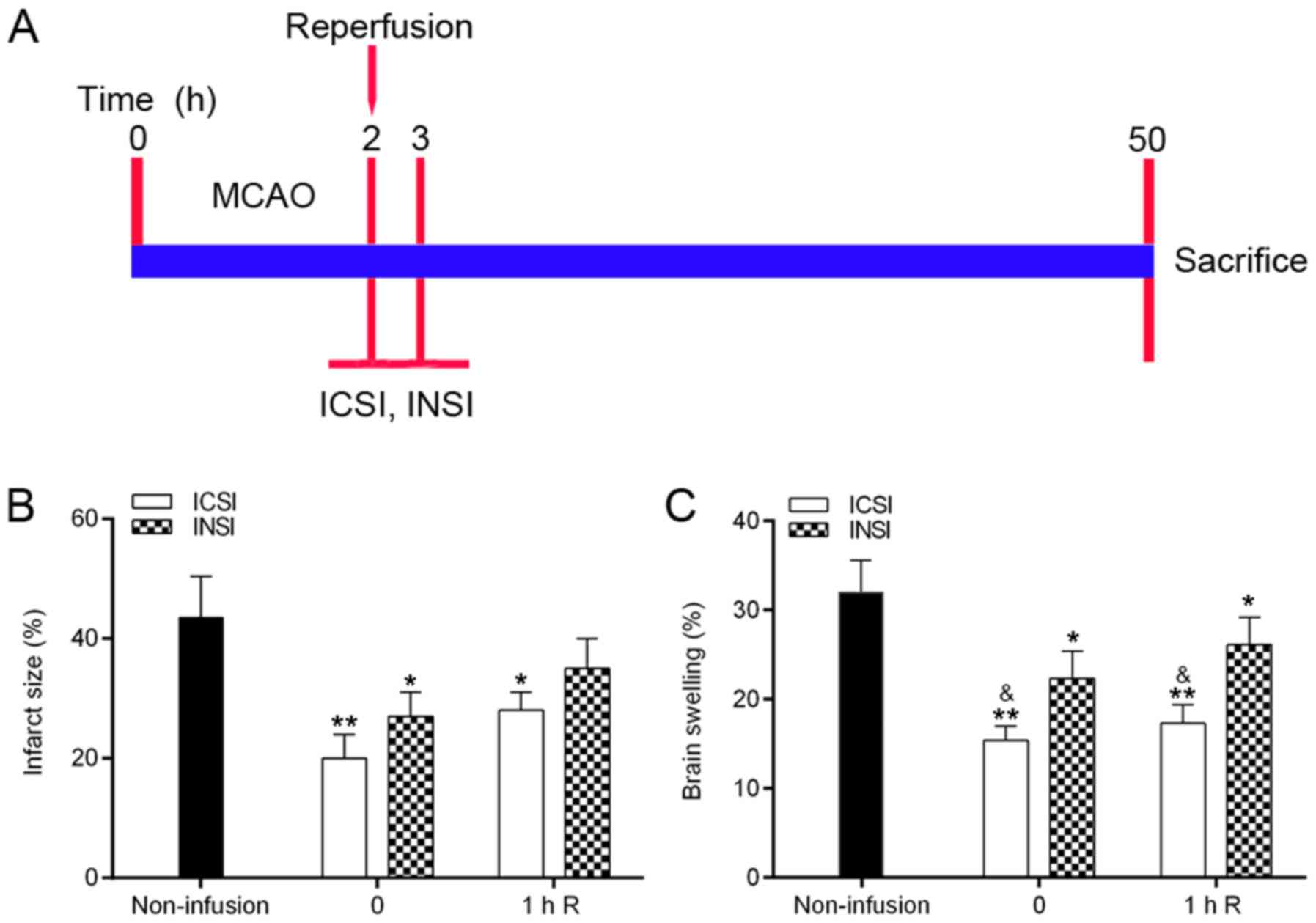 | Figure 1.ICSI decreases infarct size and brain
swelling in the rat MCAO model. Rats were randomly divided into the
MCAO group (n=24), ICSI group (n=26), INSI group (n=23),
ICSI+reperfusion group (n=25), and INSI+reperfusion group (n=23).
(A) Experimental procedure in each group of rats is shown. (B)
Infarct size and (C) brain swelling were detected in the rat MCAO
models treated with reperfusion and either ICSI or INSI.
*P<0.05, **P<0.01 (infusion groups vs. non-infusion group);
&P<0.05, ICSI vs. INSI. One-way ANOVA with
Tukey's post hoc test was performed. All experiments were repeated
three times. MCAO, middle cerebral artery occlusion; ICSI,
intracarotid cold saline infusion; INSI, intracarotid normothermic
saline infusion. 0, immediate infusion after reperfusion; 1 h R, 1
h after reperfusion. |
ICSI alleviates cell apoptosis and
nerve injury in the brain
To further explore the effects of ICSI on apoptosis
and nerve injury in the brain after reperfusion in the MCAO model,
TUNEL and Nissl staining were performed. There were significantly
fewer TUNEL-positive cells in the ICSI and ICSI-1h-R groups than
that noted in the non-infusion, INSI and INSI-1h groups (P<0.05,
P<0.01, P<0.001; Fig. 2A and
B). We also found that the relative integral optical density
(IOD) values of Nissl bodies were higher in the ICSI and ICSI-1h-R
groups than in the non-infusion group, that they were higher in the
INSI and INSI-1h groups than in the non-infusion group, and that
they were higher in the ICSI and ICSI-1h-R groups than in the INSI
and INSI-1h groups (P<0.01, P<0.001; Fig. 2A and C).
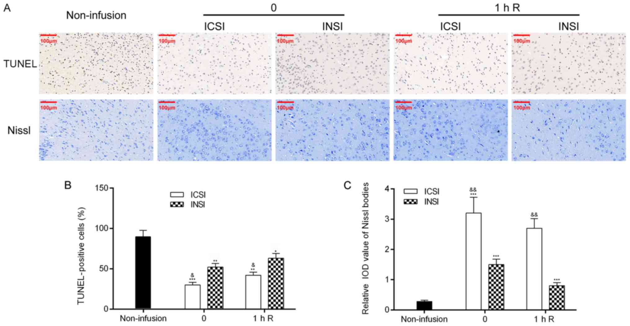 | Figure 2.ICSI inhibits apoptosis and increases
the relative IOD value of Nissl bodies in the rat MCAO model. (A)
Apoptosis and the relative IOD value of Nissl bodies were measured
using TUNEL staining and Nissl staining, respectively.
Magnification, ×100; scale bar, 100 µm. (B) TUNEL-positive cells
were analyzed. (C) Relative IOD value of Nissl bodies was
determined. *P<0.05, **P<0.01, ***P<0.001, infusion groups
vs. non-infusion group. &P<0.05,
&&P<0.01, ICSI vs. INSI group; comparisons
were carried out at the same time point. Data were evaluated using
one-way ANOVA with Tukey's post hoc test. All experiments were
repeated three times. IOD, integral optical density; MCAO, middle
cerebral artery occlusion; ICSI, intracarotid cold saline infusion;
INSI, intracarotid normothermic saline infusion. 0, immediate
infusion after reperfusion; 1 h R, 1 h after reperfusion. |
ICSI increases the expression of SGK1
and decreases the expression of the autophagy markers beclin-1 and
LC-3
To determine the expression of SGK1,
immunohistochemistry was performed. The results showed that SGK1
expression was higher in the infusion groups than that in the
non-infusion group, that it was higher in the ICSI group than in
the INSI group, and that it was higher in the ICSI-1h-R group than
in the INSI-1h-R group (Fig. 3A).
In addition, we confirmed that SGK1 expression was higher in the
infusion groups than in the non-infusion group, that it was
significantly higher in the ICSI and ICSI-1h-R groups than in the
INSI and INSI-1h-R groups. The expression of both beclin-1 and LC-3
was significantly downregulated in the infusion groups compared
with that in the non-infusion group, and it was significantly
decreased in the ICSI and ICSI-1h-R groups compared with that in
the INSI and INSI-1h-R groups (P<0.05, P<0.01, P<0.001;
Fig. 3B).
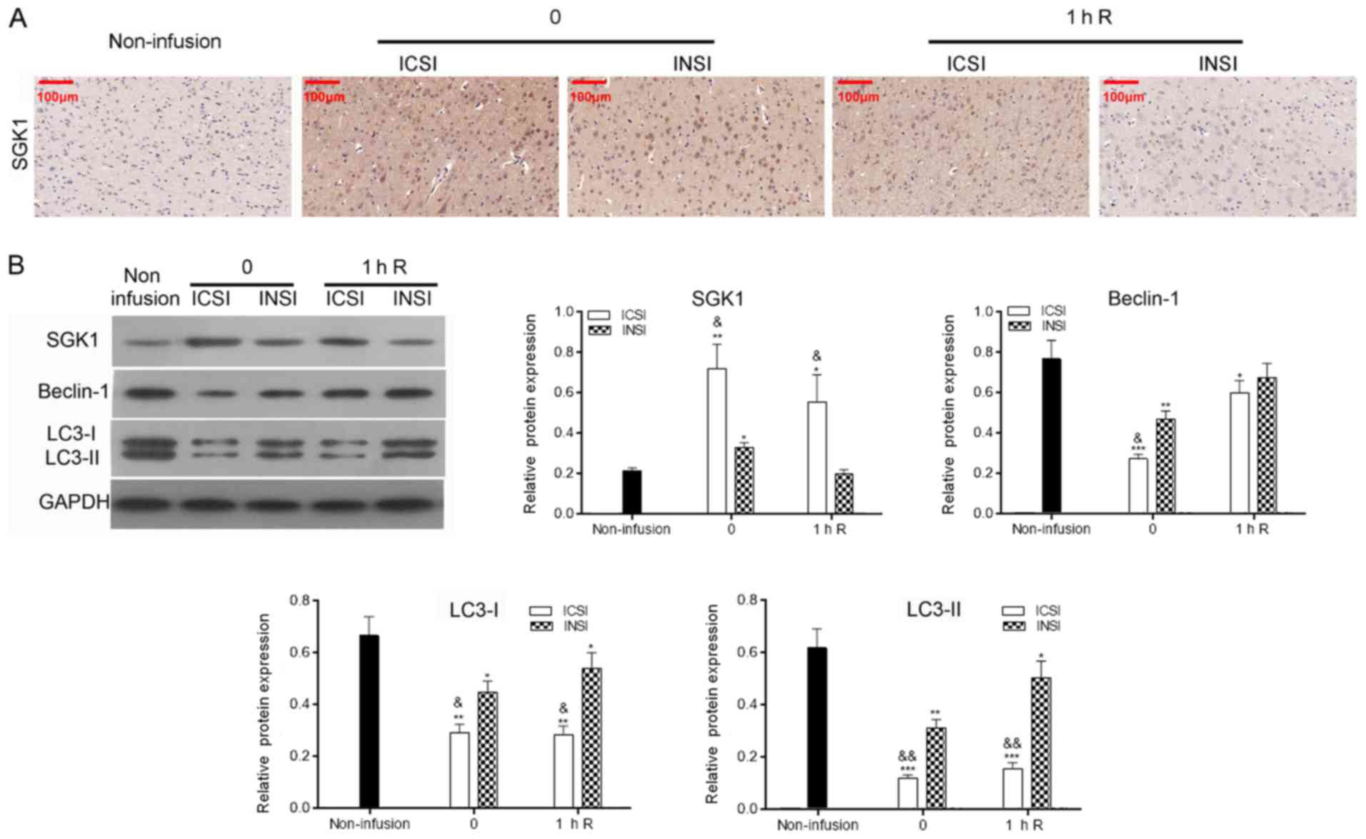 | Figure 3.ICSI upregulates SGK1 expression and
downregulates beclin-1 and LC-3 expression in the rat MCAO model.
(A) SGK1 expression was assessed using immunohistochemistry.
Magnification, ×100; scale bar, 100 µm. (B) Western blot analysis
of SGK1, beclin-1 and LC-3 expression. *P<0.05, **P<0.01,
***P<0.001, infusion groups vs. non-infusion group;
&P<0.05, &&P<0.01, ICSI vs.
INSI; comparisons were carried out using one-way ANOVA with Tukey's
post hoc test at the same time point. All experiments were repeated
three times. SGK1, serum and glucocorticoid-regulated kinase 1;
MCAO, middle cerebral artery occlusion; ICSI, intracarotid cold
saline infusion; INSI, intracarotid normothermic saline infusion.
0, immediate infusion after reperfusion; 1 h R, 1 h after
reperfusion. |
Knockdown of SGK1 decreases the
protective effect of ICSI
To investigate whether ICSI contributes to
SGK1-mediated neuroprotection in rats with ischemic stroke, animals
in the ICSI and ICSI-1h-R groups were transfected with SGK1 shRNA.
Four groups of rats were then subjected to cold saline infusion:
Rats given immediate ICSI (ICSI group), SGK1-knockdown rats given
immediate ICSI (ICSI+shSGK1), rats given ICSI starting 1 h after
reperfusion (ICSI-1h-R), and SGK1-knockdown rats given ICSI
starting 1 h after reperfusion (ICSI-1h-R+shSGK1; Fig. 4A). A western blot assay was used to
verify the knockdown effect of SGK1, and the results revealed that
SGK1 expression was significantly suppressed after transfection
with SGK1 shRNAs (P<0.01, P<0.001; Fig. 4B). TTC staining and the dry-wet
weight method were applied to measure infarct size and brain
swelling. The results revealed that knockdown of SGK1 using shRNAs
significantly increased infarct size and brain swelling, as
compared to the ICSI group (P<0.05, P<0.01; Fig. 4C and D). Therefore, we suggest that
SGK1 knockdown aggravated the process of ischemic stroke treatment
using ICSI.
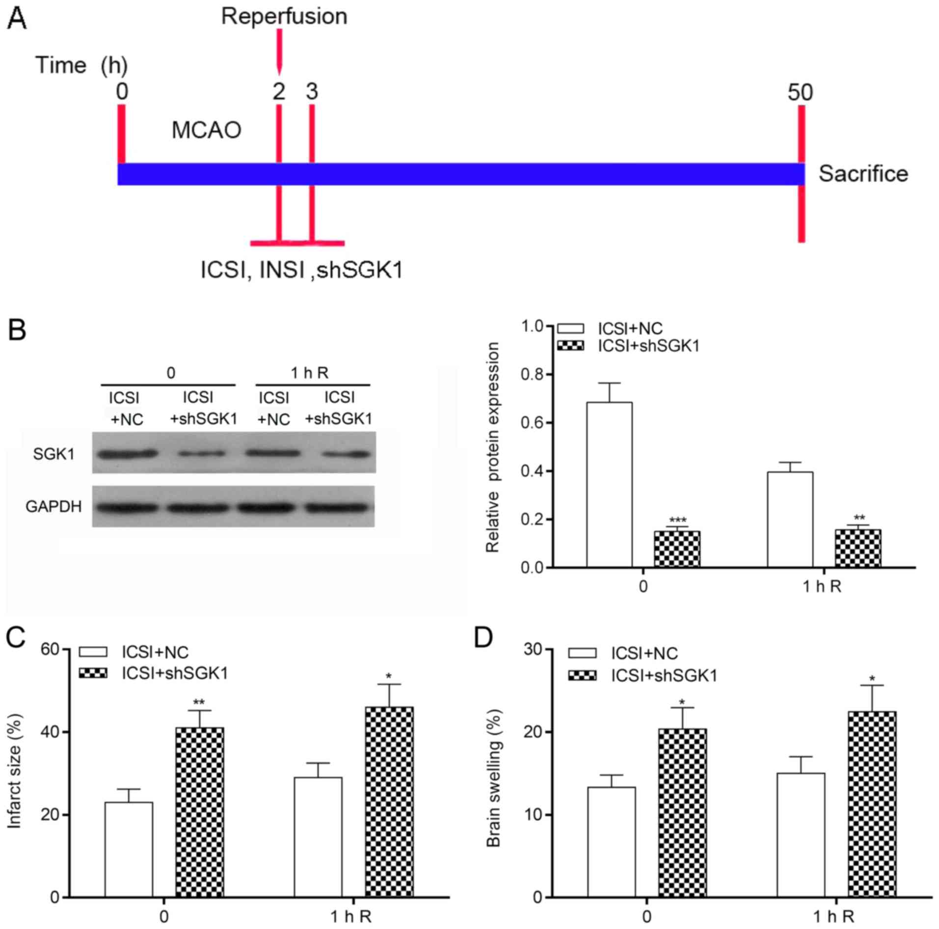 | Figure 4.SGK1 knockdown increases infarct size
and brain swelling mediated by ICSI. NC shRNAs or SGK1 shRNAs were
transfected into the rats in the ICSI and ICSI+reperfusion groups.
The rats were divided into the ICSI+NC group (n=24), ICSI+shSGK1
group (n=23), ICSI+reperfusion+NC group (n=23), and
ICSI+reperfusion+shSGK1 group (n=23). (A) Protocol of the
experiments. (B) SGK1 expression was examined by western blot
analysis in each group. (C) Infarct size was measured using TTC
staining after SGK1 knockdown in the ICSI group. (D) The brain
swelling of the ICSI group after SGK1 knockdown. *P<0.05,
**P<0.01, ***P<0.001 vs. ICSI group by Student's t-test. All
experiments were repeated three times. SGK1, serum and
glucocorticoid-regulated kinase 1; MCAO, middle cerebral artery
occlusion; ICSI, intracarotid cold saline infusion; INSI,
intracarotid normothermic saline infusion. 0, immediate infusion
after reperfusion; 1 h R, 1 h after reperfusion. |
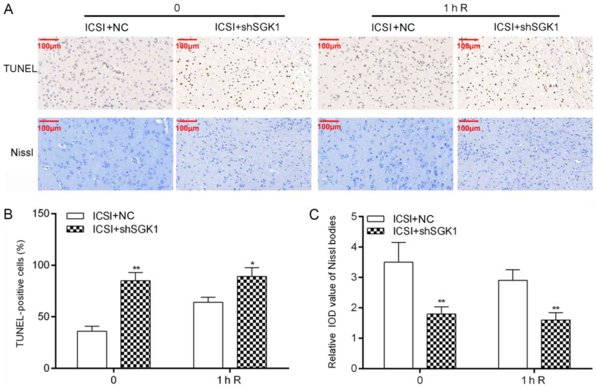
Knockdown of SGK1 decreases the
protection of neurons from apoptosis and injury
To explore whether SGK1 affects apoptosis and
ischemic stroke injury in rats treated using ICSI, TUNEL staining
and Nissl staining were performed. The results showed that SGK1
knockdown using shRNAs promoted ICSI-mediated apoptosis in the MCAO
model (P<0.05, P<0.01; Fig. 5A
and B). Meanwhile, we demonstrated that SGK1 knockdown
significantly increased the relative IOD value of Nissl bodies
mediated by ICSI in the MCAO model (P<0.01, Fig. 5A and C). Therefore, we confirmed
that SGK1 knockdown significantly accelerated apoptosis and
increased the relative IOD value of ICSI-mediated Nissl bodies in
the rat MCAO model.
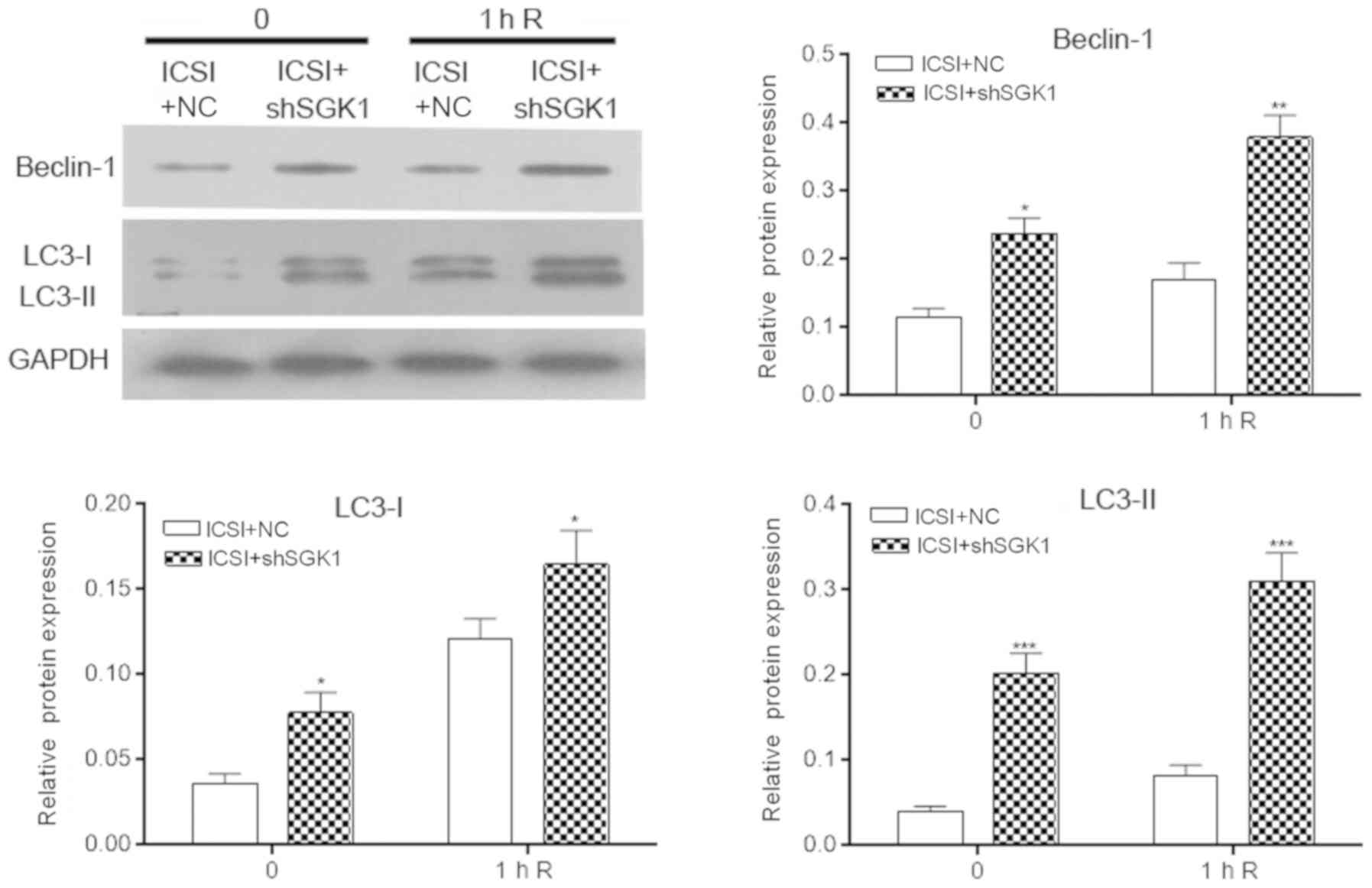
Knockdown of SGK1 upregulates
ICSI-mediated downregulation of beclin-1 and LC-3 expression
Furthermore, western blotting was performed to
analyze the influence of SGK1 knockdown on the expression of
beclin-1 and LC-3 downregulated by ICSI in the rat MCAO model.
These results indicated that the ICSI-mediated decreases in the
expression of Beclin1 and LC3 could be significantly upregulated by
the knockdown of SGK-1 (P<0.05, P<0.01, P<0.001; Fig. 6).
Discussion
Ischemic stroke involves hemiplegia and disturbance
of consciousness caused by cerebral thrombosis, cerebral artery
occlusion, and consequent cerebral infarction (25). At present, ischemic stroke is the
most common cerebrovascular disease (26). It is characterized by its high
incidence, high disability rate, high recurrence rate, and high
fatality rate, and it has become a major threat to human health
(27). Therefore, it is crucial
that researchers and clinicians discover preventative and treatment
strategies.
Therapeutic hypothermia is an effective method of
protecting brain cells, and it is widely used to treat
hypoxic-ischemic encephalopathy (28). The neuroprotective mechanisms of
low temperature include i) reducing the metabolism of nerve cells
and reducing acidosis; ii) inhibiting the production and release of
endogenous harmful substances, such as glutamic acid, aspartic acid
and serotonin; iii) protecting the blood-brain barrier and reducing
cerebral edema and intracranial pressure; iv) preventing the
proliferation of polymorphonuclear leukocytes and reducing the
intracellular inflammatory response; v) altering the transmission
of genetic information and promoting protein synthesis and
recovery; and vi) inhibiting nerve cell apoptosis (29). Intracarotid cold saline infusion
(ICSI) is a novel and selective cryogenic method (30,31).
A large number of clinical studies have shown that ICSI can rapidly
reduce brain temperature in rats, significantly protecting brain
function and ensuring clinical safety (25,32,33).
In the present study, the rat middle cerebral artery
occlusion (MCAO) model was established and treated using ICSI and
intracarotid normothermic saline infusion (INSI). TTC staining and
brain swelling measurement were used to evaluate the effects of
ICSI on local brain tissues. In the present study, ICSI was found
to alleviate infarct size and brain swelling in the MCAO model
after reperfusion. In addition, it was confirmed that ICSI
inhibited cell apoptosis and nerve injury in the brain. Therefore,
it was further demonstrated that ICSI is a fast, efficient, and
safe method of implementing selective hypothermia in the brain.
Selective cerebral hypothermia during acute ischemia has
therapeutic effects on ischemic reperfusion, including reducing
infarct volume, reducing brain swelling, and improving neurological
deficits.
Previous research has revealed that serum and
glucocorticoid-regulated kinase 1 (SGK1) protects against
ischemia-reperfusion injury. For instance, dexamethasone, which is
associated with SGK1, can protect against renal
ischemia-reperfusion injury (34).
SGK1 is involved in renal ischemia-reperfusion injury (35), while glucocorticoid affects SGK1
expression and protects against ischemia-reperfusion injury after
heart transplantation (36). SGK1
inhibits cell death and inflammation in the ischemic-reperfused
heart (37). In the present study,
it was demonstrated that ICSI increased SGK1 expression and that
SGK1 silencing increased infarct size and brain swelling,
accelerated apoptosis, and decreased the relative IOD value of
ICSI-mediated Nissl bodies. Therefore, we suggest that ICSI
protected against brain injury in MCAO-induced ischemic stroke
rats.
Autophagy is an evolutionarily conserved process in
eukaryotes that processes the turnover of intracellular substances
(16,38,39).
Many studies have shown that maladjusted autophagy regulation is
related to various diseases, such as malignant tumors, autoimmune
disease, neurodegenerative disease and pathogenic microorganism
infection (40–43). Beclin-1 and LC-3 serve as specific
markers of autophagy and have been applied in both clinical and
basic research (44,45). In the present study, it was
revealed that ICSI downregulated beclin-1 and LC-3 expression and
that knockdown of SGK1 upregulated beclin-1 and LC-3 expression.
ICSI inhibited autophagy by regulating SGK1 expression in the rat
MCAO model.
ICSI was demonstrated to have neuroprotective
potential in MCAO-induced ischemic stroke. In addition, ICSI
significantly upregulated SGK1 expression, and SGK1 silencing
promoted cerebral injury in MCAO-induced ischemic stroke.
Furthermore, it was confirmed that ICSI was conducive to the
neuroprotective efficacy of MCAO-induced ischemic stroke in rats by
SGK1 and autophagy. However, additional studies are needed to
validate the function of SGK1 and the potential mechanism of ICSI
in ischemic stroke, and we must optimize our experimental
grouping.
Acknowledgements
Not applicable.
Funding
The present study was supported by the National
Science Foundation (grant. nos. 81860321and 81460276).
Availability of data and materials
The datasets used during the present study are
available from the corresponding author upon reasonable
request.
Authors' contributions
DW, ZH, CN and WY conceived and designed the study.
DW and ZH performed the experiments. LL, YY, LX and XW analyzed the
data and were the primary contributors to the writing of the
manuscript. All authors read and approved the final manuscript.
Ethics approval and consent to
participate
The present study was approved by the Ethics
Committee of The First Affiliated Hospital of Soochow
University.
Patient consent for publication
Not applicable.
Competing interests
The authors declare that they have no competing
interests.
References
|
1
|
González-Ibarra FP, Varon J and López-Meza
EG: Therapeutic hypothermia: Critical review of the molecular
mechanisms of action. Front Neurol. 2:42011. View Article : Google Scholar : PubMed/NCBI
|
|
2
|
Froehler MT and Ovbiagele B: Therapeutic
hypothermia for acute ischemic stroke. Expert Rev Cardiovasc Ther.
8:593–603. 2010. View Article : Google Scholar : PubMed/NCBI
|
|
3
|
Choi JH, Marshall RS, Neimark MA, Konstas
AA, Lin E, Chiang YT, Mast H, Rundek T, Mohr JP and Pile-Spellman
J: Selective brain cooling with endovascular intracarotid infusion
of cold saline: A pilot feasibility study. AJNR Am J Neuroradiol.
31:928–934. 2010. View Article : Google Scholar : PubMed/NCBI
|
|
4
|
Luan X, Li J, McAllister JP II, Diaz FG,
Clark JC, Fessler RD and Ding Y: Regional brain cooling induced by
vascular saline infusion into ischemic territory reduces brain
inflammation in stroke. Acta Neuropathol. 107:227–234. 2004.
View Article : Google Scholar : PubMed/NCBI
|
|
5
|
Ji Y, Hu Y, Wu Y, Ji Z, Song W, Wang S and
Pan S: Therapeutic time window of hypothermia is broader than
cerebral artery flushing in carotid saline infusion after transient
focal ischemic stroke in rats. Neurol Res. 34:657–663. 2012.
View Article : Google Scholar : PubMed/NCBI
|
|
6
|
Cheng H, Ji X, Ding Y, Luo Y, Wang G, Sun
X, Chen J and Ling F: Focal perfusion of circulating cooled blood
reduces the infarction volume and improves neurological outcome in
middle cerebral artery occlusion. Neurol Res. 31:340–345. 2009.
View Article : Google Scholar : PubMed/NCBI
|
|
7
|
Chen J, Ji X, Ding Y, Luo Y, Cheng H and
Ling F: A novel approach to reduce hemorrhagic transformation after
interventional management of acute stroke: Catheter-based selective
hypothermia. Med Hypotheses. 72:62–63. 2009. View Article : Google Scholar : PubMed/NCBI
|
|
8
|
Linares G and Mayer SA: Hypothermia for
the treatment of ischemic and hemorrhagic stroke. Crit Care Med 37
(7 Suppl). S243–S249. 2009. View Article : Google Scholar
|
|
9
|
Liu W, Wang X, Wang Y, Dai Y, Xie Y, Ping
Y, Yin B, Yu P, Liu Z, Duan X, et al: SGK1 inhibition-induced
autophagy impairs prostate cancer metastasis by reversing EMT. J
Exp Clin Cancer Res. 37:732018. View Article : Google Scholar : PubMed/NCBI
|
|
10
|
Mason JA and Schafer ZT: SGK1 and PHLPP1:
Ras-mediated effectors during ECM-detachment. Cell Cycle.
15:2233–2234. 2016. View Article : Google Scholar : PubMed/NCBI
|
|
11
|
Zhu M, Wu G, Li YX, Stevens JK, Fan CX,
Spang A and Dong MQ: Serum- and Glucocorticoid-Inducible Kinase-1
(SGK-1) plays a role in membrane trafficking in caenorhabditis
elegans. PLoS One. 10:e01307782015. View Article : Google Scholar : PubMed/NCBI
|
|
12
|
Zuleger T, Heinzelbecker J, Takacs Z,
Hunter C, Voelkl J, Lang F and Proikas-Cezanne T: SGK1 Inhibits
autophagy in murine muscle tissue. Oxid Med Cell Longev.
2018:40437262018. View Article : Google Scholar : PubMed/NCBI
|
|
13
|
Nishida Y, Nagata T, Takahashi Y,
Sugahara-Kobayashi M, Murata A and Asai S: Alteration of
serum/glucocorticoid regulated kinase-1 (sgk-1) gene expression in
rat hippocampus after transient global ischemia. Brain Res Mol
Brain Res. 123:121–125. 2004. View Article : Google Scholar : PubMed/NCBI
|
|
14
|
Zhang W, Qian CY and Li SQ: Protective
effect of SGK1 in rat hippocampal neurons subjected to ischemia
reperfusion. Cell Physiol Biochem. 34:299–312. 2014. View Article : Google Scholar : PubMed/NCBI
|
|
15
|
Cong B, Du J, Zhu X, Lu J and Ni X:
Estrogen enhancement of SGK1 expression induced by urocortin
contributes to its cardioprotection against ischemia/reperfusion
insult. Int J Cardiol. 178:200–202. 2015. View Article : Google Scholar : PubMed/NCBI
|
|
16
|
Ashkenazi A, Bento CF, Ricketts T,
Vicinanza M, Siddiqi F, Pavel M, Squitieri F, Hardenberg MC,
Imarisio S, Menzies FM and Rubinsztein DC: Polyglutamine tracts
regulate beclin 1-dependent autophagy. Nature. 545:108–111. 2017.
View Article : Google Scholar : PubMed/NCBI
|
|
17
|
Lu J, Qian HY, Liu LJ, Zhou BC, Xiao Y,
Mao JN, An GY, Rui MZ, Wang T and Zhu CL: Mild hypothermia
alleviates excessive autophagy and mitophagy in a rat model of
asphyxial cardiac arrest. Neurol Sci. 35:1691–1699. 2014.
View Article : Google Scholar : PubMed/NCBI
|
|
18
|
Wang J, Pan XL, Ding LJ, Liu DY, Da-Peng
Lei and Jin T: Aberrant expression of Beclin-1 and LC3 correlates
with poor prognosis of human hypopharyngeal squamous cell
carcinoma. PLoS One. 8:e690382013. View Article : Google Scholar : PubMed/NCBI
|
|
19
|
Schmitz KJ, Ademi C, Bertram S, Schmid KW
and Baba HA: Prognostic relevance of autophagy-related markers LC3,
p62/sequestosome 1, Beclin-1 and ULK1 in colorectal cancer patients
with respect to KRAS mutational status. World J Surg Oncol.
14:1892014. View Article : Google Scholar
|
|
20
|
Zhang L, Niu W, He Z, Zhang Q, Wu Y, Jiang
C, Tang C, Hu Y and Jia J: Autophagy suppression by exercise
pretreatment and p38 inhibition is neuroprotective in cerebral
ischemia. Brain Res. 1587:127–132. 2014. View Article : Google Scholar : PubMed/NCBI
|
|
21
|
Zhang X, Yan H, Yuan Y, Gao J, Shen Z,
Cheng Y, Shen Y, Wang RR, Wang X, Hu WW, et al: Cerebral
ischemia-reperfusion-induced autophagy protects against neuronal
injury by mitochondrial clearance. Autophagy. 9:1321–1333. 2013.
View Article : Google Scholar : PubMed/NCBI
|
|
22
|
Vahidinia Z, Alipour N, Atlasi MA,
Naderian H, Beyer C and Azami Tameh A: Gonadal steroids block the
calpain-1-dependent intrinsic pathway of apoptosis in an
experimental rat stroke model. Neurol Res. 39:54–64. 2017.
View Article : Google Scholar : PubMed/NCBI
|
|
23
|
Seyedemadi P, Rahnema M, Bigdeli MR, Oryan
S and Rafati H: The neuroprotective effect of rosemary
(Rosmarinus officinalis L.) Hydro-alcoholic extract on
cerebral ischemic tolerance in experimental stroke. Iran J Pharm
Res. 15:875–883. 2016.PubMed/NCBI
|
|
24
|
Harawa V, Njie M, Kessler A, Choko A,
Kumwenda B, Kampondeni S, Potchen M, Kim K, Jaworowski A, Taylor T,
et al: Brain swelling is independent of peripheral plasma cytokine
levels in Malawian children with cerebral malaria. Malar J.
17:4352018. View Article : Google Scholar : PubMed/NCBI
|
|
25
|
Lazzaro MA and Prabhakaran S: Induced
hypothermia in acute ischemic stroke. Expert Opin Investig Drugs.
17:1161–1174. 2008. View Article : Google Scholar : PubMed/NCBI
|
|
26
|
Jickling GC and Sharp FR: Improving the
translation of animal ischemic stroke studies to humans. Metab
Brain Dis. 30:461–417. 2015. View Article : Google Scholar : PubMed/NCBI
|
|
27
|
Lehman LL and Rivkin MJ: Perinatal
arterial ischemic stroke: Presentation, risk factors, evaluation,
and outcome. Pediatr Neurol. 51:760–768. 2014. View Article : Google Scholar : PubMed/NCBI
|
|
28
|
Andresen M, Gazmuri JT, Marín A, Regueira
T and Rovegno M: Therapeutic hypothermia for acute brain injuries.
Scand J Trauma Resusc Emerg Med. 23:422015. View Article : Google Scholar : PubMed/NCBI
|
|
29
|
Crossley S, Reid J, McLatchie R, Hayton J,
Clark C, MacDougall M and Andrews PJ: A systematic review of
therapeutic hypothermia for adult patients following traumatic
brain injury. Crit Care. 18:R752014. View
Article : Google Scholar : PubMed/NCBI
|
|
30
|
Neimark MA, Konstas AA, Choi JH, Laine AF
and Pile-Spellman J: The role of intracarotid cold saline infusion
on a theoretical brain model incorporating the circle of willis and
cerebral venous return. Conf Proc IEEE Eng Med Biol Soc.
2007:1140–1143. 2007.PubMed/NCBI
|
|
31
|
Neimark MA, Konstas AA, Lee L, Laine AF,
Pile-Spellman J and Choi J: Brain temperature changes during
selective cooling with endovascular intracarotid cold saline
infusion: Simulation using human data fitted with an integrated
mathematical model. J Neurointerv Surg. 5:165–171. 2013. View Article : Google Scholar : PubMed/NCBI
|
|
32
|
Jaramillo A, Illanes S and Diaz V: Is
hypothermia useful in malignant ischemic stroke? Current status and
future perspectives. J Neurol Sci. 266:1–8. 2008. View Article : Google Scholar : PubMed/NCBI
|
|
33
|
Kollmar R, Blank T, Han JL, Georgiadis D
and Schwab S: Different degrees of hypothermia after experimental
stroke: Short- and long-term outcome. Stroke. 38:1585–1589. 2007.
View Article : Google Scholar : PubMed/NCBI
|
|
34
|
Rusai K, Prokai A, Juanxing C, Meszaros K,
Szalay B, Pásti K, Müller V, Heemann U, Lutz J, Tulassay T and
Szabo AJ: Dexamethasone protects from renal ischemia/reperfusion
injury: A possible association with SGK-1. Acta Physiol Hung.
100:173–185. 2013. View Article : Google Scholar : PubMed/NCBI
|
|
35
|
Rusai K, Prókai A, Szebeni B, Mészáros K,
Fekete A, Szalay B, Vannay Á, Degrell P, Müller V, Tulassay T and
Szabó AJ: Gender differences in serum and glucocorticoid regulated
kinase-1 (SGK-1) expression during renal ischemia/reperfusion
injury. Cell Physiol Biochem. 27:727–738. 2011. View Article : Google Scholar : PubMed/NCBI
|
|
36
|
Yang X, Wu X, Wu K, Yang D, Li Y, Shi J
and Liu Y: Correlation of serum- and glucocorticoid-regulated
kinase 1 expression with ischemia-reperfusion injury after heart
transplantation. Pediatr Transplant. 19:196–205. 2015. View Article : Google Scholar : PubMed/NCBI
|
|
37
|
Baban B, Liu JY and Mozaffari MS: SGK1
regulates inflammation and cell death in the ischemic-reperfused
heart: Pressure-related effects. Am J Hypertens. 27:846–856. 2014.
View Article : Google Scholar : PubMed/NCBI
|
|
38
|
Chiang WC, Wei Y, Kuo YC, Wei S, Zhou A,
Zou Z, Yehl J, Ranaghan MJ, Skepner A, Bittker JA, et al:
High-throughput screens to identify autophagy inducers that
function by disrupting Beclin 1/Bcl-2 binding. ACS Chem Biol.
13:2247–2260. 2018. View Article : Google Scholar : PubMed/NCBI
|
|
39
|
Jin Y, Lin Y, Feng JF, Jia F, Gao GY and
Jiang JY: Moderate hypothermia significantly decreases hippocampal
cell death involving autophagy pathway after moderate traumatic
brain injury. J Neurotrauma. 32:1090–1100. 2015. View Article : Google Scholar : PubMed/NCBI
|
|
40
|
Gewirtz DA: The four faces of autophagy:
Implications for cancer therapy. Cancer Res. 74:647–651. 2014.
View Article : Google Scholar : PubMed/NCBI
|
|
41
|
Kroemer G: Autophagy: A druggable process
that is deregulated in aging and human disease. J Clin Invest.
125:1–4. 2015. View Article : Google Scholar : PubMed/NCBI
|
|
42
|
Parzych KR and Klionsky DJ: An overview of
autophagy: Morphology, mechanism, and regulation. Antioxid Redox
Signal. 20:460–473. 2014. View Article : Google Scholar : PubMed/NCBI
|
|
43
|
White E, Mehnert JM and Chan CS:
Autophagy, metabolism, and cancer. Clin Cancer Res. 21:5037–5046.
2015. View Article : Google Scholar : PubMed/NCBI
|
|
44
|
Chen Y, Li X, Wu X, He C, Guo L, Zhang S,
Xiao Y, Guo W and Tan B: Autophagy-related proteins LC3 and
Beclin-1 impact the efficacy of chemoradiation on esophageal
squamous cell carcinoma. Pathol Res Pract. 209:562–567. 2013.
View Article : Google Scholar : PubMed/NCBI
|
|
45
|
Ezzeldin M, Borrego-Diaz E, Taha M,
Esfandyari T, Wise AL, Peng W, Rouyanian A, Asvadi Kermani A,
Soleimani M, Patrad E, et al: RalA signaling pathway as a
therapeutic target in hepatocellular carcinoma (HCC). Mol Oncol.
8:1043–1053. 2014. View Article : Google Scholar : PubMed/NCBI
|