Introduction
Chronic kidney disease (CKD) and end-stage renal
disease (ESRD) are global health problems worldwide (1). Pre-renal (decreased renal perfusion
pressure), intrinsic renal (pathology of the vessels, glomeruli or
tubules-interstitium) or post-renal (obstructive) are the three
disease processes, which contribute to CKD (2). It is difficult to determine the true
incidence and prevalence of CKD due to the asymptomatic nature of
early to moderate CKD. The prevalence of CKD in the general
population is ~10–14% (3).
Worldwide, CKD accounted for 2,968,600 (1%) of disability-adjusted
life-years and 2,546,700 (1–3%) of life-years lost in 2012
(4). CKD is a growing global
public health problem with notable socioeconomic burden (1). Thus, an in-depth study of CKD
pathogenesis is particularly important for its prevention and
treatment.
Podocytes are cells that encapsulate glomerular
capillaries in the Bowman's capsule of the kidney. Podocytes are
named as of their long protrusions projecting from the cells, which
are called foot processes or pedicels. Neighbouring podocytes
interdigitate to overlap the basal lamina, which is closely
associated with the glomerular capillaries. The pedicels of the
podocytes intersect, in which spaces between the cells exist, known
as slit diaphragms. These regions contain several cell-surface
proteins, including nephrin, podocin and CD2-associated protein
that cover the slits (5).
Destruction of the filtration slits or the podocytes can result in
kidney malfunction (6). Numerous
studies in the past two decades have suggested that the damage and
loss of podocytes are common clinical observations presented in the
early stages of glomerular diseases, as well as in CKDs including
minimal change disease, focal segmental glomerulosclerosis,
diabetic nephropathy, membranous nephropathy and lupus nephritis
(7,8).
Numerous lines of evidence have revealed that
nephrin and podocin play important roles in the pathogenesis of
podocyte injury. Several reports have revealed a decrease in the
expression of nephrin in various proteinuria nephropathy and animal
models of glomerular diseases (9,10).
Hence, the deficiency of nephrin is considered as a pathologic
feature of glomerular injury (11). In vivo, mild proteinuria,
foot process regression, filtration slit narrowing, mesangial
proliferation and sclerosis, glomerular basement membrane
thickening, sub-endothelial zone widening and podocyte apoptosis
were observed in mice with long-term nephrin downregulation
(12). Gene mutations coding for
nephrin can lead to congenital steroid-resistant nephrotic syndrome
of the Finnish type, which is characterised by foot process
dysmorphogenesis and the lack of inter-cellular junction formation,
and is associated with considerably severe proteinuria at birth
(13,14). In addition, podocin inactivation
may contribute to the onset of focal segmental glomerulosclerosis
and nephrotic syndrome (15); mice
with podocin deficiency develop proteinuria before delivery and
usually succumb from renal failure caused by massive mesangial
sclerosis with a median survival time of 12 weeks (16). Electron microscopy analysis of
kidney samples from these animals revealed extensive fusion of
podocyte foot processes and the lack of a slit diaphragm in the
remaining foot process junctions (17). Collectively, these studies suggest
that nephrin and podocin serve pivotal roles in the pathogenesis of
podocyte injury.
As one of the main effector hormones of the renin-
angiotensin system (RAS), angiotensin (Ang II) plays an important
role in the normal physiological maintenance of renal homeostasis
(18). Abnormal secretion of Ang
II is involved in the development of kidney diseases (19). Several studies have reported that
Ang II promotes podocyte injury (20–22).
Additionally, studies in vitro and in vivo have
revealed that podocytes present decreased nephrin expression and
increased apoptotic rates at high Ang II concentrations (23,24);
however, the mechanism for Ang II-induced podocyte injury remains
unclear. Few studies have investigated whether Ang II induces
podocyte injury via the PI3K/Akt/nuclear factor (NF)-κB pathway.
Several reports have demonstrated that the PI3K/Akt/NF-κB signaling
pathway is implicated in kidney diseases (25–29).
Hu et al (30) found that
the PI3K/Akt signaling pathway serves a pivotal role in
epithelial-mesenchymal transition of renal tubular epithelial
cells, which is induced by Ang II.
In this study, we performed in vitro
experiments to determine the effects of the PI3K/Akt/NF-κB
signaling pathway on Ang II-induced podocyte injury. In addition,
we examined the relationship between the PI3K/Akt/NF-κB signaling
pathway, and nephrin, podocin and caspase-9 synthesis in Ang
II-treated cultured mouse podocytes.
Materials and methods
Cell lines and cell culture
Mouse podocytes, which were purchased from the Cell
Center of Fudan University (FDCC-MSN059), were cultured and
differentiated as described previously (31). Briefly, the cells were cultivated
at 33°C (permissive conditions) for proliferation in RPMI-1640
medium (Gibco; Thermo Fisher Scientific, Inc.) containing 10% fetal
bovine serum (Gibco; Thermo Fisher Scientific, Inc.) with 10 U/ml
mouse recombinant γ-interferon (Invitrogen; Thermo Fisher
Scientific, Inc.). Podocytes were maintained at 37°C without
γ-interferon (non-permissive conditions) to induce differentiation
for at least 2 weeks.
Treatment of cultured podocytes at
37°C with Ang II
We treated the mouse podocytes with different
concentrations of Ang II (10−9, 10−8,
10−7 and 10−6 mol/l) for 12, 24 and 48 h for
cell viability assays, and with 10−6 mol/l of Ang II for
12, 24 and 48 h for cell apoptosis assays. Cells were also treated
with 10−6 mol/l of Ang II and or/LY294002 (inhibitor of
Akt) or 740Y-P (activator of PI3K) for 48 h (untreated cells served
as control) prior to detecting Akt, phosphorylated (phospho)-Akt,
p65 NF-κB, phospho-p65 NF-κB, nephrin, podocin and caspase-9
expression, and podocyte apoptosis.
Cell viability assay
Cell viability was examined using a Cell Counting
Kit-8 (CCK-8, Beijing Solarbio Life Sciences) assay. The podocytes
were seeded in 96-well plates at a density of 5×10³
cells per well and cultured at 37°C in 5% CO2 for 12 h,
and then treated with Ang II. After incubation for 0, 12, 24 and 48
h, 10 µl of CCK-8 solution was added to each well and incubated for
another 1–4 h at 37°C. The absorbance was measured with a
multi-mode microplate reader, TriStar LB 941 (Berthold
Technologies) at 450 nm.
Cell apoptosis assay
Apoptosis was measured using a flow cytometer
(FACSCanto II, BD Biosciences), and an Annexin V-fluorescein
isothiocyanate (FITC)/propidium iodide (PI) double-stain assay was
performed in accordance with the manufacturer's protocols
(FITC-AnnexinV/PI, BD Biosciences). After incubation of the
podocytes as described above, each supernatant was collected in the
centrifuge tubes and cells from each group were washed thrice with
PBS, trypsinized, centrifuged (400 × g at room temperature) for 5
min, adjusted to 1×10⁶/ml and suspended in binding
buffer containing Annexin V-FITC and PI. After incubation for 15
min at room temperature in the dark, the fluorescent intensity was
measured using a flow cytometer (FACSCanto II; BD Biosciences). The
apoptotic rate was calculated by summing the rate of annexin
V+/PI− and annexin
V+/PI+ cells, and was calculated using FlowJo
7.6.1 (Tree Star, Inc.).
Reverse transcription-quantitative
polymerase chain reaction (RT-qPCR) analysis
RT-qPCR analysis was performed to determine the mRNA
expression of nephrin, podocin, and caspase-9. Briefly, 2 µg of
total RNA from each sample of 5×10⁶ cells was used for
the synthesis of first strand cDNA using a FastKing RT kit (Tiangen
Biotech Co., Ltd.), according to the manufacturer's protocols.
Following the first strand cDNA synthesis, qPCR was carried out in
a 20 µl reaction volume containing 1X SuperReal PreMix Plus
(Tiangen Biotech Co., Ltd.), 0.6 µl of each of the specific forward
and reverse primers, and 2 µl of cDNA template. qPCR was performed
with an initial denaturation step at 95°C for 3 min, followed by 40
cycles of 95°C for 10 sec and 60°C for 30 sec in a
LightCycler® 96 system (Roche Diagnostics). GAPDH was
used as an endogenous housekeeping control. The mRNA expression
levels were quantified by using the 2−∆∆Cq method
(32). The data shown are
representative of the mean of three experiments. The primers for
each gene are listed in Table
I.
 | Table I.Primers of reverse
transcription-quantitative polymerase chain reaction. |
Table I.
Primers of reverse
transcription-quantitative polymerase chain reaction.
| Gene | Forward primer
(5′-3′) | Reverse primer
(5′-3′) |
|---|
| GAPDH |
AACTTTGGCATTGTGGAAGG |
GGATGCAGGGATGATGTTCT |
| Nephrin |
TCCTGCTGCGATGGTGGTTG |
GTCTGGGTTGCCTCCGATGG |
| Podocin |
TGAGGATGGCGGCTGAGAT |
GGTTTGGAGGAACTTGGGT |
| Caspase-9 |
ACCTGGTGCCTGTGGTCCTG |
GCTCCGCCAGAACCAATGTCC |
Western blot analysis
The podocytes were washed twice with PBS after
harvesting, and lysed in ice-cold radioimmunoprecipitation assay
buffer (Beyotime Institute of Biotechnology) with freshly added
0.01% protease inhibitor cocktail (CW Biotech), and were incubated
on ice for 30 min. Protein concentration was measured via a
bicinchoninic acid protein assay kit (Beyotime Institute of
Biotechnology). A total of 50 µg of protein was subjected to
electrophoresis using SDS-PAGE (8%) and was electrotransferred to a
nitrocellulose membrane (GE Healthcare). Thereafter, the membranes
were incubated for 1 h in 5% skimmed milk prepared with
Tris-buffered saline containing 0.05% Tween-20 at room temperature.
Subsequently, the membranes were incubated with primary antibodies
at 4°C overnight, including rabbit anti-nephrin (1:500; cat. no.
ab227806, Abcam), anti-podocin (1:500; cat. no. 20384-1-AP;
ProteinTech Group, Inc.), anti-Akt (1:500; cat. no. ab179463,
Abcam), anti- phospho-Akt (1:500; cat. no. ab192623), anti-p65
NF-κB (1:500; cat. no. 8242S, Cell Signalling Technology, Inc.),
anti-phospho-p65 NF-κB (1:500; cat. no. 3033S, Cell Signalling
Technology, Inc.), and anti-caspase-9 (1:500; cat. no. TA346902,
ZSBIO) which presented two bands, one indicates pro-caspase-9, with
a size of 46 KDa, whereas the other one indicates cleaved-caspase-9
with a size of 35 KDa. After washing with TBST, the membrane was
incubated with horseradish peroxidase-linked anti-rabbit secondary
antibody (cat. no. SA00001-2; ProteinTech Group, Inc.; 1:2,500) at
room temperature for 2 h and visualised for immunoreactivity.
Additionally, the membranes were processed to detect GAPDH
(1:1,000; cat. no. 6-004-1-1g, ProteinTech Group, Inc.), which
served as a loading control. Western blots were visualized using
enhanced chemiluminescence (cat. no. CW0048; CWBIO) and the images
were scanned by Quantity One software (Bio-Rad Laboratories, Inc.)
and the original intensity of each specific band was quantified
with a freeware image analysis software (ImageJ 6.0; National
Institutes of Health).
Statistical analysis
SPSS 23.0 statistical software (IBM Corp.) was used
for data analysis. Normal distribution and homogeneity of variance
were tested for all data. The data were expressed as mean ±
standard deviation. For cell viability analysis, statistical
significance was determined by multivariate analysis. One-way ANOVA
was used for analysis of variance among multiple groups; the Least
Significant Difference test was used with homogeneity of variance,
whereas a Games Howell test was used when no homogeneity in
variance was observed. Each group was repeated three times.
P<0.05 was considered to indicate a statistically significant
difference.
Results
Effects of Ang II on cell
viability
The viability of the podocytes was assessed after
treatment with Ang II, which was presumed to decrease with
increasing Ang II concentrations for 12, 24 and 48 h, as observed
by the CCK-8 assay. After 12, 24 and 48 h of incubation with Ang II
at concentrations of 10−9, 10−8,
10−7 and 10−6 mol/l, we observed a dose- and
time-dependent decrease in the cell viability compared with the
controls (Fig. 1). Therefore, this
indicated that Ang II could inhibit cell viability, and effectively
induce dose- and time-dependent cytotoxicity. Based on these
results, we decided to treat cells at a dose of 10−6
mol/l Ang II for 48 h for the following studies.
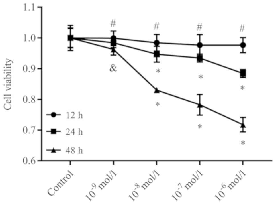 | Figure 1.Ang II suppresses the viability of
podocytes. The viability of the podocytes had a tendency to
decrease as the concentrations of Ang II increased for 12, 24 and
48 h, as determined by a Cell Counting Kit-8 assay. After 12, 24
and 48 h of incubation with Ang II at concentrations of
10−9, 10−8, 10−7 and
10−6 mol/l, we observed a dose- and time-dependent
decrease in cell viability compared with the controls.
#P>0.05, &P<0.05, *P<0.01 vs.
Control. The data are presented as the mean ± standard deviation
from three independent experiments. For cell viability analysis,
statistical significance was determined by multivariate analysis.
Ang II, angiotensin II. |
Ang II promotes podocyte
apoptosis
To determine whether the cytotoxic effect of Ang II
was due to the induction of apoptosis, the podocytes were treated
with different doses of Ang II for 48 h; 10−6 mol/l was
selected for administration at different time intervals (12, 24 and
48 h). Subsequently, cell apoptosis was determined by Annexin
V-FITC/PI staining and flow cytometry analysis. As presented in
Fig. 2, the apoptotic rate (sum of
annexin V+/PI− and annexin
V+/PI+ population/all cells events) of
podocytes were significantly increased after treatment with Ang II
from 15.80±1.12% (10−8 mol/l), 19.69±1.59%
(10−7 mol/l) to 25.17±0.91% (10−6 mol/l),
respectively, when compared with the control (9.25±0.81%). However,
no significant difference was observed between the cells incubated
with Ang II at concentrations of 10−9 mol/l (9.3%) and
the control. Additionally, the apoptotic rates of podocytes were
significantly increased after treatment with Ang II from
13.50±0.66% (12 h) and 19.80±1.08% (24 h) to 27.16±1.39% (48 h)
when compared with 0 h (8.37±0.71%). This demonstrated that Ang II
treatment induced apoptosis in these cells in a dose- and
time-dependent manner.
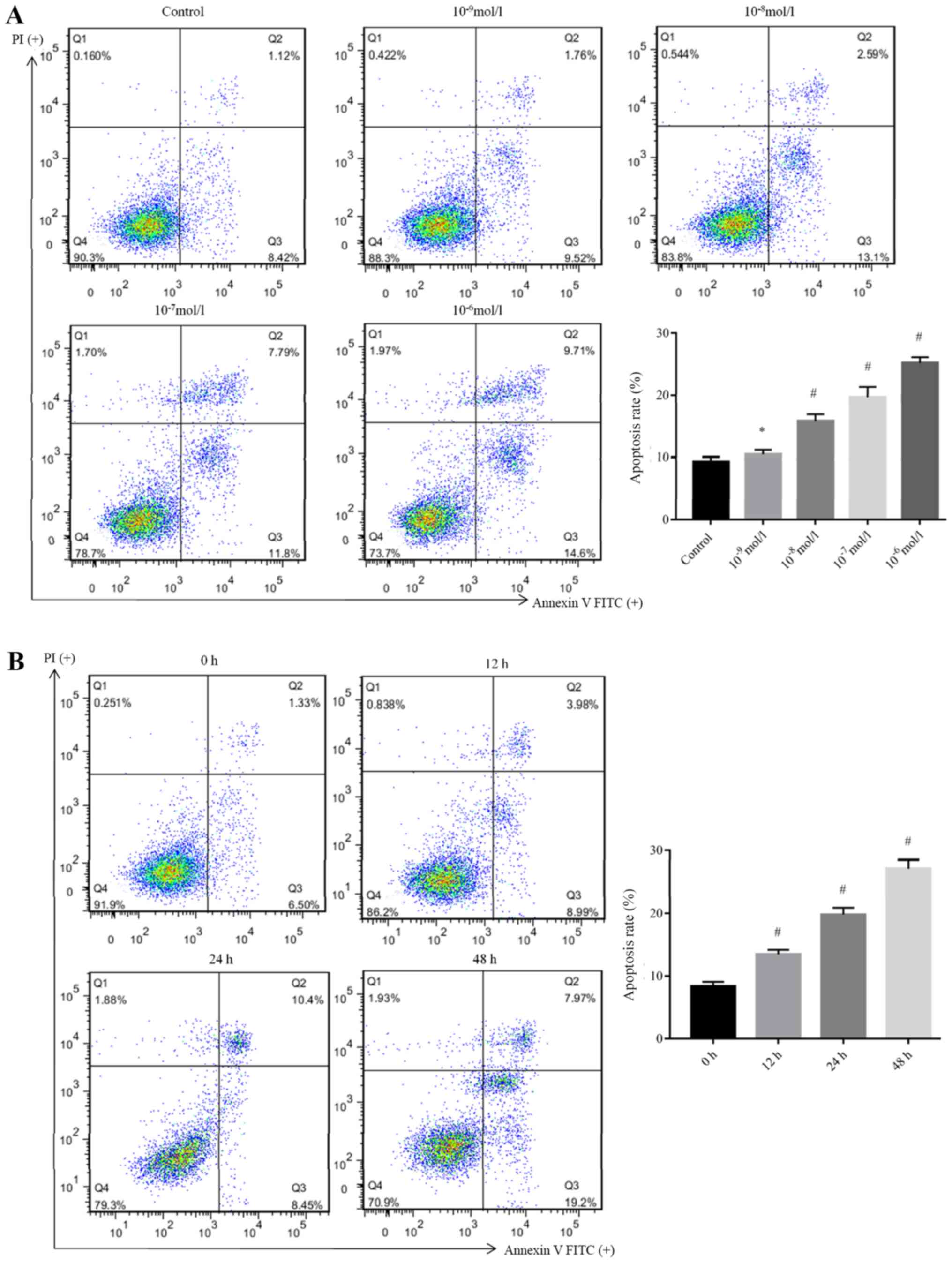 | Figure 2.Ang II promotes apoptosis of
podocytes. The apoptosis of podocytes was determined via flow
cytometry after treatment with Ang II at concentrations of
10−9, 10−8, 10−7 and
10−6 mol/l. (A) Apoptotic rates of podocytes were
significantly increased after treatment with Ang II from
10.43±0.74, 15.80±1.12, 19.69±1.59 and 25.17±0.91%, respectively,
when compared with the control (9.25±0.81%). *P>0.05,
#P<0.05 vs. Control. (B) Cell apoptosis of podocytes
after treatment with Ang II at 10−6 mol/l for different
time intervals (12, 24 and 48 h) as determined by flow cytometry.
The apoptotic rates of podocytes were significantly increased after
treatment with Ang II from 13.50±0.66% (12 h), 19.80±1.08% (24 h)
and 27.16±1.39% (48 h) when compared with 0 h (8.37±0.71%).
#P<0.05 vs. Control. The data are presented as the
mean ± standard deviation from three independent experiments. For
cell apoptosis analysis, statistical significance was determined
one-way ANOVA followed by the Least Significant Difference test.
Ang II, angiotensin II; FITC, fluorescein isothiocyanate; PI,
propidium iodide. |
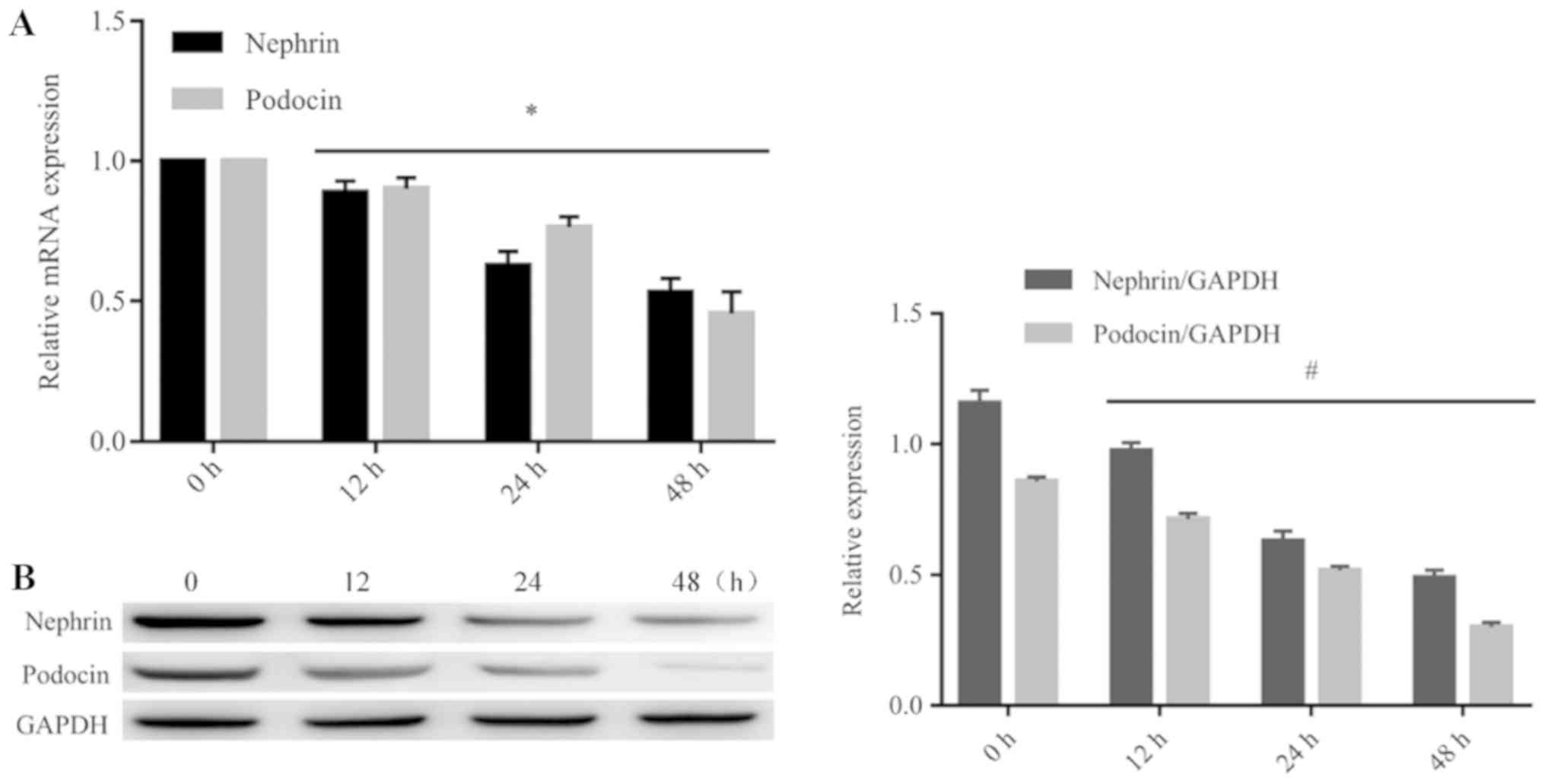
Ang II downregulates the expression of
nephrin and podocin in podocytes
Western blotting and RT-qPCR analysis were performed
to detect the expression of nephrin and podocin in podocytes after
treatment with Ang II. As presented in Fig. 3, the mRNA and protein levels of
nephrin and podocin were significantly reduced in the podocytes
after treatment with Ang II (10−6 mol/l) at different
time intervals (12, 24 and 48 h), when compared with 0 h. These
findings were consistent with cell apoptosis.
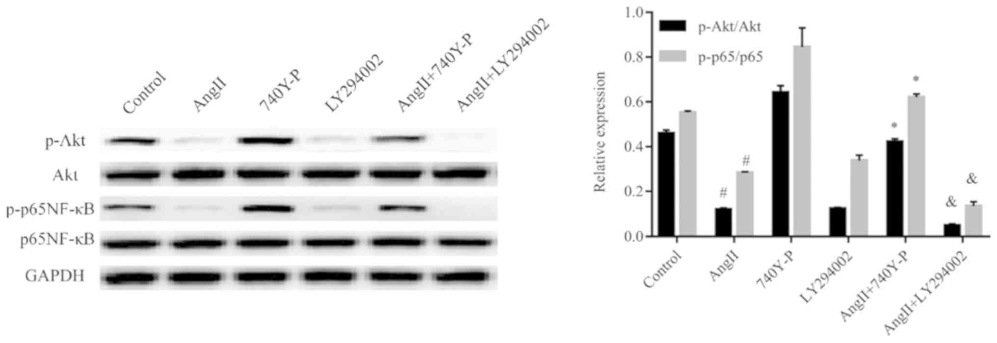
Functional mechanism of PI3K/Akt
signaling pathway in podocyte injury induced by Ang II
The PI3K/Akt pathway has been assumed to be involved
in various types of kidney diseases. Hence, we proposed that Ang II
may induce podocyte injury via the PI3K/Akt signaling pathway. To
examine our hypothesis, we used western blotting to analyse the
expression of proteins produced by genes located downstream of Ang
II-induced cells (Figs. 4–6). The results indicate that treatment
with Ang II caused a significant decrease in nephrin and podocin
expression compared with the control (Fig. 5); however, an increase in caspase-9
expression was observed (Fig. 6).
In addition, the phospho-Akt protein expression levels were
significantly reduced in the Ang II-treated group when compared
with the control levels, whereas Akt protein expression was not
significantly changed (Fig. 4).
Our results indicated that Ang II may exert its effect via
inhibition of the PI3K/Akt signaling pathway.
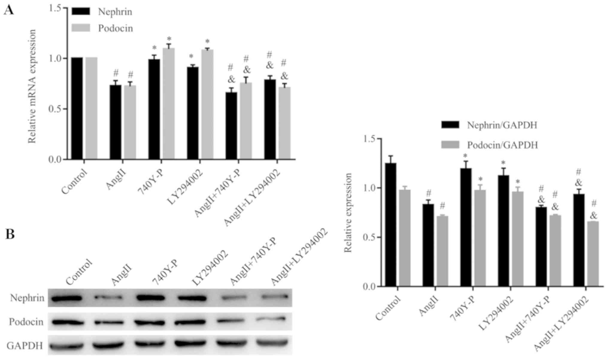
In order to confirm the contribution of the PI3K/Akt
signaling pathway in functioning of Ang II, an activator (740Y-P,
50 µg/ml) (33) and an inhibitor
(LY294002, 5 µM) (34) of the
PI3K/Akt signaling pathway were added to the cells which had been
treated with and without Ang II, respectively (Figs. 4 and 5). When compared with the control group,
740Y-P increased the expression levels of phospho-Akt. In contrast,
LY294002 inhibited the expression of phospho-Akt. Additionally,
LY294002 further decreased the expression levels of phospho-Akt in
cells, which were pre-treated with Ang II, when compared with those
treated with Ang II alone. In contrast, 740Y-P restored the
expression levels of phospho-Akt in the podocytes. Interestingly,
the expression of nephrin and podocin did not change significantly
when the cells were treated with LY294002 and 740Y-P compared with
Ang II treatment alone (Figs. 5A and
B). Our results suggested that the PI3K/Akt signaling pathway
has no effect on the expression of nephrin and podocin.
Activity of PI3K/Akt-regulated Ang
II-induced apoptosis via modulation of NF-κB activity
To investigate the mechanism of Ang II-induced
inhibition of NF-κB modulated by PI3K/Akt inhibition, the
levels of phospho-p65 NF-κB protein were analysed after Ang II
treatment followed by 740Y-P or LY294002 treatment or both.
Pre-treatment of podocytes with LY294002 significantly enhanced the
Ang II-induced decrease in the levels of phospho-p65 NF-κB. In
contrast, phospho-p65 NF-κB decreased significantly after treatment
with Ang II and increased by pre-treatment with 740Y-P. This
suggests that the PI3K/Akt signaling pathway is associated with the
regulatory activity of NF-κB (Fig.
4).
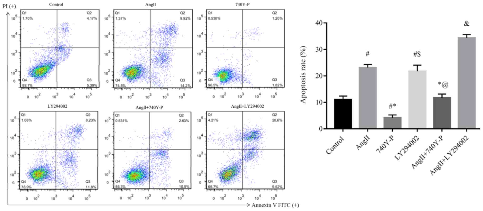
Furthermore, we focused on detecting the effect of
the PI3K/Akt/NF-κB signaling pathway on the apoptosis induced by
Ang II (Fig. 7). Compared with the
control group (11.00±1.35%), Ang II significantly stimulated
podocyte apoptosis (23.12±1.21%). Compared with the Ang II group
(23.12±1.21%), the combination of Ang II + 740Y-P reduced apoptosis
(11.73±1.43%), Ang II + LY294002 significantly increased apoptosis
(34.36±1.24%).
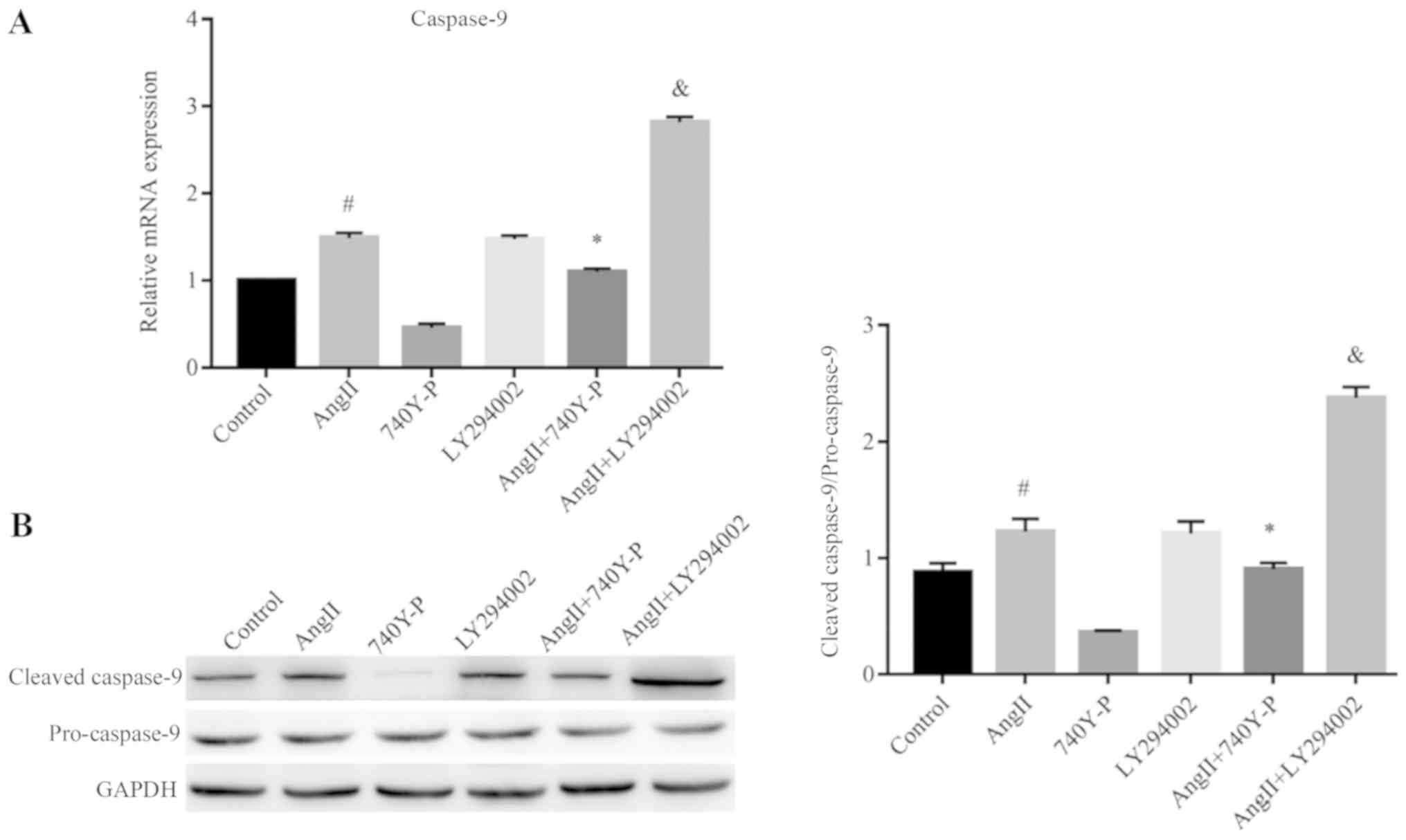
In order to provide further evidence of the role of
this pathway in the inhibition of apoptosis after Ang II treatment,
the expression levels of caspase-9 were determined by RT-qPCR and
western blotting. Ang II significantly increased the expression
levels of the caspase-9 at the mRNA and protein levels compared
with the control. This was reversed by pre-treatment with 740Y-P,
but was significantly increased by pre-treatment with LY294002
compared with Ang II treatment alone (Fig. 6), suggesting their essential role
in promoting cells for apoptosis following Ang II treatment.
Thus, these preliminary results confirmed that Ang
II could regulate the expression of nephrin, podocin and caspase-9
at least partly through the PI3K/Akt/NF-κB signaling pathway in
podocytes, and may play a crucial role in podocyte injury.
Discussion
Podocytes, which surround the capillaries of the
glomerulus constitute one of the main components of the glomerular
blood filtration barrier; the unique actin-based morphological
aspect of the foot processes are the most common features of kidney
diseases (35). Podocyte injury
and dysfunction are the major reasons for the pathogenesis of
proteinuria and glomerulosclerosis (36). Thus, decoding the complex molecular
and signaling mechanisms that are involved in podocyte dysfunction,
is an important way to develop a treatment for proteinuria. In our
present research, we investigated the involvement of PI3K/Akt/NF-κB
signaling pathway in podocytes to Ang II-induced injury. Our
results revealed a possible mechanism underlying podocyte injury.
The expression levels of nephrin and podocin, two podocyte-specific
markers, were downregulated, and the expression level of caspase-9,
a pro-apoptotic factor, was upregulated in AngII-induced podocytes
injury. However, the PI3K/Akt/NF-κB signaling pathway may not be
involved in this process. When the physical and chemical
environments are altered, cells adapt by changing several cellular
processes including cell survival, proliferation, differentiation
and apoptosis (37). As the
central product of the RAS, Ang II promotes the progression of
glomerular injury via its hemodynamic and/or non-hemodynamic
effects (38). Ang II induces cell
apoptosis and growth, and activates multiple signaling pathways in
renal cells (39). Within the
renal system, Ang II mediates various pathological processes
including apoptosis via the Ang II type 1 receptor, which results
in the development and progression of renal hypertrophy,
extracellular matrix accumulation, and proteinuria (40). Several reports have confirmed that
Ang II can promote apoptosis and suppress the proliferation of
podocytes via different pathways (21,22,41,42).
In accordance with other studies, in our study, we treated
podocytes with different doses of Ang II for various time
intervals. The results revealed that Ang II suppressed the
viability and the promoted the apoptosis of podocytes in a dose-
and time-dependent manner.
Protein kinase B, also referred to as Akt, is a
serine/threonine-specific protein kinase that plays a crucial role
in various cellular processes including apoptosis, cell
proliferation, transcription, and cell migration (43). Akt activation regulates the
maintenance and survival status of cellular stress fibres through
the phosphorylation of various substrates (43). Lin et al (44) found that activation of the PI3K/Akt
signaling pathway could inhibit kidney cell apoptosis and block the
formation of interstitial fibrosis. Akt is activated by PI3K, and
the PI3K/Akt signaling pathway plays a crucial role in the
resistance of podocytes to apoptosis (45). In puromycin aminonucleoside
(PAN)-induced podocyte injury models, Akt activity was markedly
decreased, and dexamethasone inhibited podocyte apoptosis by
stabilising the PI3K/Akt signaling pathway (46). Another study reported that PAN
reduced Akt phosphorylation levels, while LY294002 could further
promote podocyte apoptosis induced by PAN (47). The anti-apoptotic effects of
PI3K/Akt on podocyte apoptosis were further supported by
observations in which reduced Akt phosphorylation caused podocyte
apoptosis (48).
A previous study reported that the local production
of Ang II results in the progression of podocyte injury (49). It was also reported that Ang II
promotes podocyte apoptosis in the cultured podocytes (21). In our study, phospho-Akt was
downregulated in Ang II-treated podocytes, implying that
phospho-Akt could be involved in Ang II-induced podocyte injury.
Notably, LY294002 alone induced podocyte apoptosis and further
promoted podocyte apoptosis induced by Ang II. In contrast, 740Y-P
alleviated podocyte apoptosis induced by Ang II. Therefore, Ang II
is likely to induce apoptosis by affecting the Akt activity in
podocytes.
Downstream in the PI3K/Akt signaling pathway, NF-κB
is a protein complex, which plays an important role in controlling
the DNA transcription, cytokine production, and cell survival
(50). Active NF-κB promotes cell
proliferation and protects them from conditions that would lead to
apoptosis. Defects in the NF-κB machinery may lead an increased
susceptibility to apoptosis and cell death (51). Studies have suggested that NF-κB is
involved in the intensity of proteinuria and is restricted to
diseases of the renal tubules, as well as glomerular diseases
(52–54).
Studies have reported that Akt can interfere with
cell apoptosis via regulating apoptosis-related proteins, such as
caspase-9, as well as NF-Κb (55,56).
It was reported that the activation of Akt and NF-κB could promote
the proliferation of human mesangial cells (57). In addition, several studies
demonstrated that NF-κB is involved in podocyte injury (58,59);
however, its role in the glomerulus and in the pathophysiology of
podocyte diseases remains largely unexplored. To clarify the
mechanism mediating the inhibitory effects of Ang II on NF-κB
activation, we investigated the PI3K/Akt signaling pathway; we
demonstrated that NF-κB inactivation was promoted in podocytes
treated with Ang II, but was significantly enhanced by blockage of
the PI3K/Akt pathway with LY249002 and restored by the activator of
the PI3K/Akt pathway, 740Y-P. This suggested that inhibition of the
PI3K/Akt/NF-κB pathway may represent one of the primary mechanisms
for Ang II-induced apoptosis in podocytes.
Caspase-9 is an enzyme, which is encoded by the
caspase 9 gene in humans. It is an initiator caspase, critical to
the apoptotic pathway found in several tissues (60). To further investigate the apoptosis
mechanism of podocytes induced by Ang II, the expression of
caspase-9 was detected. The results indicated that treatment of
podocytes with Ang II could increase the apoptotic rate of
podocytes, as well as the expression of caspase-9. Furthermore,
when compared with the treatment of podocytes with Ang II, the
amount of caspase-9 was decreased after co-treating the cells with
Ang II and 740Y-P; however, caspase-9 expression was increased
after the cells were co-treated with Ang II and LY294002. This
indicated that 740Y-P could restore the inhibitory effect and that
LY294002 further promoted the activation effect of Ang II based on
the levels of caspase-9. Collectively, it appears that Ang II could
promote the expression of mRNA and protein of caspase-9 to induce
apoptosis of podocytes. Our results are in accordance with some
previous studies in which the activation of Akt inhibited apoptosis
by regulating the apoptotic initiation protein, caspase-9 (61–63).
Therefore, our findings suggest that a
PI3K/Akt/NF-κB signaling pathway in podocytes may be responsible
for promoting apoptosis following treatment with Ang II, which is
also linked to impaired upregulation of caspase-9. The results
suggest that these molecular protein mediators are necessary for
the involvement of the PI3K/Akt/NF-κB signaling pathway in relation
to podocyte damage induced by Ang II.
Nephrin and podocin are the main structural
components of the slit diaphragm. They work closely at the outer
membrane of the podocyte foot processes and regulate the normal
relationship between the podocytes of the epithelial cells and the
basement membrane. As the structural components of the slit
diaphragm, they play an important role in the appropriate
functioning of the renal filtration barrier (5). Nephrin regulates several pathways in
the podocyte including suppression of cell death and forming a
complex with podocin (64).
Mutational analysis reveals that abnormal or inefficient signaling
via the nephrin-podocin complex results in the development of
podocyte dysfunction and proteinuria (16). In addition, studies have indicated
that decreased levels of nephrin and podocin are related to the
degradation of the foot processes (64), and this may be involved in several
kidney diseases including lupus nephritis (65,66).
Some researchers have reported the potential
mechanism by which Ang II decreases the nephrin and podocin
expression. In order to explore the mechanism of podocyte injury
induced by Ang II, Yu et al (67) treated human podocytes with various
concentrations of Ang II type I receptor agonistic autoantibody
(AT1-AA); the results revealed that AT1-AA decreased the expression
of nephrin in a dose-dependent manner and the underlying mechanism
might involve activation of the transient receptor potential cation
channer subfamily C member 6-calcium/calcineurin pathway (67). A similar conclusion was drawn in
another study of Zhao et al (68). Additionally, Zhao et al
(41) found that the activation of
NOD-like receptor 3 inflammasome and mitochondrial dysfunction are
related to the loss of nephrin and podocin, which was induced by
Ang II. In the present study, we found that nephrin and podocin are
impaired in Ang II-treated cultured podocytes; however, we were
unable to reveal whether Akt is involved in the expression of
nephrin and podocin proteins in our experimental model. Several
other studies present a tentative link between nephrin, podocin and
the PI3K/Akt signaling pathway. Yang et al (69) confirmed that Akt is a downstream
intermediate of nephrin signaling. In addition, it has been
reported that nephrin and podocin interact with a subunit of PI3K
and subsequently activate the Akt kinase pathway, which is
necessary for the regulation of actin dynamics and the cell
survival (69–71). This suggests that downregulation of
nephrin can regulate Akt inactivation and podocyte injury; however,
the molecular mechanism of nephrin-Akt signal transduction requires
further investigation.
However, there are some limitations to this study.
We were unable to identify whether Akt signaling was involved in
regulating the expression of nephrin and podocin proteins in our
experimental model, and how Ang II induces the injury of podocytes
in vivo was not determined. The relationship between the
PI3K/Akt/NF-κB signaling pathway and nephrin and podocin proteins,
and in vivo analysis will be investigated in our future
studies.
In summary, the present study demonstrated that Ang
II could induce podocyte damage via the PI3K/Akt/NF-κB signaling
pathway, which could promote the development of podocyte injury
following treatment with Ang II. In addition, activation of the
PI3K/Akt/NF-κB survival axis may be a novel therapeutic strategy
for treating Ang II-induced podocyte injury.
Acknowledgements
Not applicable.
Funding
The present study was supported by the National
Natural Science Foundation of China (grant nos. 81560271 and
81860296), Key Project of Scientific Research of the Guangxi
Colleges and Universities (grant no. KY2015ZD092), and the Program
of Natural Science Foundation of Guangxi (grant nos.
2017GXNSFDA198005 and 2018GXNSFAA281038).
Availability of data and materials
All data generated or analysed during the current
study are included in this published article.
Authors' contributions
JW, SS and DF carried out the experimental work. YY
and JW participated in the design of the study, and together with
DF, who performed the statistical analysis. YY drafted the
manuscript. All the authors read and approved the final
manuscript.
Ethics approval and consent to
participate
Not applicable.
Patient consent for publication
Not applicable.
Competing interests
The authors declare that they have no competing
interests.
Glossary
Abbreviations
Abbreviations:
|
Ang II
|
angiotensin II
|
|
PI3K
|
phosphatidylinositol 3-kinase
|
|
NF-κB
|
nuclear factor-κB
|
|
RAS
|
renin-angiotensin system
|
|
PAN
|
puromycin aminonucleoside
|
References
|
1
|
Nath JD and Kashem A: Etiology and
frequency of hospital admissions in maintenance hemodialysis
patients in chronic kidney disease. Saudi J Kidney Dis Transpl.
30:508–512. 2019. View Article : Google Scholar : PubMed/NCBI
|
|
2
|
Vaidya SR and Aeddula NR: Chronic Renal
FailureStatPearls. StatPearls Publishing LLC.; Treasure Island, FL:
2019
|
|
3
|
Coresh J, Astor BC, Greene T, Eknoyan G
and Levey AS: Prevalence of chronic kidney disease and decreased
kidney function in the adult US population: Third National Health
and Nutrition Examination Survey. Am J Kidney Dis. 41:1–12. 2003.
View Article : Google Scholar : PubMed/NCBI
|
|
4
|
Webster AC, Nagler EV, Morton RL and
Masson P: Chronic kidney disease. Lancet. 389:1238–1252. 2017.
View Article : Google Scholar : PubMed/NCBI
|
|
5
|
Reiser J and Altintas MM: Podocytes.
F1000Res. 5:F10002016. View Article : Google Scholar : PubMed/NCBI
|
|
6
|
Vivarelli M, Massella L, Ruggiero B and
Emma F: Minimal change disease. Clin J Am Soc Nephrol. 12:332–345.
2017. View Article : Google Scholar : PubMed/NCBI
|
|
7
|
Lal MA and Patrakka J: Understanding
podocyte biology to develop novel kidney therapeutics. Front
Endocrinol (Lausanne). 9:4092018. View Article : Google Scholar : PubMed/NCBI
|
|
8
|
Asanuma K: The role of podocyte injury in
chronic kidney disease. Nihon Rinsho Meneki Gakkai Kaishi.
38:26–36. 2015.(In Japanese). View Article : Google Scholar : PubMed/NCBI
|
|
9
|
Doublier S, Salvidio G, Lupia E,
Ruotsalainen V, Verzola D, Deferrari G and Camussi G: Nephrin
expression is reduced in human diabetic nephropathy: Evidence for a
distinct role for glycated albumin and angiotensin II. Diabetes.
52:1023–1030. 2003. View Article : Google Scholar : PubMed/NCBI
|
|
10
|
Bertuccio CA: Relevance of VEGF and
nephrin expression in glomerular diseases. J Signal Transduct.
2011:7186092011. View Article : Google Scholar : PubMed/NCBI
|
|
11
|
Verma R, Venkatareddy M, Kalinowski A, Li
T, Kukla J, Mollin A, Cara-Fuentes G, Patel SR and Garg P: Nephrin
is necessary for podocyte recovery following injury in an adult
mature glomerulus. PLoS One. 13:e01980132018. View Article : Google Scholar : PubMed/NCBI
|
|
12
|
Li X, Chuang PY, D'Agati VD, Dai Y, Yacoub
R, Fu J, Xu J, Taku O, Premsrirut PK, Holzman LB and He JC: Nephrin
preserves podocyte viability and glomerular structure and function
in adult kidneys. J Am Soc Nephrol. 26:2361–2377. 2015. View Article : Google Scholar : PubMed/NCBI
|
|
13
|
Aya K, Tanaka H and Seino Y: Novel
mutation in the nephrin gene of a Japanese patient with congenital
nephrotic syndrome of the Finnish type. Kidney Int. 57:401–404.
2000. View Article : Google Scholar : PubMed/NCBI
|
|
14
|
Kestilä M, Lenkkeri U, Männikkö M,
Lamerdin J, McCready P, Putaala H, Ruotsalainen V, Morita T,
Nissinen M, Herva R, et al: Positionally cloned gene for a novel
glomerular protein-nephrin is mutated in congenital nephrotic
syndrome. Mol Cell. 1:575–582. 1998. View Article : Google Scholar : PubMed/NCBI
|
|
15
|
Mollet G, Ratelade J, Boyer O, Muda AO,
Morisset L, Lavin TA, Kitzis D, Dallman MJ, Bugeon L, Hubner N, et
al: Podocin inactivation in mature kidneys causes focal segmental
glomerulosclerosis and nephrotic syndrome. J Am Soc Nephrol.
20:2181–2189. 2009. View Article : Google Scholar : PubMed/NCBI
|
|
16
|
Tabatabaeifar M, Wlodkowski T, Simic I,
Denc H, Mollet G, Weber S, Moyers JJ, Brühl B, Randles MJ, Lennon
R, et al: An inducible mouse model of podocin-mutation-related
nephrotic syndrome. PLoS One. 12:e01865742017. View Article : Google Scholar : PubMed/NCBI
|
|
17
|
Roselli S, Heidet L, Sich M, Henger A,
Kretzler M, Gubler MC and Antignac C: Early glomerular filtration
defect and severe renal disease in podocin-deficient mice. Mol Cell
Biol. 24:550–560. 2004. View Article : Google Scholar : PubMed/NCBI
|
|
18
|
Lewko B, Maryn A, Latawiec E, Daca A and
Rybczynska A: Angiotensin II modulates podocyte glucose transport.
Front Endocrinol (Lausanne). 9:4182018. View Article : Google Scholar : PubMed/NCBI
|
|
19
|
Remuzzi G, Benigni A and Remuzzi A:
Mechanisms of progression and regression of renal lesions of
chronic nephropathies and diabetes. J Clin Invest. 116:288–296.
2006. View
Article : Google Scholar : PubMed/NCBI
|
|
20
|
Yu S: Role of nephrin in podocyte injury
induced by angiotension II. J Recept Signal Transduct Res. 36:1–5.
2016. View Article : Google Scholar : PubMed/NCBI
|
|
21
|
Cardoso VG, Gonçalves GL, Costa-Pessoa JM,
Thieme K, Lins BB, Casare FAM, de Ponte MC, Camara NOS and
Oliveira-Souza M: Angiotensin II-induced podocyte apoptosis is
mediated by endoplasmic reticulum stress/PKC-δ/p38 MAPK pathway
activation and trough increased Na+/H+
exchanger isoform 1 activity. BMC Nephrol. 19:1792018. View Article : Google Scholar : PubMed/NCBI
|
|
22
|
Zhang L, Ren Z, Yang Q and Ding G: Csk
regulates angiotensin II-induced podocyte apoptosis. Apoptosis.
21:846–855. 2016. View Article : Google Scholar : PubMed/NCBI
|
|
23
|
Jia J, Ding G, Zhu J, Chen C, Liang W,
Franki N and Singhal PC: Angiotensin II infusion induces nephrin
expression changes and podocyte apoptosis. Am J Nephrol.
28:500–507. 2008. View Article : Google Scholar : PubMed/NCBI
|
|
24
|
Ding G, Reddy K, Kapasi AA, Franki N,
Gibbons N, Kasinath BS and Singhal PC: Angiotensin II induces
apoptosis in rat glomerular epithelial cells. Am J Physiol Renal
Physiol. 283:F173–F180. 2002. View Article : Google Scholar : PubMed/NCBI
|
|
25
|
Chen F, Sun Z, Zhu X and Ma Y: Astilbin
inhibits high glucose-induced autophagy and apoptosis through the
PI3K/Akt pathway in human proximal tubular epithelial cells. Biomed
Pharmacother. 106:1175–1181. 2018. View Article : Google Scholar : PubMed/NCBI
|
|
26
|
Zhang Y, Wang B, Guo F, Li Z and Qin G:
Involvement of the TGFβ1-ILK-Akt signaling pathway in the effects
of hesperidin in type 2 diabetic nephropathy. Biomed Pharmacother.
105:766–772. 2018. View Article : Google Scholar : PubMed/NCBI
|
|
27
|
Hong J, Wang X, Zhang N, Fu H and Li W:
D-ribose induces nephropathy through RAGE-dependent NF-κB
inflammation. Arch Pharm Res. 41:838–847. 2018. View Article : Google Scholar : PubMed/NCBI
|
|
28
|
Li X, Wang M, Hong H, Luo C, Liu Z and
Yang R: Sophocarpine attenuates murine lupus nephritis via
inhibiting NLRP3 inflammasome and NF-κB activation. Immunol Res.
66:521–527. 2018. View Article : Google Scholar : PubMed/NCBI
|
|
29
|
Lin N, Ji Z and Huang C: Smad7 alleviates
glomerular mesangial cell proliferation via the ROS-NF-κB pathway.
Exp Cell Res. 361:210–216. 2017. View Article : Google Scholar : PubMed/NCBI
|
|
30
|
Hu H, Hu S, Xu S, Gao Y, Zeng F and Shui
H: miR-29b regulates Ang II-induced EMT of rat renal tubular
epithelial cells via targeting PI3K/AKT signaling pathway. Int J
Mol Med. 42:453–460. 2018.PubMed/NCBI
|
|
31
|
Mundel P, Reiser J, Zúñiga Mejía Borja A,
Pavenstädt H, Davidson GR, Kriz W and Zeller R: Rearrangements of
the cytoskeleton and cell contacts induce process formation during
differentiation of conditionally immortalized mouse podocyte cell
lines. Exp Cell Res. 236:248–258. 1997. View Article : Google Scholar : PubMed/NCBI
|
|
32
|
Livak KJ and Schmittgen TD: Analysis of
relative gene expression data using real-time quantitative PCR and
the 2(-Delta Delta C(T)) method. Methods. 25:402–408. 2001.
View Article : Google Scholar : PubMed/NCBI
|
|
33
|
Derossi D, Williams EJ, Green PJ, Dunican
DJ and Doherty P: Stimulation of mitogenesis by a cell-permeable PI
3-kinase binding peptide. Biochem Biophys Res Commun. 251:148–152.
1998. View Article : Google Scholar : PubMed/NCBI
|
|
34
|
Ha TS, Park HY, Seong SB and Ahn HY:
Angiotensin II induces endoplasmic reticulum stress in podocyte,
which would be further augmented by PI3-kinase inhibition. Clin
Hypertens. 21:132015. View Article : Google Scholar : PubMed/NCBI
|
|
35
|
Martin CE and Jones N: Nephrin signaling
in the podocyte: An updated view of signal regulation at the slit
diaphragm and beyond. Front Endocrinol (Lausanne). 9:3022018.
View Article : Google Scholar : PubMed/NCBI
|
|
36
|
Reiser J and Sever S: Podocyte biology and
pathogenesis of kidney disease. Annu Rev Med. 64:357–366. 2013.
View Article : Google Scholar : PubMed/NCBI
|
|
37
|
Gong JH, Dong JY, Xie T and Lu SL: The
influence of AGEs environment on proliferation, apoptosis,
homeostasis, and endothelial cell differentiation of human adipose
stem cells. Int J Low Extrem Wounds. 16:94–103. 2017. View Article : Google Scholar : PubMed/NCBI
|
|
38
|
Ren Z, Liang W, Chen C, Yang H, Singhal PC
and Ding G: Angiotensin II induces nephrin dephosphorylation and
podocyte injury: Role of caveolin-1. Cell Signal. 24:443–450. 2012.
View Article : Google Scholar : PubMed/NCBI
|
|
39
|
Kim S and Iwao H: Molecular and cellular
mechanisms of angiotensin II-mediated cardiovascular and renal
diseases. Pharmacol Rev. 52:11–34. 2000.PubMed/NCBI
|
|
40
|
Mezzano SA, Ruiz-Ortega M and Egido J:
Angiotensin II and renal fibrosis. Hypertension. 38:635–638. 2001.
View Article : Google Scholar : PubMed/NCBI
|
|
41
|
Zhao M, Bai M, Ding G, Zhang Y, Huang S,
Jia Z and Zhang A: Angiotensin ii stimulates the nlrp3 inflammasome
to induce podocyte injury and mitochondrial dysfunction. Kidney Dis
(Basel). 4:83–94. 2018. View Article : Google Scholar : PubMed/NCBI
|
|
42
|
Yang Y, Yang Q, Yang J, Ma Y and Ding G:
Angiotensin II induces cholesterol accumulation and injury in
podocytes. Sci Rep. 7:106722017. View Article : Google Scholar : PubMed/NCBI
|
|
43
|
Yang ZZ, Tschopp O, Baudry A, Dummler B,
Hynx D and Hemmings BA: Physiological functions of protein kinase
B/Akt. Biochem Soc Trans. 32:350–354. 2004. View Article : Google Scholar : PubMed/NCBI
|
|
44
|
Lin X, Jiang C, Luo Z and Qu S: Protective
effect of erythropoietin on renal injury induced in rats by four
weeks of exhaustive exercise. BMC Nephrol. 14:1302013. View Article : Google Scholar : PubMed/NCBI
|
|
45
|
Zhang Y, Chen X, Yuan L, Zhang Y, Wu J,
Guo N, Chen X and Liu J: Down-regulation of IRAK1 attenuates
podocyte apoptosis in diabetic nephropathy through PI3K/Akt
signaling pathway. Biochem Biophys Res Commun. 506:529–535. 2018.
View Article : Google Scholar : PubMed/NCBI
|
|
46
|
Yu-Shengyou and Li Y: Dexamethasone
inhibits podocyte apoptosis by stabilizing the PI3K/Akt signal
pathway. BioMed Res Int. 2013:3269862013. View Article : Google Scholar : PubMed/NCBI
|
|
47
|
Ren Q and You Yu S: CD2-associated protein
participates in podocyte apoptosis via PI3K/Akt signaling pathway.
J Recept Signal Transduct Res. 36:288–291. 2016. View Article : Google Scholar : PubMed/NCBI
|
|
48
|
Wang XM, Yao M, Liu SX, Hao J, Liu QJ and
Gao F: Interplay between the Notch and PI3K/Akt pathways in high
glucose-induced podocyte apoptosis. Am J Physiol Renal Physiol.
306:F205–F213. 2014. View Article : Google Scholar : PubMed/NCBI
|
|
49
|
Fukuda A, Wickman LT, Venkatareddy MP,
Sato Y, Chowdhury MA, Wang SQ, Shedden KA, Dysko RC, Wiggins JE and
Wiggins RC: Angiotensin II-dependent persistent podocyte loss from
destabilized glomeruli causes progression of end stage kidney
disease. Kidney Int. 81:40–55. 2012. View Article : Google Scholar : PubMed/NCBI
|
|
50
|
Xu P, Wang J, Yang ZW, Lou XL and Chen C:
Regulatory roles of the PI3K/Akt signaling pathway in rats with
severe acute pancreatitis. PLoS One. 8:e817672013. View Article : Google Scholar : PubMed/NCBI
|
|
51
|
Seitz CS, Freiberg RA, Hinata K and
Khavari PA: NF-kappaB determines localization and features of cell
death in epidermis. J Clin Invest. 105:253–260. 2000. View Article : Google Scholar : PubMed/NCBI
|
|
52
|
Markó L, Vigolo E, Hinze C, Park JK, Roël
G, Balogh A, Choi M, Wübken A, Cording J, Blasig IE, et al: Tubular
epithelial NF-κB activity regulates ischemic AKI. J Am Soc Nephrol.
27:2658–2669. 2016. View Article : Google Scholar : PubMed/NCBI
|
|
53
|
Silva GE, Costa RS, Ravinal RC, Ramalho
LZ, Dos Reis MA, Coimbra TM and Dantas M: NF-κB expression in IgA
nephropathy outcome. Dis Markers. 31:9–15. 2011. View Article : Google Scholar : PubMed/NCBI
|
|
54
|
Sun F, Teng J, Yu P, Li W, Chang J and Xu
H: Involvement of TWEAK and the NF-κB signaling pathway in lupus
nephritis. Exp Ther Med. 15:2611–2619. 2018.PubMed/NCBI
|
|
55
|
Tang B, Tang F, Wang Z, Qi G, Liang X, Li
B, Yuan S, Liu J, Yu S and He S: Upregulation of
Akt/NF-κB-regulated inflammation and Akt/Bad-related apoptosis
signaling pathway involved in hepatic carcinoma process:
Suppression by carnosic acid nanoparticle. Int J Nanomedicin.
11:6401–6420. 2016. View Article : Google Scholar
|
|
56
|
Liu CJ, Lo JF, Kuo CH, Chu CH, Chen LM,
Tsai FJ, Tsai CH, Tzang BS, Kuo WW and Huang CY: Akt mediates
17beta-estradiol and/or estrogen receptor-alpha inhibition of
LPS-induced tumor necresis factor-alpha expression and myocardial
cell apoptosis by suppressing the JNK1/2-NFkappaB pathway. J Cell
Mol Med. 13:3655–3667. 2009. View Article : Google Scholar : PubMed/NCBI
|
|
57
|
Zheng N, Wang D, Ming H, Zhang H and Yu X:
BAFF promotes proliferation of human mesangial cells through
interaction with BAFF-R. BMC Nephrol. 16:722015. View Article : Google Scholar : PubMed/NCBI
|
|
58
|
Ory V, Fan Q, Hamdaoui N, Zhang SY,
Desvaux D, Audard V, Candelier M, Noel LH, Lang P, Guellaën G, et
al: c-mip down-regulates NF-κB activity and promotes apoptosis in
podocytes. Am J Pathol. 180:2284–2292. 2012. View Article : Google Scholar : PubMed/NCBI
|
|
59
|
Li S, Liu X, Lei J, Yang J, Tian P and Gao
Y: Crocin protects podocytes against oxidative stress and
inflammation induced by high glucose through inhibition of NF-κB.
Cell Physiol Biochem. 42:1481–1492. 2017. View Article : Google Scholar : PubMed/NCBI
|
|
60
|
Druskovic M, Suput D and Milisav I:
Overexpression of caspase-9 triggers its activation and apoptosis
in vitro. Croat Med J. 47:832–840. 2006.PubMed/NCBI
|
|
61
|
Song G, Ouyang G and Bao S: The activation
of Akt/PKB signaling pathway and cell survival. J Cell Mol Med.
9:59–71. 2005. View Article : Google Scholar : PubMed/NCBI
|
|
62
|
Shiojima I and Walsh K: Role of Akt
signaling in vascular homeostasis and angiogenesis. Circ Res.
90:1243–1250. 2002. View Article : Google Scholar : PubMed/NCBI
|
|
63
|
Yuan J, Deng Y, Zhang Y, Gan X, Gao S, Hu
H, Hu S, Hu J, Liu H, Li L and Wang J: Bmp4 inhibits goose
granulosa cell apoptosis via PI3K/AKT/Caspase-9 signaling pathway.
Anim Reprod Sci. 200:86–95. 2019. View Article : Google Scholar : PubMed/NCBI
|
|
64
|
Huber TB, Kottgen M, Schilling B, Walz G
and Benzing T: Interaction with podocin facilitates nephrin
signaling. J Biol Chem. 276:41543–41546. 2001. View Article : Google Scholar : PubMed/NCBI
|
|
65
|
Perysinaki GS, Moysiadis DK, Bertsias G,
Giannopoulou I, Kyriacou K, Nakopoulou L, Boumpas DT and Daphnis E:
Podocyte main slit diaphragm proteins, nephrin and podocin, are
affected at early stages of lupus nephritis and correlate with
disease histology. Lupus. 20:781–791. 2011. View Article : Google Scholar : PubMed/NCBI
|
|
66
|
Eichinger A, Ponsel S, Bergmann C,
Günthner R, Hoefele J, Amann K and Lange-Sperandio B: Cyclosporine
A responsive congenital nephrotic syndrome with single heterozygous
variants in NPHS1, NPHS2, and PLCE1. Pediatr Nephrol. 33:1269–1272.
2018. View Article : Google Scholar : PubMed/NCBI
|
|
67
|
Yu Y, Zhang L, Xu G, Wu Z, Li Q, Gu Y and
Niu J: Angiotensin II type I receptor agonistic autoantibody
induces podocyte injury via activation of the
TRPC6-calcium/calcineurin pathway in pre-eclampsia. Kidney Blood
Press Res. 43:1666–1676. 2018. View Article : Google Scholar : PubMed/NCBI
|
|
68
|
Zhao Y, Wu J, Zhang M, Zhou M, Xu F, Zhu
X, Zhou X, Lang Y, Yang F, Yun S, et al: Angiotensin II induces
calcium/calcineurin signaling and podocyte injury by downregulating
microRNA-30 family members. J Mol Med (Berl). 95:887–898. 2017.
View Article : Google Scholar : PubMed/NCBI
|
|
69
|
Yang Q, Ma Y, Liu Y, Liang W, Chen X, Ren
Z, Wang H, Singhal PC and Ding G: Angiotensin II down-regulates
nephrin-Akt signaling and induces podocyte injury: Role of c-Abl.
Mol Biol Cell. 27:197–208. 2016. View Article : Google Scholar : PubMed/NCBI
|
|
70
|
Zhu J, Sun N, Aoudjit L, Li H, Kawachi H,
Lemay S and Takano T: Nephrin mediates actin reorganization via
phosphoinositide 3-kinase in podocytes. Kidney Int. 73:556–566.
2008. View Article : Google Scholar : PubMed/NCBI
|
|
71
|
Liu J, Zhang YD, Chen XL, Zhu XL, Chen X,
Wu JH and Guo NF: The protective effect of the EP2 receptor on
TGF-β1 induced podocyte injury via the PI3K/Akt signaling pathway.
PLoS One. 13:e01971582018. View Article : Google Scholar : PubMed/NCBI
|