Introduction
Breast cancer (BC) has a high mortality rate, which
is largely due to development of metastases (1). The epithelial-to-mesenchymal
transition (EMT) serves a central role in metastasis formation, and
is thus an important treatment target (2,3).
Many signaling pathways are known to regulate the process of EMT
(4). Among these pathways, the
transforming growth factor (TGF)-β-dependent pathway has received
relatively more research attention given its greater potency in
inducing EMT (5). TGF-β1 binds to
its receptors TβRI and TβRII, resulting in the phosphorylation of
TβRII, which then activates TβRI to stimulate receptor-associated
Smad 2/3 in the cytoplasm. Thereafter, phosphorylated Smad 2/3
(activated Smad 2/3) can form a stable complex with Smad4 and then
regulate the transcription of target genes (6,7).
Lipid rafts are rich in sphingolipids and
cholesterol, and serve numerous roles in physiological and
biochemical processes as a signaling platform (8–10).
The signal transduction of TGF-β receptors are depended on the
lipid rafts (11,12) and we recently reported that
increasing the content of sphingomyelin, a type of sphingolipid, in
lipid rafts inhibited the process of EMT in breast cancer cells by
suppressing the TGF-β/Smad signaling pathway (13). However, the effect of cholesterol,
another important component of lipid rafts (14), on the EMT and its underlying
mechanism remain to be elucidated. Therefore, the present study
sought to explore the effects of the change in cholesterol in lipid
rafts in the development of EMT regulated by the TGF-β/Smad
signaling pathway. Toward accomplish this,
hydroxypropyl-β-cyclodextrin (HP-β-CD) was used for in vitro
treatment of MDA-MB-231 cells to deplete cholesterol in lipid rafts
(15).
The endoplasmic reticulum (ER) is the main site of
protein folding, calcium homeostasis, and thus also participates in
regulating various intracellular signaling pathways (16). When the integrity of the ER is
disturbed by adverse conditions, misfolded proteins will accumulate
in the ER, giving rise to misfolded protein response, or ER stress,
which is associated with many cellular biological functions,
including EMT (17,18). In addition, a definite association
has demonstrated that the TGF-β/Smad signaling pathway can regulate
ER stress in lung cancer cells (19), podocytes (20) and even breast cancer cells
(21). Therefore, it was
hypothesized that HP-β-CD could regulate ER stress via TGF-β/Smad
signaling pathway to influence EMT in MDA-MB-231 cells. To examine
this hypothesis, the cells were treated with or without HP-β-CD and
then stimulated with TGF-β1 or the ER stress inhibitor sodium
4-phenylbutyrate (4-PBA) to explore the effect of cholesterol in
lipid rafts on the TGF-β/Smad pathway. These findings may provide
novel insight into the mechanism of metastasis progression in
breast cancer and in the meantime highlight new treatment
targets.
Materials and methods
Cell culture and treatment
MDA-MB-231 cells (The Cell Bank of Type Culture
Collection of the Chinese Academy of Sciences) were incubated in
Dulbecco's modified Eagle's medium or RPMI-1640 medium (Beijing
Solarbio Science & Technology Co., Ltd.) with penicillin (100
U/ml), 10% FBS and streptomycin 100 µg/ml, and cultured at 37°C in
an atmosphere of 90% relative humidity and 5% CO2.
HP-β-CD (BBI Life Sciences, Shanghai, China) was dissolved in
phosphate-buffered saline (PBS) then filtered with a syringe-driven
filter (Guangzhou Jet Bio-Filtration Co., Ltd.). The cells were
treated with HP-β-CD diluted to the desired concentrations with
complete medium. To choose the optimal concentration and treatment
time of HP-β-CD, a previous study was referred to and cells treated
with different concentrations (0, 2.5, 5, 10 mmol/l) for 48 h
(22). Then the expression of EMT
markers vimentin and E-cadherin was detected by western blotting.
Cells were stimulated with 10 ng/ml TGF-β1 (13) (cat. no. 10804-HNAC; Sino Biological
Inc.) dissolved in PBS for 48 h and the same amount of PBS was
added to control group. For inhibition of ER stress, 5 mmol/l 4-PBA
(23) (Shanghai Macklin
Biochemical Co., Ltd.) was dissolved in DMSO and then diluted to
the desired concentrations with complete medium; DMSO (<0.1%)
was then added to the culture medium.
MTT assay
Untreated MDA-MB-231 cells were seeded on 96-well
plates and incubated. When the cells reached 50% confluence,
different concentrations (0, 2.5, 5, 10 mmol/l) of HP-β-CD were
added to the medium. After 48 h, 20 µl MTT (5 mg/ml; cat. no.
M8180; Beijing Solarbio Science & Technology Co., Ltd.) was
added to each plate and after 4 h, the medium containing MTT was
removed from 96-well plates and 200 µl DMSO was added to dissolve
the formazan. Finally, the absorbance was measured at a wavelength
of 490 nm; the experiment was performed in triplicate.
Wound healing assay
MDA-MB-231 cells were seeded on 12-well plates and
incubated for 48 h at 37°C. When the cells reached 90% confluence,
a straight line was drawn by a sterile 200 µl pipette tip
perpendicular to a sterilized ruler in the middle of each well. The
cells were then treated with 5 mmol/l HP-β-CD followed by 10 ng/ml
TGF-β1 or 5 mmol/l 4-PBA in serum-free medium; untreated cells
served as the control group. This time point was taken as 0 h, and
then images of wound closure were acquired with a phase contrast
inverted microscope after 48 h (Olympus IX71; Olympus Corporation;
magnification, ×4).
Transwell invasion assay
The effects of HP-β-CD on the invasive ability of
the breast cancer cells were evaluated with a Transwell assay (24
wells; cat. no. 353097; Corning Life Sciences). In brief,
2×10⁵ cells cultured with 10% fetal calf
serum-containing medium (cat. no. TBD11HT; Tianjin Haoyang
Biological Products Technology Co., Ltd.) were plated in the top
chamber of a Transwell plate pre-coated with Matrigel for 6 h at
37°C (cat. no. 356234; Corning Inc.) for 24 h. Then serum-free
medium was filled in the upper chamber, along with HP-β-CD alone or
in combination with TGF-β1 or 4-PBA. After 48 h, cells that did not
emerge from the pores were removed by dabbing lightly with cotton
swabs. Those cells that had migrated to the lower chamber were
fixed in 4% paraformaldehyde for 30 min at room temperature and
then stained with 1% crystal violet for 5 min at room temperature
(Beijing Solarbio Science & Technology Co., Ltd.). The number
of invading cells was counted under the same microscope as above
(magnification, ×4).
Western blot analysis
Western blotting was used to assess the changes of
biomarkers in the process of apoptosis, EMT, TGF-β/Smad signaling
pathway and ER stress. Following treatment of the cells as
described above, 2×10⁶ cells were washed with pre-cooled
PBS, and the total proteins were extracted with Protein Extraction
Reagent (cat. no. WLA019b; Wanleibio Co., Ltd.). The 60 µg proteins
were separated on 10–12% SDS-PAGE gels and then transferred on to a
polyvinylidene difluoride membrane. After blocking with 10% skimmed
milk for 2 h at room temperature, the membranes were incubated
overnight at 4°C with constant agitation and the following
antibodies: B-cell lymphoma 2 (Bcl-2; cat. no. BS-0032R;
Polyclonal; 1:1,000; BIOSS), Bcl-2-associated X protein (Bax; cat.
no. 60267-1-Ig; Monoclonal; 1:2,000; ProteinTech Group, Inc.,
Chicago, IL, USA), vimentin antibody (cat. no. 60330-1-Ig;
Monoclonal; 1:4,000; ProteinTech Group, Inc.), E-cadherin antibody
(cat. no. 20874-1-AP; Polyclonal; 1:4,000; ProteinTech Group,
Inc.), phosphorylated-Smad2 antibody (cat. no. AF3450; 1:2,000;
Polyclonal; Affinity Biosciences), Smad2 antibody (cat. no.
WL02286; 1:2,000; Polyclonal; Wanleibio Co., Ltd.), TβRI antibody
(cat. no. AF5347; 1:2,000; Polyclonal; Affinity Biosciences),
binding immunoglobulin protein antibody (BIP/GRP78; cat. no.
66574-1-Ig; 1:4,500; Monoclonal; ProteinTech Group, Inc.),
activating transcription factor 6 (ATF6) antibody (cat. no.
WL02407; 1:2,000; Polyclonal; Wanleibio Co., Ltd.), and GAPDH
antibody (cat. no. HRP-60004; 1:12,000; Monoclonal; ProteinTech
Group, Inc.). Secondary anti-rabbit antibodies (cat. no. SA00001-2;
1:10,000; ProteinTech Group, Inc.) or anti-mouse antibodies (cat.
no. SA00001-1; 1:12,000 ProteinTech Group, Inc.) were used to
incubate the membranes for 1 h at room temperature with constant
agitation. Proteins were visualized with an enhanced
chemiluminescence reagent (Wanleibio Co., Ltd.) and a
Chemiluminescence Detection System (Image Lab version 5.1; Bio-Rad
Laboratories, Inc.); the GAPDH signal was used as a loading
control. Each assay was repeated at least three times.
Immunofluorescence and confocal
microscopy
A total of 1×10⁴ cells were placed on
coverslips in a 48-well plate and then treated with HP-β-CD alone
or in combination with TGF-β1 for 48 h at 37°C. The cells were
rinsed in PBS and then fixed in 4% paraformaldehyde for 30 min at
room temperature, followed by another three washes in PBS. Then the
cells were treated with 0.5% Triton X-100 in PBS to increase the
permeability, and the coverslips were immersed in 5% bovine serum
albumin (cat. no. A8020; Beijing Solarbio Science & Technology
Co., Ltd.) at room temperature in PBS for 1 h. The cells were
incubated with anti-TβRI antibodies (cat. no. AF5347; 1:100;
Affinity Biosciences) overnight at 4°C, and then incubated with
fluorescein isothiocyanate-conjugated goat anti-rabbit antibody
(cat. no. SF134; 1:400; Beijing Solarbio Science & Technology
Co., Ltd.) for 60 min at room temperature. DAPI (Wuhan Boster
Biological Technology, Ltd.) was used to stain the nuclei for 10
min at room temperature, and the cells were rinsed with PBS. The
cells were observed under a confocal microscope (Leica SP8; Leica
Microsystems, Ltd.; magnification, ×40), and the fluorescence of
TβRI was analyzed with ImageJ software (ver. 2.1; National
Institutes of Health).
Statistical analysis
All data were analyzed using GraphPad Prism 6.0
software (GraphPad Software, Inc.). Results are expressed as the
mean ± standard deviation. The differences between groups were
analyzed using one-way analysis of variance followed by a Dunnett's
post hoc test. All experiments were repeated ≥3 times. P<0.05
was considered to indicate a statistically significant
difference.
Results
HP-β-CD suppresses EMT features in
MDA-MB-231 cells
First, MTT and western blotting was used to test the
cytotoxic ability of HP-β-CD. MTT demonstrated that HP-β-CD
exhibited no significant cytotoxicity in the cells up to 5 mmol/l
concentration (Fig. 1A). Western
blotting demonstrated that the expression of Bax/Bcl-2 had no
significant change up to 5 mmol/l concentration (Fig. 1B). However, HP-β-CD was found to
significantly attenuate cellular growth at a concentration above 5
mmol/l in MTT (Fig. 1A) and
western blotting demonstrated the same result (Fig. 1B).
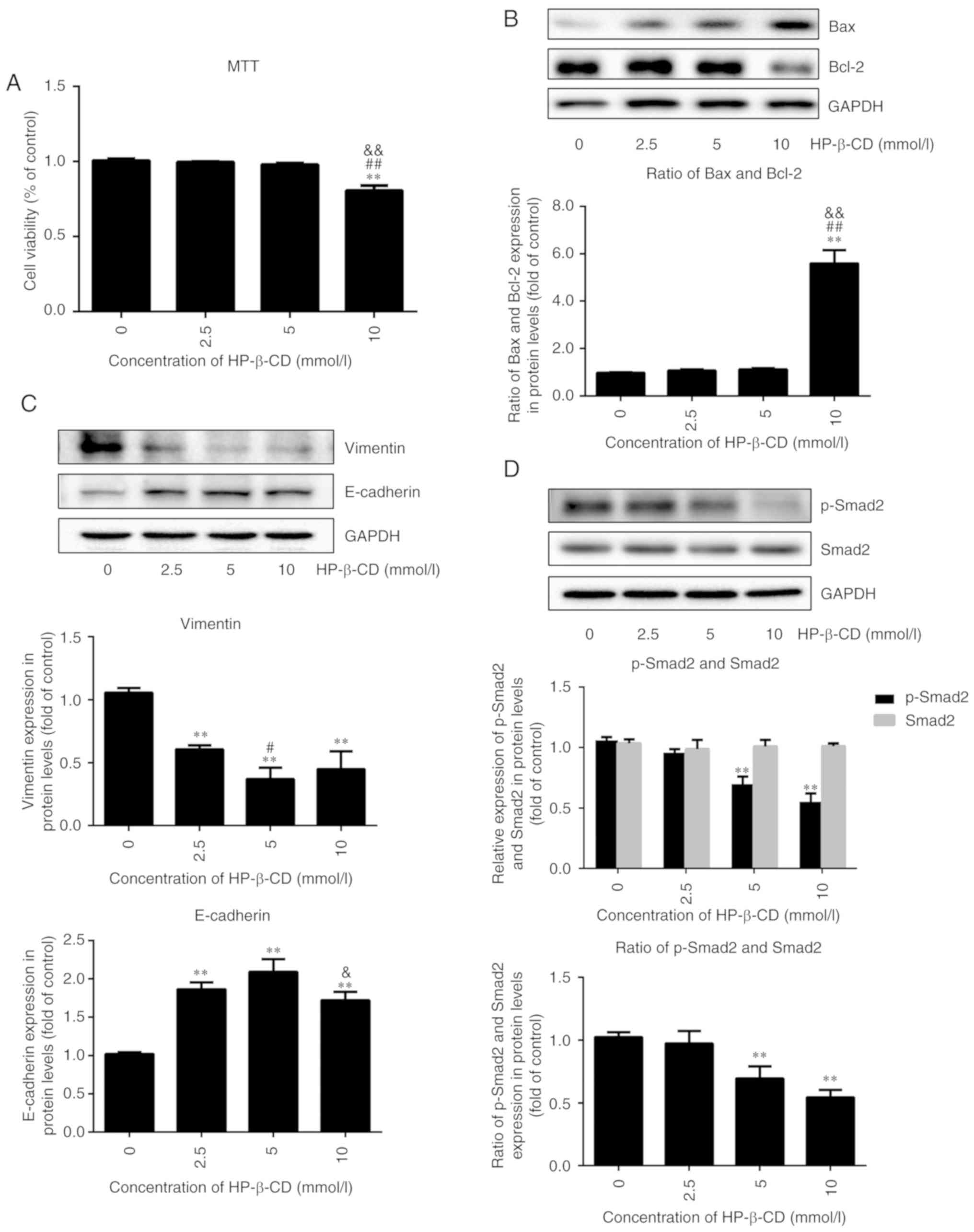 | Figure 1.HP-β-CD suppresses EMT features and
the TGF-β/Smad signaling pathway in MDA-MB-231 breast cancer cells.
MDA-MB-231 cells were incubated in the presence of HP-β-CD at
different concentrations (0, 2.5, 5 and 10 mmol/l). MTT was used to
detect the cell viability after 48 h, and western blotting was used
to measure the expression level of apoptosis, EMT and TGF-β/Smad
pathway-related proteins. (A) Viability of MDA-MB-231 cells. (B)
Expression of Bax and Bcl-2 in cells. (C) Expression of vimentin
and E-cadherin in cells. (D) Expression of p-Smad2 and Smad2 in
cells. Values are shown as the mean ± standard deviation (n=6 or
n=3). **P<0.01 vs. the control group; #P<0.05,
##P<0.01 vs. 2.5 mmol/l HP-β-CD group;
&P<0.05, &&P<0.01 vs. 5
mmol/l HP-β-CD group. HP-β-CD, hydroxypropyl-β-cyclodextrin; EMT,
epithelial-to-mesenchymal transition; TGF, transforming growth
factor; p-, phosphorylated. |
HP-β-CD inhibited EMT in breast cancer cells in a
dose-dependent pattern, with the greatest effect observed with
treatment of 5 mmol/l HP-β-CD, resulting in a 64% decrease in the
expression of vimentin and a 105% increase in E-cadherin expression
(Fig. 1C; P<0.001; n=3).
Therefore, 5 mmol/l HP-β-CD was chosen as the appropriate
concentration to influence EMT for subsequent experiments. In
addition, the expression of p-Smad2 was downregulated by HP-β-CD
(Fig. 1D; P<0.001; n=3), but
this had no effect on Smad2 expression. These results suggested
that p-Smad2 might be involved in regulating the HP-β-CD-induced
suppression of EMT in MDA-MB-231 cells.
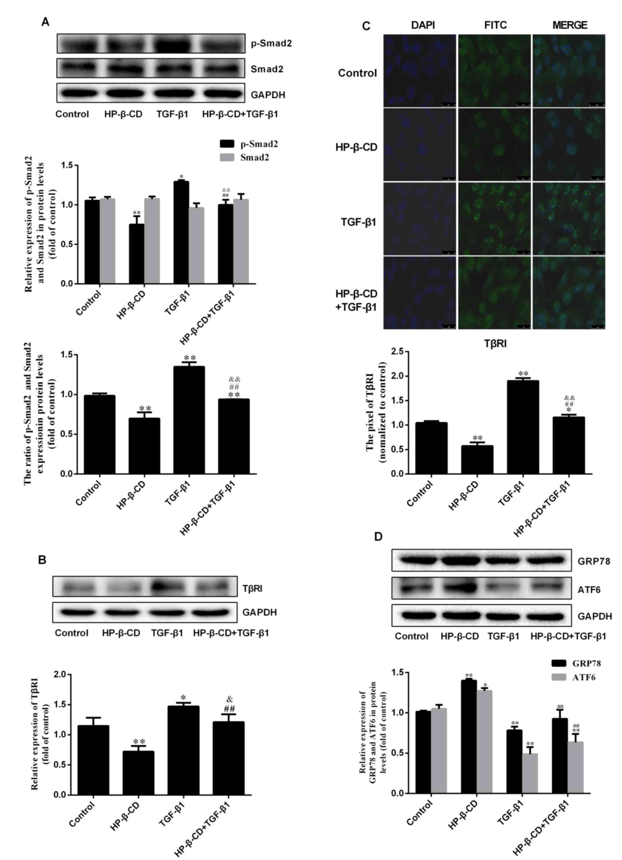
HP-β-CD can inhibit the TGF-β/Smad
signaling pathway
Consistent with the results above, treatment of 5
mmol/l HP-β-CD for 48 h significantly downregulated p-Smad2
(Fig. 2A; P<0.001; n=3)
expression but demonstrated no significant effect on Smad2
expression. However, TGF-β1 treatment upregulated p-Smad2
expression by 70.7%, with no significant changes in Smad2 (Fig. 2A; P<0.001; n=3). Western
blotting demonstrated that TGF-β1 treatment increased the
expression of TβRI by 28.3%; however, the combination of HP-β-CD
and TGF-β1 treatment diminished the expression of TβRI by 17.8%
compared to TGF-β1 treatment alone (Fig. 2B; P<0.05; n=3), which was
confirmed by immunofluorescence and confocal laser microscopy
(Fig. 2C; P<0.001; n=3).
Treatment of TGF-β1 alone downregulated the expression of the ER
stress markers GRP78 and ATF6, and HP-β-CD treatment reversed these
effects, increasing the expression of GRP78 and ATF6 by 18.8 and
37.6% respectively (Fig. 2D;
P<0.05; n=3). These results indicated that HP-β-CD regulates ER
stress.
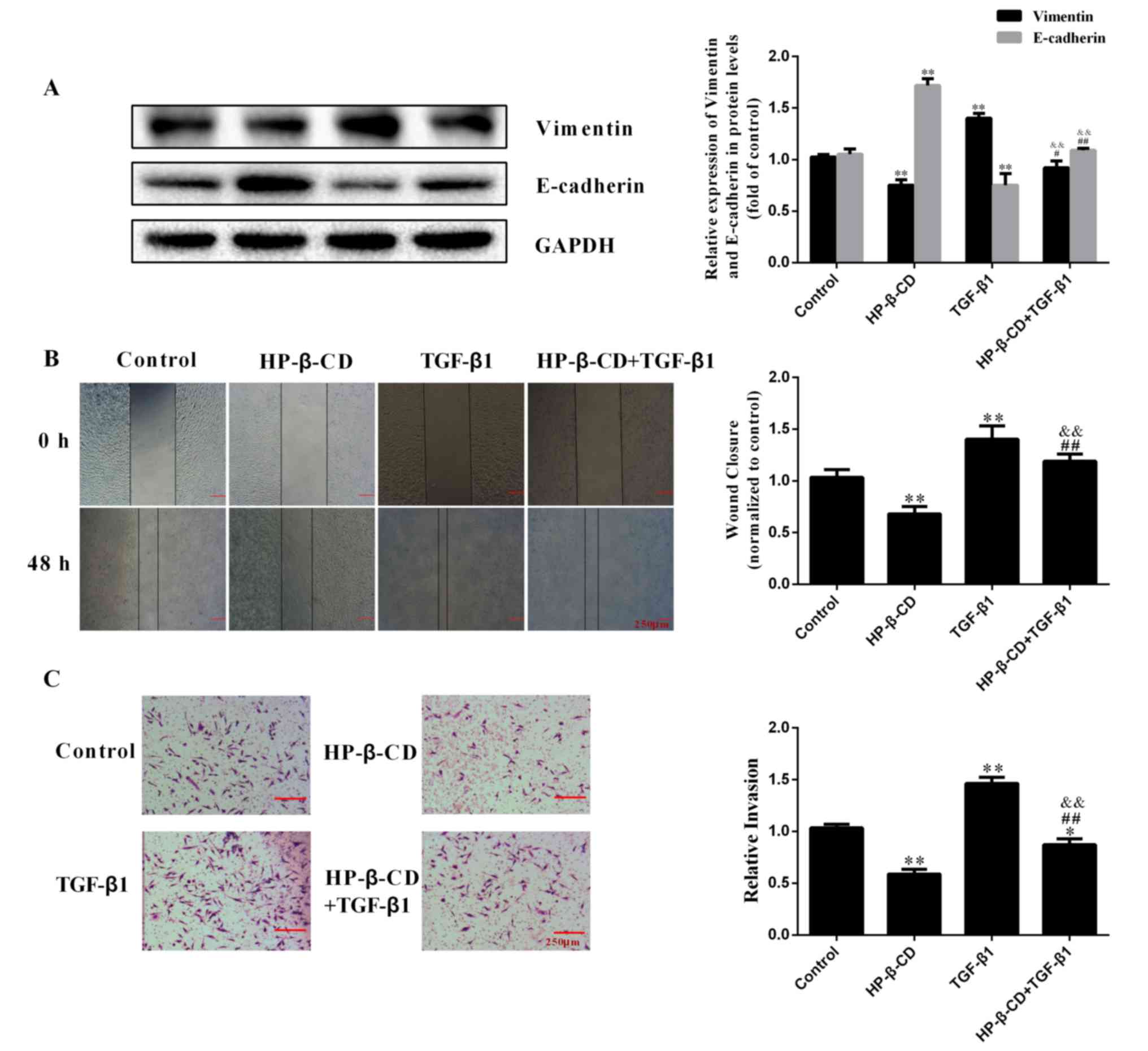
HP-β-CD inhibits the TGF-β1-induced
migration and invasion of MDA-MB-231 cells
HP-β-CD reversed the effects of TGF-β1 on EMT,
shifting MDA-MB-231 cells to a more epithelial phenotype,
downregulating vimentin expression by 35.9% while upregulating
E-cadherin expression by 28.5% (Fig.
3A; P<0.001; n=3). The wound healing assay demonstrated that
TGF-β1 treatment significantly increased the migration ability of
cells by 41.2%, which was significantly inhibited by HP-β-CD
(Fig. 3B). The invasive ability of
MDA-MB-231 cells was increased after 48 h of TGF-β1 treatment and
this effect was significantly inhibited with HP-β-CD pre-treatment
(Fig. 3C).
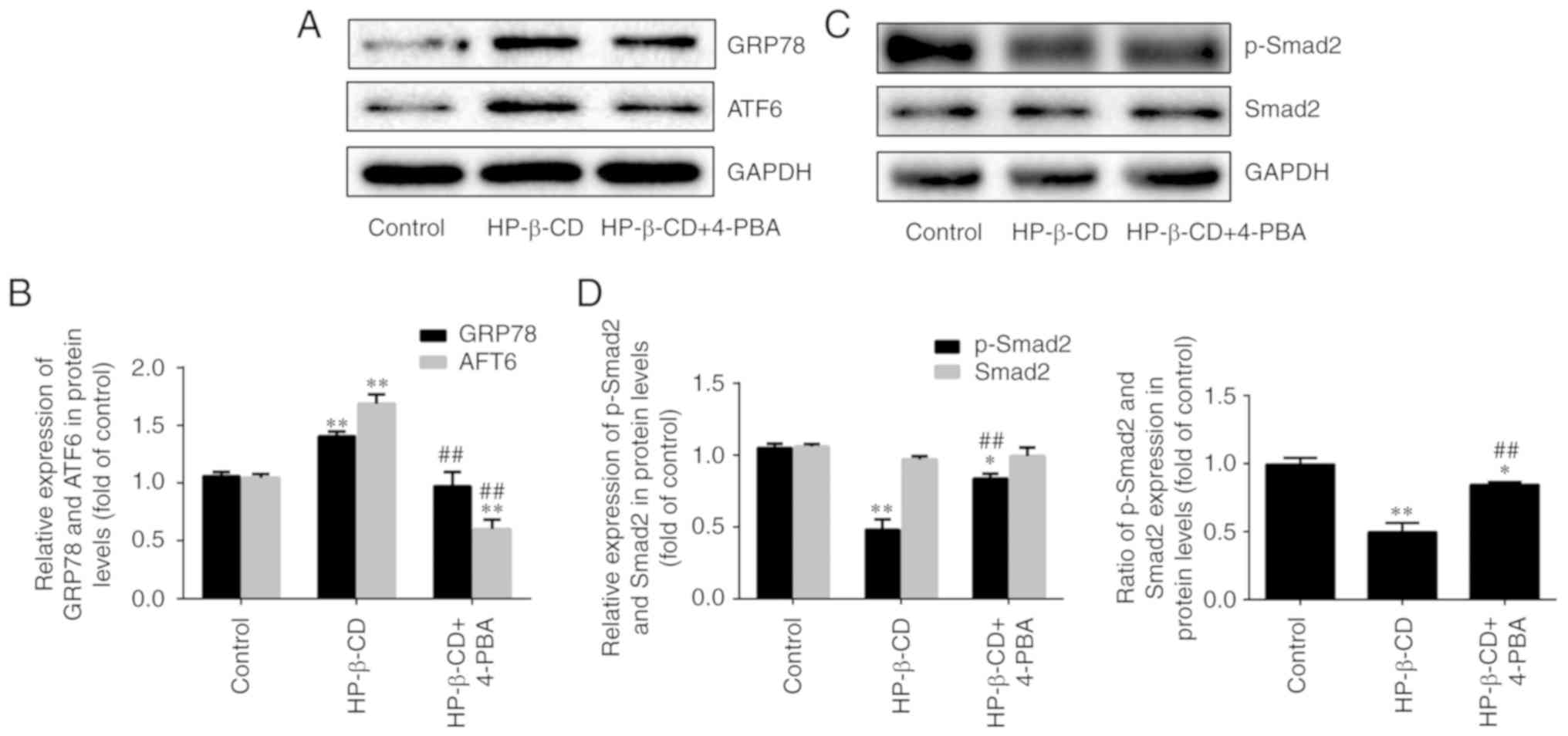
HP-β-CD activates ER stress in
MDA-MB-231 cells
Treatment of cells with HP-β-CD alone upregulated
the expression of ER stress-related proteins GRP78 and ATF6 by 37.7
and 61.8% respectively (Fig. 4A and
B; P<0.001; n=3). However, treatment with the ER stress
inhibitor 4-PBA blocked these effects, downregulating GRP78 and
ATF6 expression by 30.7 and 64.2% respectively (Fig. 4A and B; P<0.001; n=3). In
addition, p-Smad2 was downregulated by 54.3% after cells were
treated with HP-β-CD alone. Whereas after the combination of
HP-β-CD and 4-PBA treatment, it was upregulated by 77.1%.
Consistent with the experiments described above, no significant
changes in the expression of Smad2 were observed (Fig. 4C and D; P<0.001; n=3). These
data confirmed that HP-β-CD can activate ER stress.
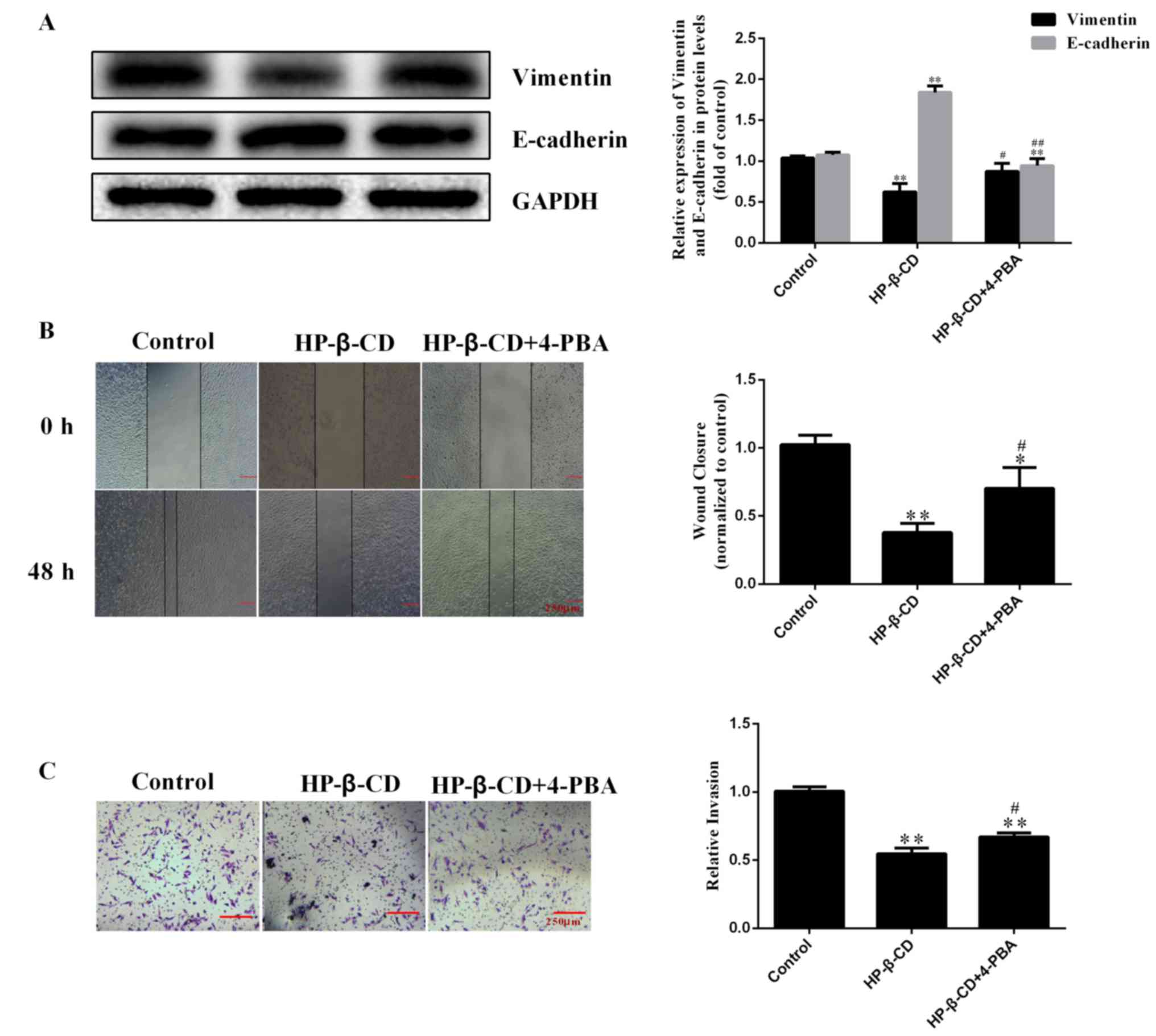
HP-β-CD attenuates the EMT via
activating ER stress in MDA-MB-231 cells
Inhibition of ER stress with 4-PBA also reversed the
HP-β-CD-induced inhibition of EMT, resulting in upregulation of
vimentin and downregulation of E-cadherin (Fig. 5A). Furthermore, the migratory
ability of cells was increased after 4-PBA treatment compared with
that detected under HP-β-CD treatment alone (Fig. 5B), and the inhibition of the
invasive ability of MDA-MB-231 cells was also reversed by 4-PBA
(Fig. 5C).
Discussion
The present study demonstrated that HP-β-CD can
suppress the EMT of breast cancer cells by activating ER stress.
MTT analysis demonstrated that 5 mmol/l HP-β-CD has no significant
effects on the viability of MDA-MB-231 cells, with no significant
change in the relative expression of Bax/Bcl-2. Meanwhile, it was
found that HP-β-CD suppressed EMT features in MDA-MB-231 cells by
downregulating TβRI expression, interfering with TGF-β1-induced
Smad2 phosphorylation, and thereby increasing the expression of
E-cadherin and decreasing that of vimentin. In addition, HP-β-CD
inhibited the migratory and invasive capacities of MDA-MB-231
cells. These effects were mediated through the induction of ER
stress by HP-β-CD, since blocking ER stress with 4-PBA, an
inhibitor of ER stress, promoted the migratory and invasive
capability of cells treated with HP-β-CD. Notably, TGF-β1 decreased
the expression levels of ER stress marker proteins, and 4-PBA
increased the expression of p-Smad2. Overall, these results
demonstrated that depletion of cholesterol in lipid rafts could
inhibit TGF-β1-induced EMT in MDA-MB-231 cells, which could
suppress metastasis.
Lipid raft is a signaling platform (8–10)
and can regulate many signaling pathways, therefore it serves vital
roles in many biological processes, including cytoskeleton
rearrangement, proliferation, and the migration and apoptosis of
various types of cancer cells (24–26).
We previously identified that sphingomyelin synthase 1 (SMS1)
overexpression could inhibit TGF-β1-induced EMT by increasing the
content of sphingomyelin in lipid rafts (13). Thus, the present study focused on
the effects of another important lipid raft component, cholesterol,
in the context of breast cancer. HP-β-CD can deplete cholesterol in
lipid rafts and this may affect the construction of lipid rafts and
then cancer cell viability by regulating the transmembrane
signaling (15,27). For example, Alawin et al
(28) found that antiproliferative
effects of γ-tocotrienol are associated with lipid raft disruption
in HER2-positive human breast cancer cells and Badana et al
(29). demonstrated that lipid
rafts disruption induces apoptosis by attenuating expression of
LRP6 and survivin in triple negative breast cancer. Although many
reports have suggested that the transmembrane signaling of the
TGF-β/Smad signaling pathway is dependent on lipid raft (30–33),
to the best of the authors' knowledge, this is the first study to
demonstrate its role in EMT regulated by TGF-β/Smad signaling
pathway.
Multiple signaling pathways are known to mediate
EMT, including TGF-β, Notch-, Hedgehog- and mitogen-activated
protein kinase-dependent pathways (34,35).
Among these pathways, the TGF-β-dependent pathway is considered to
be a primary inducer of EMT (5).
TGF-β receptors are internalized by clathrin-dependent endocytosis
as a key regulatory event in signal transduction (30,36).
Therefore, changing the main components of lipid rafts can
influence the localization of TGF-β receptors in the lipid
rafts/caveolae and the consequent signal transduction, as confirmed
in our previous study (13). The
present study further demonstrated that HP-β-CD negatively
regulated TβRI expression, probably by influencing the distribution
of TβRI in lipid rafts. These changes would then block the
TGF-β1-induced activation of the TGF-β/Smad signaling pathway to
suppress TGF-β1-induced EMT, as well as the corresponding migration
and invasion abilities of the cancer cells. Lipid composition and
membrane biophysical properties serve an important role in cell
movement (37). Notably, our
previous findings and those of the present study demonstrated that
changing the composition of lipid rafts by using HP-β-CD to deplete
cholesterol and overexpressing SMS1 to increase the content of
sphingomyelin had the same effect on EMT, which means that
cholesterol and sphingomyelin may serve opposite roles in the
process of EMT. It also identified that cholesterol and
sphingomyelin had the opposite roles in regulating the endothelial
dysfunction (data not shown). Therefore, the detailed mechanism
needs further investigations.
In the present study, TGF-β1 decreased the
expression levels of ER stress markers, which could be reversed by
HP-β-CD pre-treatment. In addition, 4-PBA activated the TGF-β/Smad
signaling pathway and inhibited the effects of HP-β-CD on EMT. The
role of ER stress in EMT is somewhat controversial. Huang et
al (21) demonstrated that
pterostilbene-induced ER stress could inhibit EMT in breast cancer,
and recent studies have pointed to a relationship between the
TGF-β/Smad signaling pathway and ER stress in a number of cell
lines, including A549 cells and podocytes (19,38)
suggesting that TGF-β1 can activate ER stress to promote the
process of EMT. The present study demonstrated that HP-β-CD could
block both the TGF-β/Smad signaling pathway and EMT, revealing a
positive association between TGF-β/Smad signaling pathway and EMT,
in line with the studies of Moon et al (38) and Yamashita et al (19). However, in contrast to these
reports, the present study found that HP-β-CD could induce ER
stress to attenuate the EMT, indicating a negative association, and
this is in line with Huang et al (21) and Dasgupta et al (39) who found that pterostilbene and
AECHL-1 suppressed the EMT by activating ER stress in breast
cancer. These apparent contradictory findings highlight the cell
type specificity of the effects of ER stress on EMT. In addition,
other ER stress inducers, such as tunicamycin, can activate ER
stress and then regulate the process of EMT; and inducing the ER
stress can alter many other signaling pathways, including the AKT
and MAPK signaling pathways (40)
and the Wnt/β-catenin signaling pathway (41), therefore, further studies are
required to compliment the present study. Although there are
several studies that do not include a 4-PBA alone group (41–44),
it would have been better if the present study had added a 4-PBA
group to overcome the influence of 4-PBA on the experimental
results. Collectively, the results of the present study suggested
that HP-β-CD regulates the EMT through inhibition of the TGF-β/Smad
signaling pathway, which then activates ER stress in MDA-MB-231
cells (Fig. 6). Since this can
attenuate EMT in MDA-MB-231 cells, this mechanism holds promise as
a treatment target to suppress the progression of breast cancer
prior to metastasis formation.
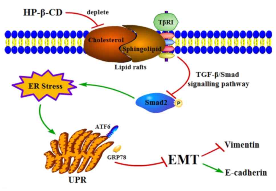 | Figure 6.Proposed mechanism of how HP-β-CD
attenuates EMT via ER stress in MDA-MB-231 breast cancer cells.
HP-β-CD can deplete cholesterol in lipid rafts then regulates TβRI
expression negatively to attenuate the TGF-β/Smad signaling
pathway, which then activates ER stress, thereby leading to
suppression of EMT. HP-β-CD, hydroxypropyl-β-cyclodextrin; EMT,
epithelial-to-mesenchymal transition; ER, endoplasmic reticulum;
TβRI, TGF receptor; TGF, transforming growth factor; GRP78, binding
immunoglobulin protein antibody; ATF6, activating transcription
factor 6; UPR, unfolded protein response. |
Acknowledgements
Not applicable.
Funding
The present study was supported by grants from The
National Natural Science Foundation of China (grant no. 81560151)
and The Jiangxi Provincial Department of Science and Technology
(grant no. 20181BAB205022).
Availability of data and materials
The datasets used and/or analyzed during the current
study are available from the corresponding author on reasonable
request.
Authors' contributions
YW and YZ performed the experiments, analyzed data
and were major contributors in writing the manuscript. LC, XH and
ZH also analyzed and interpreted the data of the manuscript. TW and
LW were responsible for the design and drafting of the manuscript.
NY conceived the study and designed the experiments. All authors
read and approved the final manuscript.
Ethics approval and consent to
participate
Not applicable.
Patient consent for publication
Not applicable.
Competing interests
The authors declare that they have no competing
interests.
References
|
1
|
Wittkowski KM, Dadurian C, Seybold MP, Kim
HS, Hoshino A and Lyden D: Complex polymorphisms in endocytosis
genes suggest alpha-cyclodextrin as a treatment for breast cancer.
PLoS One. 13:e01990122018. View Article : Google Scholar : PubMed/NCBI
|
|
2
|
Li L and Li W: Epithelial-mesenchymal
transition in human cancer: Comprehensive reprogramming of
metabolism, epigenetics, and differentiation. Pharmacol Ther.
150:33–46. 2015. View Article : Google Scholar : PubMed/NCBI
|
|
3
|
Ruben B and Gerhard C: The relevance of
EMT in breast cancer metastasis: Correlation or causality? FEBS
Lett. 589:1577–1587. 2015. View Article : Google Scholar : PubMed/NCBI
|
|
4
|
Das V, Bhattacharya S, Chikkaputtaiah C,
Hazra S and Pal M: The basics of epithelial-mesenchymal transition
(EMT): A study from a structure, dynamics, and functional
perspective. J Cell Physiol. Feb 5–2019.(Epub ahead of print).
View Article : Google Scholar
|
|
5
|
Heldin CH, Landström M and Moustakas A:
Mechanism of TGF-beta signaling to growth arrest, apoptosis, and
epithelial-mesenchymal transition. Curr Opin Cell Biol. 21:166–176.
2009. View Article : Google Scholar : PubMed/NCBI
|
|
6
|
Lamouille S, Xu J and Derynck R: Molecular
mechanisms of epithelial-mesenchymal transition. Nat Rev Mol Cell
Biol. 15:178–196. 2014. View
Article : Google Scholar : PubMed/NCBI
|
|
7
|
Sánchez-Tilló E, Liu Y, de Barrios O,
Siles L, Fanlo L, Cuatrecasas M, Darling DS, Dean DC, Castells A
and Postigo A: EMT-activating transcription factors in cancer:
Beyond EMT and tumor invasiveness. Cell Mol Life Sci. 69:3429–3456.
2012. View Article : Google Scholar : PubMed/NCBI
|
|
8
|
Beloribi-Djefaflia S, Vasseur S and
Guillaumond F: Lipid metabolic reprogramming in cancer cells.
Oncogenesis. 5:e1892016. View Article : Google Scholar : PubMed/NCBI
|
|
9
|
Kraft ML: Plasma membrane organization and
function: Moving past lipid rafts. Mol Biol Cell. 24:2765–2768.
2013. View Article : Google Scholar : PubMed/NCBI
|
|
10
|
Li YC, Park MJ, Ye SK, Kim CW and Kim YN:
Elevated levels of cholesterol-rich lipid rafts in cancer cells are
correlated with apoptosis sensitivity induced by
cholesterol-depleting agents. Am J Pathol. 168:1107–1118. 2006.
View Article : Google Scholar : PubMed/NCBI
|
|
11
|
Razani B, Zhang XL, Bitzer M, von
Gersdorff G, Böttinger EP and Lisanti MP: Caveolin-1 regulates
transforming growth factor (TGF)-beta/SMAD signaling through an
interaction with the TGF-beta type I receptor. J Biol Chem.
276:6727–6738. 2001. View Article : Google Scholar : PubMed/NCBI
|
|
12
|
Le Roy C and Wrana JL: Clathrin- and
non-clathrin-mediated endocytic regulation of cell signalling. Nat
Rev Mol Cell Biol. 6:112–126. 2005. View
Article : Google Scholar : PubMed/NCBI
|
|
13
|
Liu S, Hou H, Zhang P, Wu Y, He Q, Li H
and Yan N: Sphingomyelin synthase 1 regulates the
epithelialtomesenchymal transition mediated by the TGF-β/Smad
pathway in MDA-MB-231 cells. Mol Med Rep. 19:1159–1167.
2019.PubMed/NCBI
|
|
14
|
Maxfield FR and Tabas I: Role of
cholesterol and lipid organization in disease. Nature. 438:612–621.
2005. View Article : Google Scholar : PubMed/NCBI
|
|
15
|
Kline MA, O'Connor Butler ES, Hinzey A,
Sliman S, Kotha SR, Marsh CB, Uppu RM and Parinandi NL: A simple
method for effective and safe removal of membrane cholesterol from
lipid rafts in vascular endothelial cells: Implications in
oxidant-mediated lipid signaling. Methods Mol Biol. 610:201–2011.
2010. View Article : Google Scholar : PubMed/NCBI
|
|
16
|
Engin F and Hotamisligil GS: Restoring
endoplasmic reticulum function by chemical chaperones: An emerging
therapeutic approach for metabolic diseases. Diabetes Obes Metab.
12:108–115. 2010. View Article : Google Scholar : PubMed/NCBI
|
|
17
|
Ron D and Walter P: Signal integration in
the endoplasmic reticulum unfolded protein response. Nat Rev Mol
Cell Biol. 8:519–529. 2007. View
Article : Google Scholar : PubMed/NCBI
|
|
18
|
Wang S and Kaufman R: The impact of the
unfolded protein response on human disease. J Cell Biol.
197:857–867. 2012. View Article : Google Scholar : PubMed/NCBI
|
|
19
|
Yamashita M, Ogasawara M, Kawasaki Y,
Niisato M, Saito H, Kasai S, Maesawa C, Maemondo M and Yamauchi K:
Deficiency of protein-L-isoaspartate (D-aspartate)
O-methyl-transferase expression under endoplasmic reticulum stress
promotes epithelial mesenchymal transition in lung adenocarcinoma.
Oncotarget. 9:13287–13300. 2018. View Article : Google Scholar : PubMed/NCBI
|
|
20
|
Chen CA, Chang JM, Chang EE, Chen HC and
Yang YL: Crosstalk between transforming growth factor-beta1 and
endoplasmic reticulum stress regulates alpha-smooth muscle cell
actin expression in podocytes. Life Sci. 209:9–14. 2018. View Article : Google Scholar : PubMed/NCBI
|
|
21
|
Huang Y, Du J, Mi Y, Li T, Gong Y, Ouyang
H and Hou Y: Long non-coding RNAs contribute to the inhibition of
proliferation and EMT by pterostilbene in human breast cancer. Fron
Oncol. 8:6292018. View Article : Google Scholar
|
|
22
|
Badana A, Chintala M, Varikuti G, Pudi N,
Kumari S, Kappala RV and Malla RR: Lipid raft integrity is required
for survival of triple negative breast cancer cells. J Breast
Cancer. 19:372–384. 2016. View Article : Google Scholar : PubMed/NCBI
|
|
23
|
Lee HM, Kang JH, Shin JM, Lee SA and Park
HO: Chemical chaperone of endoplasmic reticulum stress inhibits
epithelial-mesenchymal transition induced by TGF-β1 in airway
epithelium via the c-src pathway. Mediators Inflamm. 2017:1–9.
2017. View Article : Google Scholar
|
|
24
|
Deans JP, Li H and Polyak MJ:
CD20-mediated apoptosis: Signalling through lipid rafts.
Immunology. 107:176–182. 2010. View Article : Google Scholar
|
|
25
|
Gómez-Llobregat J, Buceta J and Reigada R:
Interplay of cytoskeletal activity and lipid phase stability in
dynamic protein recruitment and clustering. Sci Rep. 3:26082013.
View Article : Google Scholar : PubMed/NCBI
|
|
26
|
Wang R, Bi J, Ampah KK, Ba X, Liu W and
Zeng X: Lipid rafts control human melanoma cell migration by
regulating focal adhesion disassembly. Biochim Biophys Acta.
1833:3195–3205. 2013. View Article : Google Scholar : PubMed/NCBI
|
|
27
|
Head BP, Patel HH and Insel PA:
Interaction of membrane/lipid rafts with the cytoskeleton: Impact
on signaling and function: Membrane/lipid rafts, mediators of
cytoskeletal arrangement and cell signaling. Biochim Biophys Acta.
1838:532–545. 2014. View Article : Google Scholar : PubMed/NCBI
|
|
28
|
Alawin OA, Ahmed RA, Ibrahim BA, Briski KP
and Sylvester PW: Antiproliferative effects of γ-tocotrienol are
associated with lipid raft disruption in HER2-positive human breast
cancer cells. J Nutr Biochem. 27:266–277. 2015. View Article : Google Scholar : PubMed/NCBI
|
|
29
|
Badana AK, Chintala M, Gavara MM, Naika S,
Kumari S, Kappala VR, Iska BR and Malla RR: Lipid rafts disruption
induces apoptosis by attenuating expression of LRP6 and survivin in
triple negative breast cancer. Biomed Pharmacother. 97:359–369.
2017. View Article : Google Scholar : PubMed/NCBI
|
|
30
|
Mitchell H, Choudhury A, Pagano RE and
Leof EB: Ligand-dependent and -independent transforming growth
factor-beta receptor recycling regulated by clathrin-mediated
endocytosis and Rab11. Mol Biol Cell. 15:4166–4178. 2004.
View Article : Google Scholar : PubMed/NCBI
|
|
31
|
Le Roy C and Wrana JL: Clathrin- and
non-clathrin-mediated endocytic regulation of cell signalling. Nat
Rev Mol Cell Biol. 6:112–126. 2005. View
Article : Google Scholar : PubMed/NCBI
|
|
32
|
Midgley AC, Rogers M, Hallett MB, Clayton
A, Bowen T, Phillips AO and Steadman R: Transforming growth
factor-β1 (TGF-β1) stimulated fibroblast to myofibroblast
differentiation is mediated by hyaluronan (HA)-facilitated
epidermal growth factor receptor (EGFR) and CD44 co-localization in
lipid rafts. J Biol Chem. 288:14824–14838. 2013. View Article : Google Scholar : PubMed/NCBI
|
|
33
|
Chen YG: Endocytic regulation of TGF-beta
signaling. Cell Res. 19:58–70. 2009. View Article : Google Scholar : PubMed/NCBI
|
|
34
|
Derynck R, Muthusamy BP and Saeteurn KY:
Signaling pathway cooperation in TGF-β-induced
epithelial-mesenchymal transition. Curr Opin Cell Biol. 31:56–66.
2014. View Article : Google Scholar : PubMed/NCBI
|
|
35
|
Takebe N, Warren RQ and Ivy SP: Breast
cancer growth and metastasis: Interplay between cancer stem cells,
embryonic signaling pathways and epithelial-to-mesenchymal
transition. Breast Cancer Res. 13:211. 2011. View Article : Google Scholar : PubMed/NCBI
|
|
36
|
Conner SD and Schmid SL: Regulated portals
of entry into the cell. Nature. 422:37–44. 2003. View Article : Google Scholar : PubMed/NCBI
|
|
37
|
Ewers H and Helenius A: Lipid-mediated
endocytosis. Cold Spring Harb Perspect Biol. 3:a0047212011.
View Article : Google Scholar : PubMed/NCBI
|
|
38
|
Moon SY, Kim HS, Nho KW, Jang YJ and Lee
SK: Endoplasmic reticulum stress induces epithelial-mesenchymal
transition through autophagy via activation of c-Src kinase.
Nephron. Exp Nephrol. 126:127–140. 2014. View Article : Google Scholar
|
|
39
|
Dasgupta A, Sawant MA, Kavishwar G,
Lavhale M and Sitasawad S: AECHL-1 targets breast cancer
progression via inhibition of metastasis, prevention of EMT and
suppression of cancer stem cell characteristics. Sci Rep.
6:380452016. View Article : Google Scholar : PubMed/NCBI
|
|
40
|
Huang S, Wang D, Zhang S, Huang X, Wang D,
Ljaz M and Shi Y: Tunicamycin potentiates paclitaxel-induced
apoptosis through inhibition of PI3K/AKT and MAPK pathways in
breast cancer. Cancer Chemother Pharmacol. 80:685–696. 2017.
View Article : Google Scholar : PubMed/NCBI
|
|
41
|
He Z, He X, Liu M, Hua L, Wang T, Liu Q,
Chen L and Yan N: Simvastatin attenuates H2O2-induced endothelial
cell dysfunction by reducing endoplasmic reticulum stress.
Molecules. 24(pii): E17822019. View Article : Google Scholar : PubMed/NCBI
|
|
42
|
Wu F, Xu K, Liu L, Zhang K, Xia L, Zhang
M, Teng C, Tong H, He Y, Xue Y, et al: Vitamin B12 enhances nerve
repair and improves functional recovery after traumatic brain
injury by inhibiting ER stress-induced neuron injury. Front
Pharmacol. 10:4062019. View Article : Google Scholar : PubMed/NCBI
|
|
43
|
Wang EH, Yao SQ, Tao L and Xi JY: Sodium
4-phenylbutyrate attenuates high-fat diet-induced impaired
spermatogenesis. Biomed Environ Sci. 12:876–882. 2018.
|
|
44
|
Guo Q, Hu H, Zhou Y, Yan Y, Wei X, Fan X,
Yang D, He H, Oh Y, Chen K, et al: Glucosamine induces increased
musclin gene expression through endoplasmic reticulum
stress-induced unfolding protein response signaling pathways in
mouse skeletal muscle cells. Food Chem Toxicol. 125:95–105. 2018.
View Article : Google Scholar : PubMed/NCBI
|