Introduction
Hepatocellular carcinoma (HCC) is a primary liver
malignancy accounting for 80–90% of all liver cancer (1). According to the latest global cancer
data, the incidence of HCC has rapidly risen with ~782,000 deaths
in 2018 (2). The majority of HCC
is resistant to treatment as it is often diagnosed at advanced
stages of disease, so the overall median survival is <1 year
(3). To the best of the authors'
knowledge, sorafenib and levatinib are the only first-line systemic
therapeutic agents available for the treatment of advanced HCC.
Several second-line drugs, including regorafenib, ramucirumab and
cabozantinib, have been approved for patients with HCC who have
previously been treated with sorafenib, but the overall survival
(OS) rate remains poor (4,5).
Immune checkpoint blockade therapies have emerged as
potential treatments in cancer immunotherapy (6). The cytotoxic T-cell mediated immune
response is accompanied by co-stimulatory signals and co-inhibitory
signals or immune checkpoints (7,8). The
immune checkpoint molecules are often overexpressed or activated in
the tumor microenvironment, thus resulting in immune evasion by
tumor cells (7–9). Immune checkpoint inhibitors (ICIs)
that block programmed cell death protein-1 (PD-1) or its ligand,
programmed death receptor ligand (PD-L)1, have shown therapeutic
potential in several cancer types, including HCC (10,11).
Anti-PD-1 ICIs, nivolumab and pembrolizumab, have been approved by
The Food and Drug Administration for the treatment of HCC in
patients where treatment using sorafenib has failed (12). There are a number of ongoing
clinical trials on ICIs targeting PD-1 (nivolumab and
pembrolizumab) and PD-L1 (atezolizumab) in HCC (7,13).
Due to the success of anti-PD-1/PD-L1 immunotherapy clinical
studies, understanding the underlying mechanisms regulating PD-L1
expression has attracted increasing attention. Previous research by
the authors and other researchers demonstrated an increased
expression of other immune checkpoint molecules in HCC (8,14).
However, the mechanisms regulating the expression of these and
other immune checkpoint molecules in HCC remains unclear (8).
A key process in HCC progression is
epithelial-to-mesenchymal transition (EMT). EMT is a complex
molecular and cellular process that allows epithelial cells to gain
mesenchymal features, including migration, invasiveness and
increased resistance to immune evasion (15,16).
Mesenchymal-to-epithelial transition (MET) is the reverse process
of EMT that offers phenotypic plasticity for conversion of
mesenchymal cells to epithelial derivatives (15). EMT status is closely associated
with cancer metastasis, stemness, immune escape and drug resistance
in HCC (17,18). It was previously identified that
EMT is involved in the regulation of PD-L1 in several cancer types,
including breast cancer (19,20),
lung cancer (17,21), pancreatic cancer (22), esophageal cancer (23) and salivary adenoid cystic carcinoma
(24). The authors previously
demonstrated that EMT is associated with PD-L1 expression in
patients with HCC (8).
In the present study, it was hypothesized that EMT
is associated with the ability of HCC tumor cells to escape
detection and destruction by the immune response, by regulating the
expression of immune checkpoint molecules, such as PD-L1. In the
present study, the changes in immune checkpoint expression during
the EMT process were examined by incorporating a reversible model
of EMT based on tumor necrosis factor (TNF)-α treatment of the HCC
cell lines Hep3B and PLC/PRF/5. TNF-α was selected as an inducer of
EMT, as it has been previously demonstrated to play an important
role in promoting EMT in HCC (25). This in vitro system may
provide an improved understanding of the modulation of immune
checkpoints during both EMT and MET.
Materials and methods
Cell culture and reagents
The human HCC cell line Hep3B was provided by
Professor V. Nathan Subramaniam, The Queensland University of
Technology. The human HCC cell line PLC/PRF/5 was purchased from
CellBank Australia (cat. no. 85061113). Both cell lines were
mycoplasma-tested using the MycoAlert™ mycoplasma detection kit
(Lonza Group, Ltd.) and cultured in DMEM (Thermo Fisher Scientific,
Inc.) with 10% FBS (Gibco; Thermo Fisher Scientific, Inc.) and 1%
penicillin/streptomycin (Thermo Fisher Scientific, Inc.), and
incubated at 37°C under a humidified atmosphere with 5%
CO2 in air (26). The
cytokine TNF-α was purchased from PeproTech, Inc.
EMT reversal assay
An EMT reversal assay was performed to determine the
association between EMT and immune checkpoint expression. Firstly,
EMT was induced by using culture medium with 20 ng/ml TNF-α for 3
days at 37°C and then reversal of EMT was induced by changing the
culture medium without TNF-α for the next 3 days.
RNA extraction and cDNA synthesis
RNA was isolated from Hep3B and PLC/PRF/5 cells, as
previously described, using Isolate II Bioline RNA synthesis kit
(Bioline), according to the manufacturer's protocol (26). The quantity and purity of RNA was
confirmed using the NanoDrop™ 2000c spectrophotometer (Thermo
Fisher Scientific, Inc.). cDNA was synthesized by reverse
transcribing 1 µg RNA into cDNA using the Bioline SensiFAST cDNA
synthesis kit (Bioline) with the following thermocycling
conditions: Primer annealing at 25°C for 10 min, reverse
transcription at 42°C for 15 min, inactivation at 85°C for 5 min
and a final extension 4°C for 30 min.
Reverse transcription-quantitative
(RT-q)PCR
RT-qPCR was performed using SensiFast™
SYBR® Lo-ROX kit (Bioline) on a ViiA7 Real-Time PCR
system (Applied Biosystems; Thermo Fisher Scientific, Inc.), as
previously described (26). A
three-step cycle procedure was applied with 40 cycles of the
following conditions: 95°C for 5 sec, 63°C for 20 sec and 75°C for
20 sec. β-actin was used as an internal control. The primer
sequences used are listed in Table
I. Data were analyzed using the 2−ΔΔCq method where
β-actin was assigned as the housekeeping gene. The results are
expressed as relative mRNA expression to the control (27).
 | Table I.List of primers for reverse
transcription-quantitative PCR. |
Table I.
List of primers for reverse
transcription-quantitative PCR.
| Gene | Primer sequence,
5′-3′ |
|---|
| β-actin | F:
CCAACCGCGAGAAGATGA |
|
| R:
CCAGAGGCGTACAGGGATAG |
| E-cadherin | F:
AGGCCAAGCAGCAGTACATT |
|
| R:
ATTCACATCCAGCACATCCA |
| N-cadherin | F:
TCCTTGCTTCTGACAATGGA |
|
| R:
TTCGCAAGTCTCTGCCTCTT |
| Occludin | F:
TAGTCAGATGGGGGTGAAGG |
|
| R:
CATTTATGATGAGCAGCCCC |
| Vimentin | F:
CTTCAGAGAGAGGAAGCCGA |
|
| R:
ATTCCACTTTGCGTTCAAGG |
| Snai2 | F:
TGGTTGCTTCAAGGACACAT |
|
| R:
GTTGCAGTGAGGGCAAGAA |
| Fibronectin | F:
CAGTGGGAGACCTCGAGAAG |
|
| R:
TCCCTCGGAACATCAGAAAC |
| PD-L1 | F:
TGCCGACTACAAGCGAATTACTG |
|
| R:
CTGCTTGTCCAGATGACTTCGG |
| PD-L2 | F:
ACCGTGAAAGAGCCACTTTG |
|
| R:
GCGACCCCATAGATGATTATGC |
| CD73 | F:
TTGGAAATTTGGCCTCTTTG |
|
| R:
ACTTCATGAACGCCCTGC |
| B7-H3 | F:
CTGGCTTTCGTGTGCTGGAGAA |
|
| R:
GCTGTCAGAGTGTTTCAGAGGC |
| VISTA | F:
AGATGCACCATCCAACTGTGTGG |
|
| R:
AGGCAGAGGATTCCTACGATGC |
| VTCN1 | F:
CTCACAGATGCTGGCACCTACA |
|
| R:
GCAAGGTCTCTGAGCTGGCATT |
Western blot analysis
Cells were seeded (2×10⁵ cells/well) in
6-well plates and subsequently lysed using RIPA buffer (Thermo
Fisher Scientific, Inc.) with complete protease inhibitors and
PhosSTOP™ phosphatase inhibitors (both purchased from Roche
Diagnostics) on ice to extract the total protein. The total protein
was measured with a Pierce™ BCA protein assay kit (Thermo Fisher
Scientific, Inc.). A total of 10 µg protein was used for 4%
SDS-PAGE, following which proteins were transferred to a PVDF
membrane. After blocking with 5% skimmed milk in Tris-buffered
saline containing 0.1% Tween-20 at room temperature for 1 h, the
membranes were incubated with primary antibodies at 4°C overnight.
An enhanced chemiluminescence reagent, SuperSignal™ West Femto
Maximum Sensitivity Substrate (Thermo Fisher Scientific, Inc.), was
used to detect the protein adhering to the membranes following
incubation with horseradish peroxidase-conjugated secondary
antibodies at room temperature for 1 h. All antibodies used are
listed in Table II, including the
dilutions, cat. nos. and suppliers. GAPDH was selected as the
housekeeping control in each group. The images were captured with
ImageQuant™ LAS 500 and quantified with Image Studio™ Lite v5.2
software (LI-COR Biosciences).
| Table II.List of antibodies. |
Table II.
List of antibodies.
| A, Primary
antibodies |
|---|
|
|---|
|
|
|
| Dilution |
|---|
|
|
|
|
|
|---|
| Name | Cat. no. | Manufacturer | Western
blotting |
Immuno-fluorescence |
|---|
| E-cadherin | ab76055 | Abcam | 1:1,000 | 1:100 |
| N-cadherin | SC-53488 | Santa Cruz
Biotechnology, Inc. | 1:200 | – |
| Occludin | SC-133255 | Santa Cruz
Biotechnology, Inc. | 1:200 | 1:50 |
| Vimentin | ab92547 | Abcam | 1:1,000 | 1:600 |
| CD73 | ab133582 | Abcam | 1:250 |
|
| CD73 | ab175396 | Abcam | – | 1:100 |
| PD-L1 | ab238697 | Abcam | 2 µg/ml | 2 µg/ml |
| B7-H3 | 14058S | Cell Signaling
Technology, Inc. | 1:1,000 | – |
| PD-L2 | 82723S | Cell Signaling
Technology, Inc. | 1:1,000 | – |
| GAPDH | MAB376 | EMD Millipore |
1:100,000 | – |
|
| B, Secondary
antibody |
|
|
|
|
|
Dilution |
|
|
|
|
|
| Name | Cat.
no. |
Manufacturer | Western
blotting |
Immuno-fluorescence |
|
| Goat anti-mouse
HRP | 6421040 | Invitrogen; Thermo
Fisher Scientific, Inc. | 1:50,000 | – |
| Goat anti-rabbit
HRP | 656120 | Invitrogen; Thermo
Fisher Scientific, Inc. | 1:50,000 | – |
| Alexafluor 488
anti-mouse | A21202 | Thermo Fisher
Scientific, Inc. | – | 1:200 |
| Alexafluor 594
anti-rabbit | A21207 | Thermo Fisher
Scientific, Inc. | – | 1:200 |
Immunofluorescence
Cells (5×10³ cells/well) were cultured in
8-well tissue culture treated chamber slides. Cells were washed
with 1X PBS and then fixed with 4% paraformaldehyde (Thermo Fisher
Scientific, Inc.) at room temperature for 20 min followed by
treatment with 0.1% Triton X-100 (Sigma-Aldrich; Merck KGaA). The
cells were then blocked with 5% FBS at room temperature for 1 h,
followed by overnight incubation with primary antibodies at 4°C.
The cells were incubated with secondary antibodies at room
temperature for 1 h, followed by 10 min incubation with DAPI
(Thermo Fisher Scientific, Inc.) at room temperature. All
antibodies used are listed in Table
II, including the dilutions, cat. nos. and suppliers. The
slides were then mounted in ProLong® Diamond (Thermo
Fisher Scientific, Inc.) for observation with a Nikon C2 confocal
microscope (magnification, ×40), and captured and analyzed with
NIS-Elements-AR software (version 5.01.00; Nikon Corporation).
SurvExpress bioinformatics tool
An online tool, SurvExpress, was used to generate
survival analysis using datasets of patients with HCC, as
previously described (8,28) A total of six publically available
datasets of patients with HCC were used: Hoshida Golub Liver
GSE10143 (162 patients), Tsuchiya Rusyn Liver GSE17856 (95
patients), Hoshida Golub Liver GSE10186 (118 patients), Liver
Hepatocellular Carcinoma The Cancer Genome Atlas (TCGA; 12
patients), TCGA-Liver-Cancer (422 patients) and LIHC-TCGA-Liver
hepatocellular carcinoma June 2016 (361 patients) (8,28–31).
The gene expression of immune checkpoint molecules in combination
with TNF-α were examined and their association with the survival of
patients was analyzed (Cox regression analyses) using the patient
database with patient survival information (8). The survival times were estimated
using Kaplan-Meier curves (8).
Statistical analysis
All experiments were repeated at least three times
and representative results are presented. Statistical analyses were
performed using Prism software version 8.00 (GraphPad Software,
Inc.). Data for dose concentration and time course experiments were
analyzed using one-way ANOVA followed by Dunnet's multiple
comparisons test. Comparisons of TNF-α-induced EMT and MET
following reversal assay were performed using ANOVA followed by
Sidak's multiple comparisons test. Gene expression differences
between control and TNF-α treated cells were analyzed using
Student's t-test. The results are presented as the mean ± SEM.
Error bars indicate SEM. P<0.05 was considered to indicate a
statistically significant difference.
For survival analyses using SurvExpress, a log-rank
test was used for testing the P-value of survival curves, and
deviance residuals were applied for the correlation coefficient
(8,32). Moreover, the hazard ratio (HR)
between the groups were estimated using another Cox model (8).
Results
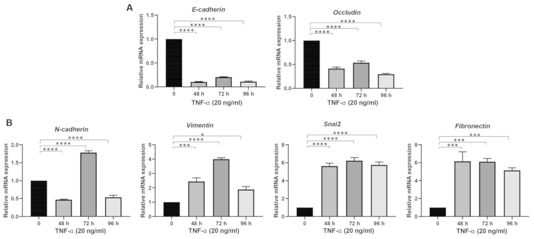
TNF-α induces EMT in human HCC
cells
Several cytokines are known to induce EMT in HCC
cells, including TNF-α. In order to induce EMT, Hep3B and PLC/PRF/5
cells were treated with various concentrations of TNF-α for 72 h to
assess EMT induction. EMT induced by TNF-α in Hep3B cells was
determined by downregulation of epithelial markers (E-cadherin and
Occludin) and upregulation of mesenchymal markers [N-cadherin,
Vimentin, snail family transcriptional repressor 2 (Snai2) and
Fibronectin]. The optimal EMT effects of TNF-α were observed at 20
ng/ml (Fig. 1). Thus, the
concentration of 20 ng/ml was selected for all further studies.
Furthermore, Hep3B cells were treated with 20 ng/ml TNF-α at
various time-points to observe optimal EMT induction. The qPCR
results showed that TNF-α treatment at various time-points was able
to induce EMT in Hep3B cells (Fig.
2). At 72 h, robust EMT marker changes were observed;
therefore, this time-point was selected for further study.
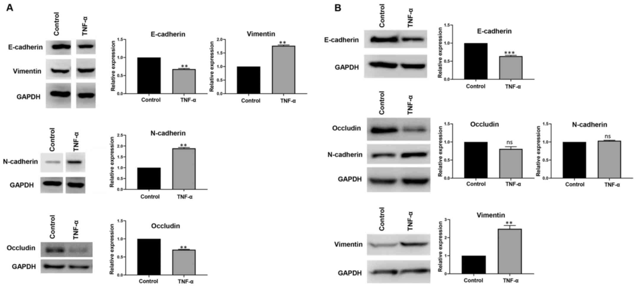
Similarly, robust EMT in PLC/PRF/5 cells treated with 20 ng/ml of
TNF-α at 72 h was observed. EMT induction was demonstrated by the
downregulation of epithelial markers (E-cadherin and Occludin) and
an upregulation of mesenchymal markers (N-cadherin and Vimentin;
Fig. S1). No expression of Snai2
and Fibronectin was detected in this cell line. The ability of
TNF-α to induce EMT in HCC was further validated at the protein
level by western blot analysis in Hep3B (Fig. 3A) and PLC/PRF/5 (Fig. 3B) cells.
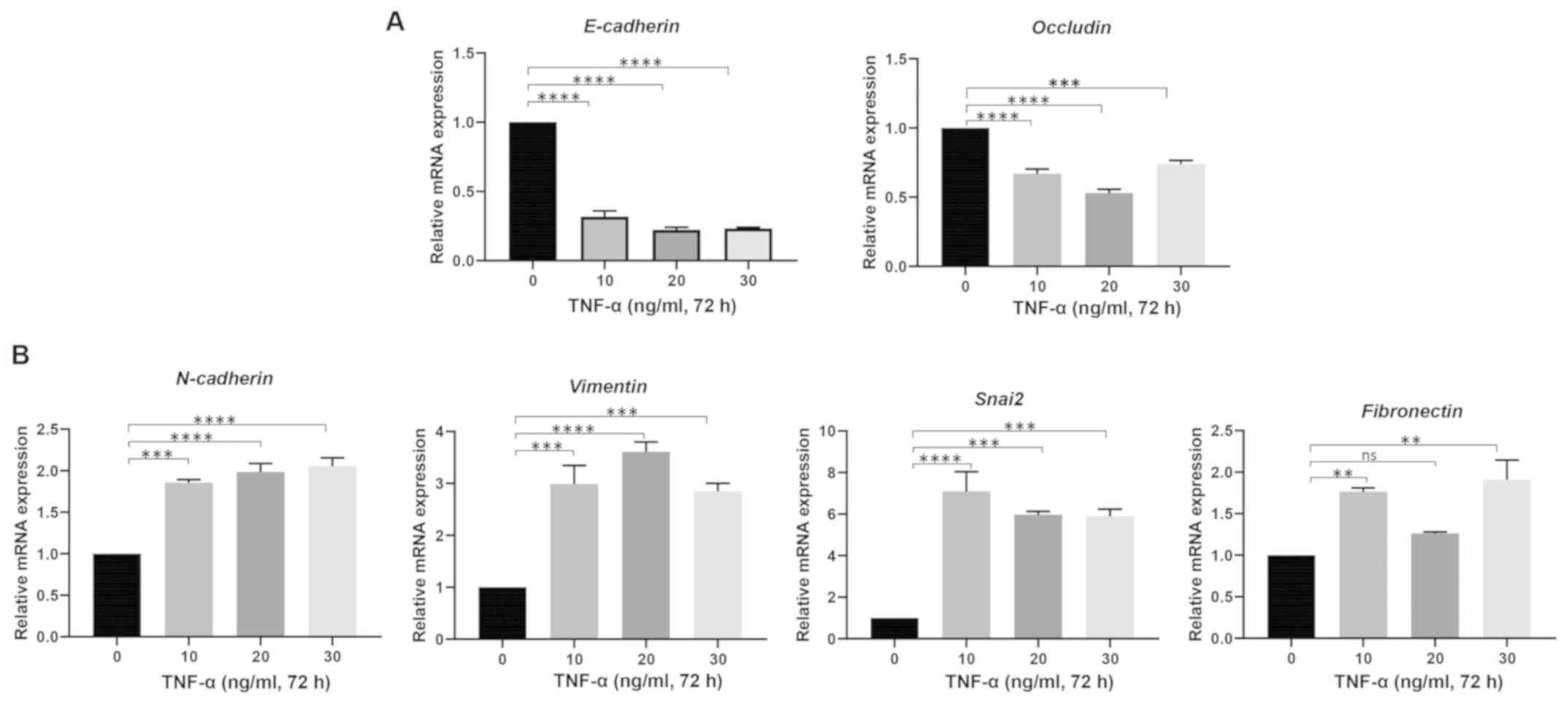 | Figure 1.TNF-α induces
epithelial-to-mesenchymal transition in Hep3B cells at various
doses. Reverse transcription-quantitative PCR analysis demonstrated
(A) lower expression of E-cadherin and Occludin, and (B) higher
expression of N-cadherin, Vimentin, Snai2 and Fibronectin upon
treatment with TNF-α at concentrations of 0, 10, 20 and 30 ng/ml
for 72 h. n=3. **P<0.01, ***P<0.005, ****P<0.001. TNF-α,
tumor necrosis factor-α; Snai2, snail family transcriptional
repressor 2; ns, not significant. |
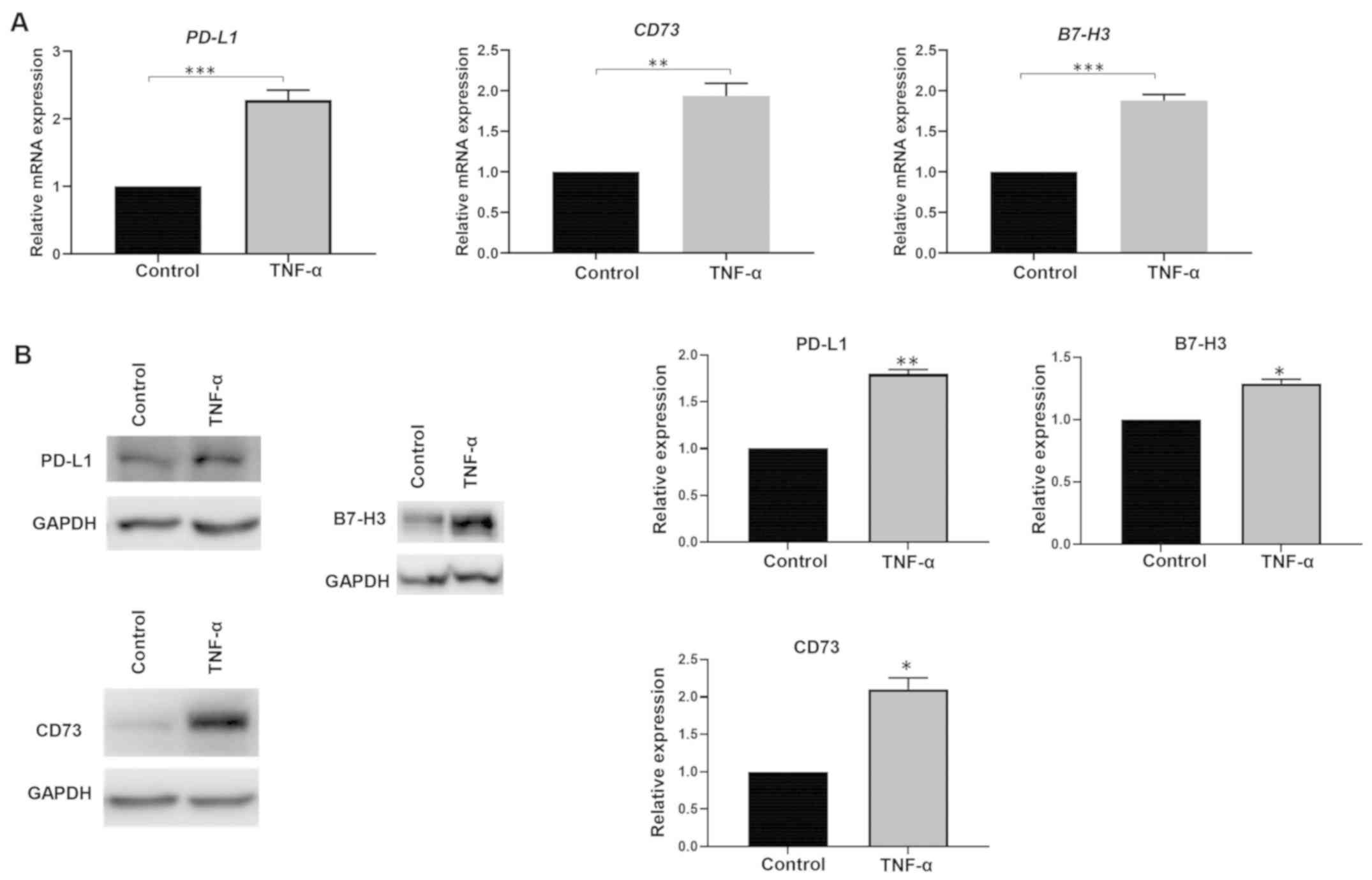
TNF-α-mediated EMT upregulates immune
checkpoint expression
To examine whether TNF-α-mediated EMT influenced the
expression of immune modulators, Hep3B and PLC/PRF/5 cells were
treated with 20 ng/ml TNF-α for 72 h. The cells were then evaluated
for expression of immune modulators PD-L1, PD-L2, CD73, B7-H3,
V-set domain-containing T-cell activation inhibitor 1 (VTCN1) and
V-domain immunoglobulin suppressor of T-cell activation (VISTA).
These immune regulators were assessed as our group previously
demonstrated that these immune checkpoint molecules are associated
with a poor prognosis in patients with HCC (8). The qPCR results revealed upregulation
of four immune modulators (PD-L1, PD-L2, CD73 and B7-H3) and
downregulation of two immune modulators (VTCN1 and VISTA) upon
treatment with TNF-α in Hep3B cells (Fig. 4A and B). The upregulation of immune
checkpoints by TNF-α-mediated EMT in Hep3B cells was validated at
the protein level by western blot analysis (Fig. 4C).
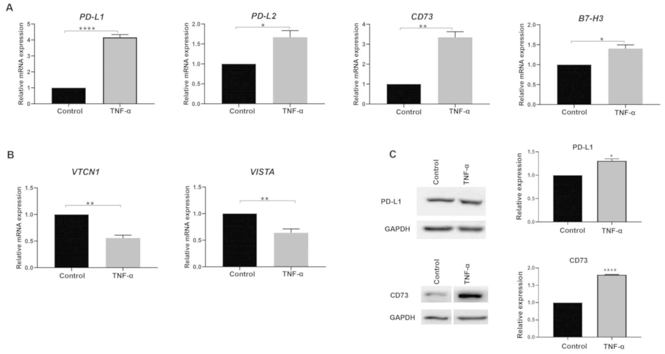 | Figure 4.TNF-α-induced
epithelial-to-mesenchymal transition regulates expression of immune
checkpoint molecules in Hep3B cells. Reverse
transcription-quantitative PCR analysis demonstrated (A) higher
expression of PD-L1, PD-L2, CD73 and B7-H3, and (B) lower
expression of VTCN1 and VISTA upon treatment with 20 ng/ml TNF-α
for 72 h. n=3. *P<0.05, **P<0.01, ****P<0.001. (C) Western
blot analysis demonstrated upregulation of PD-L1 and CD73 in Hep3B
cells upon treatment with 20 ng/ml TNF-α for 72 h. n=3. *P<0.05,
****P<0.001 vs. control. GAPDH was used as the loading control.
TNF-α, tumor necrosis factor-α; VTCN1, V-set domain-containing
T-cell activation inhibitor 1; VISTA, V-domain immunoglobulin
suppressor of T-cell activation; PD-L, programmed death receptor
ligand. |
Similarly, upregulation of immune modulators PD-L1,
CD73 and B7-H3 was observed upon treatment with TNF-α in PLC/PRF/5
cells (Fig. 5A). PD-L2, VTCN1 and
VISTA were not detected in PLC/PRF/5 cells. This upregulation of
immune modulators by TNF-α in PLC/PRF/5 cells was further confirmed
by western blot analysis (Fig.
5B). This demonstrated that TNF-α has a potentially important
role in modulating immune checkpoints and EMT in HCC.
Reversal of TNF-α-mediated EMT
reverses immune checkpoint expression
In order to assess the association between
TNF-α-mediated EMT and immune modulator expression, an EMT reversal
assay was performed. The induction of MET was evidenced by the
increase in epithelial markers (E-cadherin and Occludin) and a
decrease in mesenchymal markers (N-cadherin, Vimentin, Snai2 and
Fibronectin) in Hep3B cells (Fig. 6A
and B). Similar MET was observed in PLC/PRF/5 cells, with an
increase in epithelial markers (E-cadherin and Occludin) and a
decrease in mesenchymal markers (N-cadherin and Vimentin; Fig. 6C and D). The changes in the EMT
status observed by the reversal assay in Hep3B cells was further
confirmed by immunofluorescence and western blot analysis (Fig. 7). The reversal of EMT status in the
reversal assay in PLC/PRF/5 cells was also demonstrated by
immunofluorescence and western blot analysis (Fig. S2).
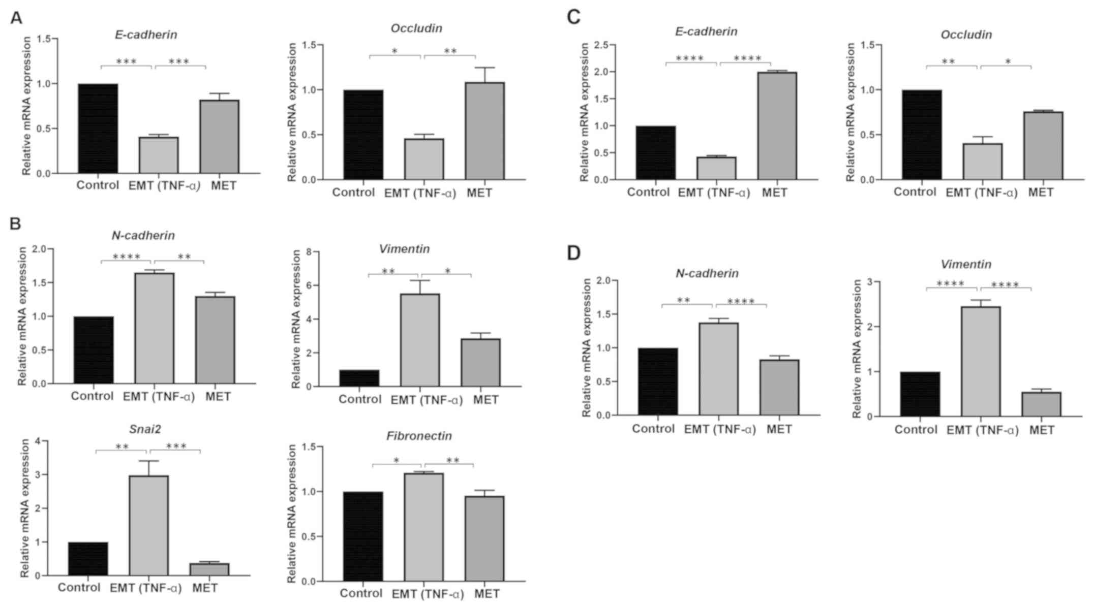 | Figure 6.TNF-α-induced EMT in hepatocellular
carcinoma cells is reversible. RT-qPCR analysis demonstrated (A)
higher expression of E-cadherin and Occludin, and (B) lower
expression of N-cadherin, Vimentin, Snai2 and Fibronectin in TNF-α
treated Hep3B cells following reversal assay. n=3. *P<0.05,
**P<0.01, ***P<0.005, ****P<0.001. RT-qPCR analysis
presented (C) higher expression of E-cadherin and Occludin, and (D)
lower expression of N-cadherin and Vimentin in TNF-α treated
PLC/PRF/5 cells following reversal assay. n=3. *P<0.05,
**P<0.01, ***P<0.005, ****P<0.001. EMT,
epithelial-to-mesenchymal transition; TNF-α, tumor necrosis
factor-α; RT-qPCR, reverse transcription-quantitative PCR; Snai2,
snail family transcriptional repressor 2; MET,
mesenchymal-to-epithelial transition. |
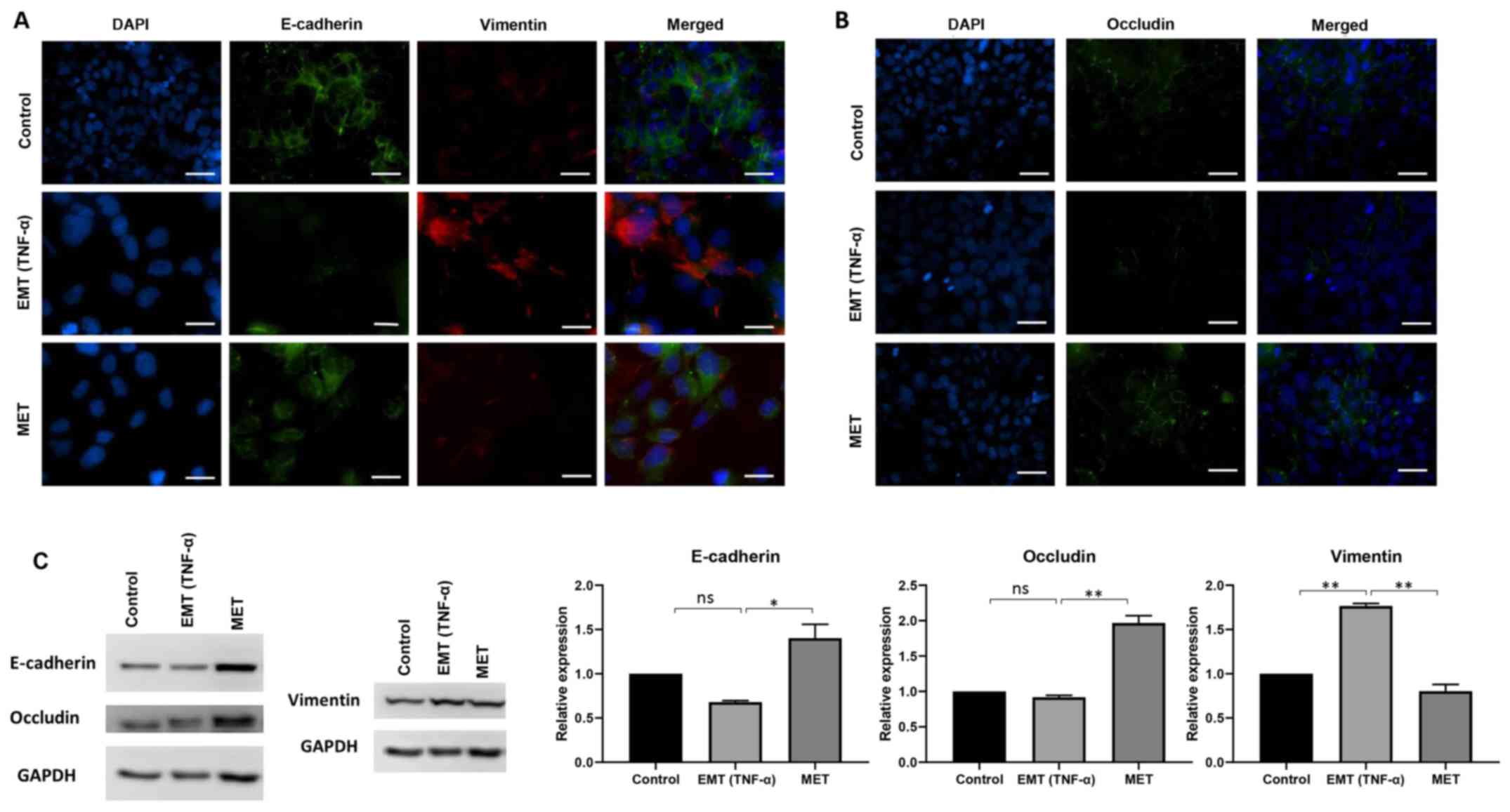 | Figure 7.TNF-α-induced EMT in Hep3B cells is
reversible. Fluorescence microscopy identified (A) suppression of
E-cadherin and upregulation of Vimentin during EMT, and
upregulation of E-cadherin and decreased expression of Vimentin
during MET. (B) During EMT, repression of Occludin was observed and
during MET, higher expression of Occludin was observed. Scale bar,
100 µm. Magnification, ×40. (C) Western blot analysis demonstrated
upregulation of E-cadherin and Occludin, and downregulation of
Vimentin during MET. n=3. *P<0.05, **P<0.01. GAPDH was used
as loading control. EMT, epithelial-to-mesenchymal transition;
TNF-α, tumor necrosis factor-α; MET, mesenchymal-to-epithelial
transition; ns, not significant. |
The expression of immune checkpoints was also
detected following the reversal assay. Downregulation of PD-L1,
PD-L2, CD73 and B7-H3 was observed in Hep3B cells upon removal of
TNF-α (Fig. 8A). Downregulation of
PD-L1, CD73 and B7-H3 was additionally observed in PLC/PRF/5 cells
upon removal of TNF-α (Fig. 8B).
The reversal in immune checkpoint expression upon reversal of
TNF-α-mediated EMT suggested that TNF-α is involved in regulation
of immune checkpoint expression.
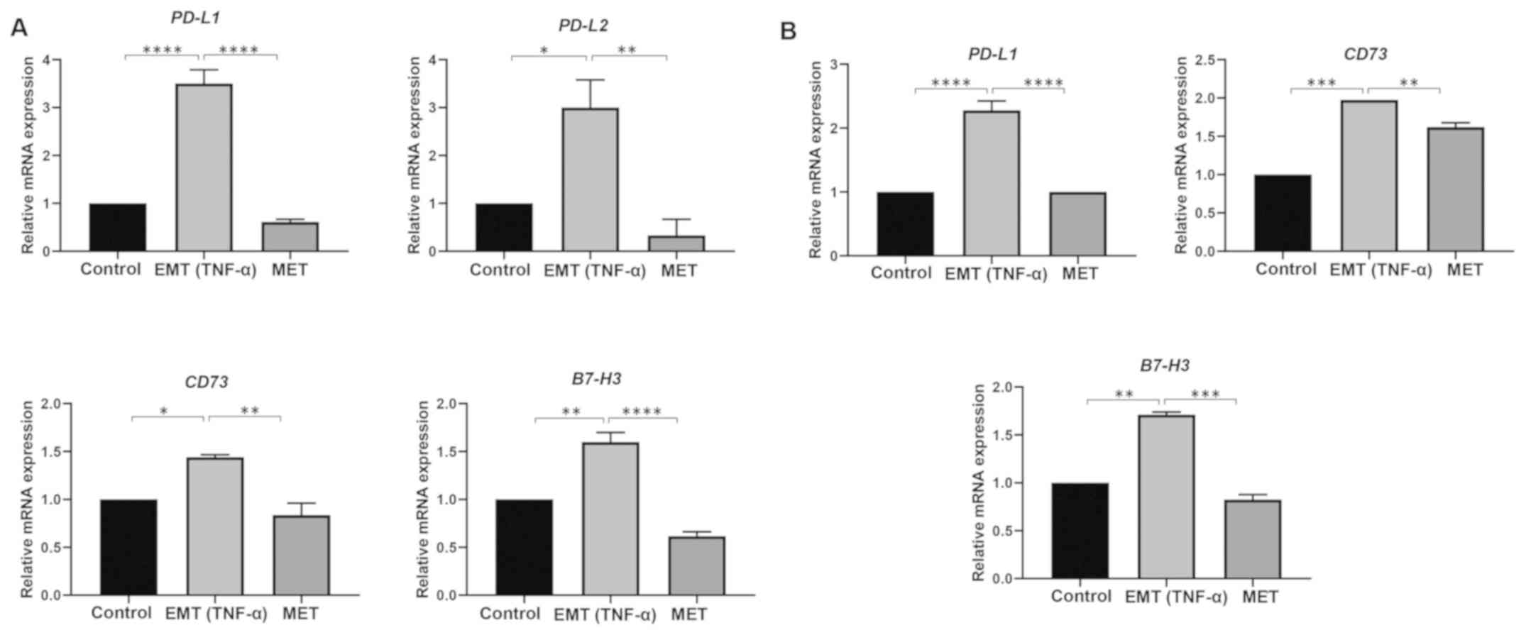 | Figure 8.Reversal of EMT returns immune
checkpoint expression to control levels in hepatocellular carcinoma
cells. Reverse transcription-quantitative PCR analysis demonstrated
(A) lower expression of immune checkpoint molecules PD-L1, PD-L2,
CD73 and B7-H3 in EMT induced Hep3B cells and (B) lower expression
of immune checkpoint molecules PD-L1, CD73 and B7-H3 in EMT induced
PLC/PRF/5 cells following reversal assay. n=3. *P<0.05,
**P<0.01, ***P<0.005, ****P<0.001. EMT,
epithelial-to-mesenchymal transition; TNF-α, tumor necrosis
factor-α; MET, mesenchymal-to-epithelial transition; PD-L,
programmed death receptor ligand. |
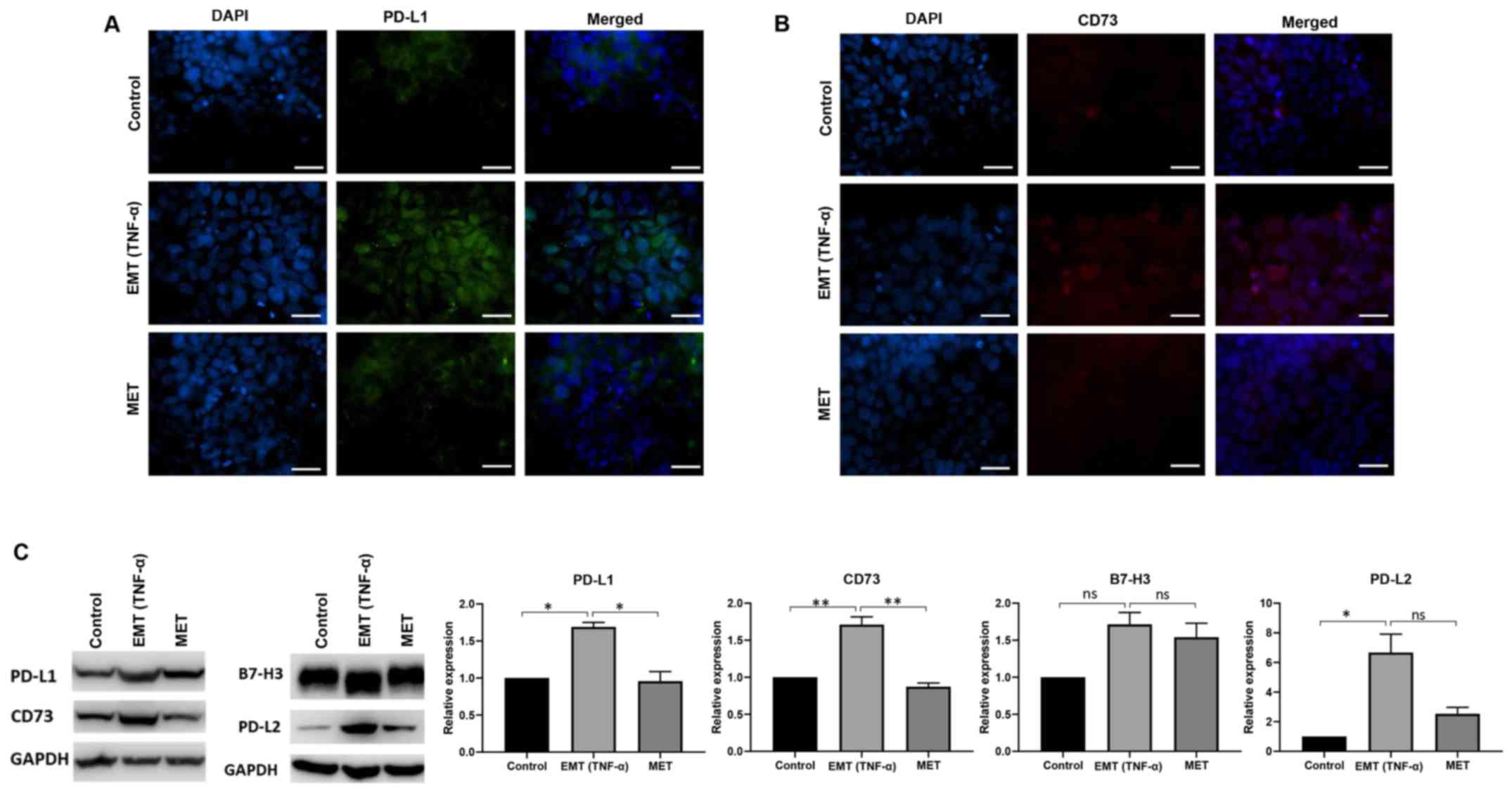
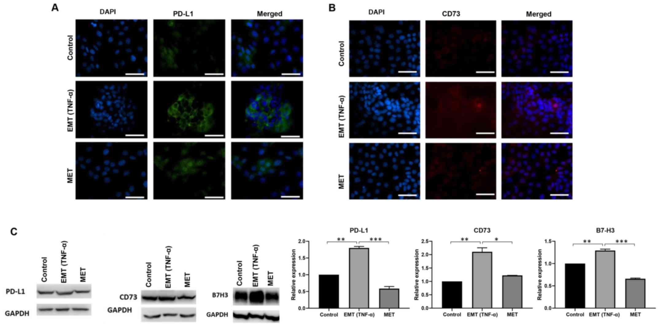
To further investigate the association between
TNF-α-mediated EMT and PD-L1 expression, immunofluorescence and
western blot analysis were performed upon TNF-α treatment followed
by the reversal assay. The immunofluorescence staining demonstrated
upregulated expression of PD-L1 and CD73 during TNF-α-induced EMT
and decreased expression upon reversal of EMT in Hep3B cells
(Fig. 9A and B). In addition,
western blot analysis showed increased expression of PD-L1, CD73,
B7-H3 and PD-L2 during TNF-α-induced EMT and decreased expression
upon reversal of EMT in Hep3B cells (Fig. 9C).
Furthermore, the immunofluorescence staining showed
elevated expression of PD-L1 and CD73 during TNF-α-induced EMT and
decreased expression upon reversal of EMT in PLC/PRF/5 cells
(Fig. 10A and B). Moreover,
western blot analysis showed increased expression of PD-L1, CD73
and B7-H3 during TNF-α-induced EMT and decreased expression upon
reversal of EMT in PLC/PRF/5 cells (Fig. 10C). Overall, the present data
demonstrated that TNF-α simultaneously induced EMT and immune
checkpoint expression in HCC cells.
 | Figure 10.Reversal of EMT reverses immune
checkpoint expression in PLC/PRF/5 cells. Fluorescence microscopy
revealed (A) higher expression of PD-L1 upon EMT induction and
lower expression of PD-L1 following reversal assay, and (B) higher
expression of CD73 upon EMT induction and lower expression of CD73
following reversal assay in PLC/PRF/5 cells. Scale bar, 100 µm.
Magnification, ×40. (C) Western blot analysis demonstrated higher
expression of PD-L1, CD73 and B7-H3 upon EMT induction and lower
expression following reversal assay in PLC/PRF/5 cells. n=3.
*P<0.05, **P<0.01, ***P<0.005. GAPDH was used as the
loading control. EMT, epithelial-to-mesenchymal transition; TNF-α,
tumor necrosis factor-α; MET, mesenchymal-to-epithelial transition;
PD-L, programmed death receptor ligand. |
Coordinate expression of TNF-α and
immune modulators in HCC
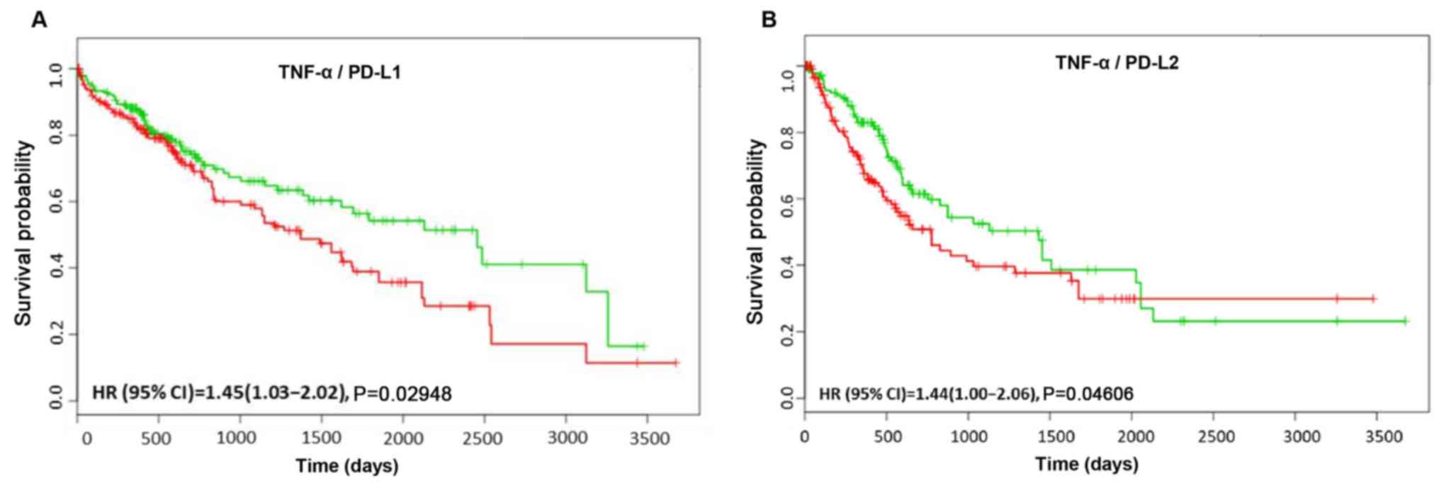
A total of six different HCC datasets within
SurvExpress were used to analyze the recurrence free survival and
OS of immune checkpoint molecules in combination with TNF-α in HCC
patients. It was observed that coordinate expression of PD-L1 in
combination with TNF-α resulted in a significantly worse OS in 422
patients [HR: 1.45; confidence interval (CI): 1.03–2.02; Log-Rank
Equal Curves P=0.02948; Fig.
11A]. Similarly, PD-L2 when combined with TNF-α showed a
significantly worse recurrence free survival (HR: 1.44; CI:
1.00–2.06; Log-Rank Equal Curves P=0.04606; Fig. 11B). There was no significant
difference in recurrence free survival and OS in patients with HCC
who showed coordinate expression of TNF-α with CD73 or B7-H3 (data
not shown). The present data suggested a significant association
between TNF-α and immune checkpoint expression in patients with
HCC, as they showed an adverse prognosis with this profile.
Discussion
In the present study, an association between EMT and
immune checkpoint molecules in HCC was identified. It was
demonstrated that TNF-α simultaneously induced EMT and the
expression of immune checkpoint molecules PD-L1, PD-L2, CD73 and
B7-H3 in Hep3B cells, along with increased expression of PD-L1,
CD73 and B7-H3 in PLC/PRF/5 cells. Moreover, it was demonstrated
that EMT status is closely associated with immune checkpoint
upregulation as MET induced by the reversal of TNF-α-mediated EMT
attenuated the expression of immune checkpoints. Notably, a
significant association between TNF-α and immune checkpoint levels
was identified in patients with HCC. Patients with increased
expression of both TNF-α and PD-L1 showed poor OS, whereas patients
with high expression of TNF-α and PD-L2 showed high rates of
recurrence.
Various systems have been utilized to induce EMT in
human HCC cell lines (33,34). Stimulation of HCC cells with TNF-α
leads to the induction of EMT (35). TNF-α is known to induce EMT alone
or in combination with other cytokines such as transforming growth
factor (TGF)-β in several cancer types, such as breast cancer, lung
cancer and HCC (19,21,25).
In the present study, a TNF-α based in vitro model was used
to induce EMT in a reversible manner. To the best of the authors'
knowledge, the present study is the first study to evaluate the
role of EMT in the regulation of immune checkpoint expression in
HCC.
Despite the promising results of ICIs from clinical
trials, these therapies have failed in several instances due to
mutations that alter immunogenicity, expression of alternative
immune checkpoint molecules and dysregulated T-cell infiltration
(36). Understanding the
underlying molecular biological mechanisms regulating immune
checkpoints may result in developing new and effective treatment
strategies. In the present study it was noted that the expression
of immune checkpoint molecules PD-L1, PD-L2, CD73 and B7-H3 was
upregulated following TNF-α-induced EMT. PD-L1 is an essential and
widely studied immunomodulatory ligand that is aberrantly
upregulated in several cancer types, has roles in promoting tumor
escape and is associated with poor prognosis (37–41).
Our group and other researchers have reported the association
between upregulation of PD-L1 and poor survival in patients with
HCC (8,42). Several previous studies have
reported different mechanisms involved in the regulation of PD-L1
expression in cancer cells (19,43–45).
However, the mechanism of PD-L1 expression in HCC still remains
unclear (46).
In previous years, previous studies have
demonstrated cytokine-induced EMT, in particular TNF-α, TGF-β and
interferon-γ are responsible for elevating the expression of PD-L1
in cancer, such as non-small cell lung carcinoma and breast cancer
(17,19,21).
The present finding that TNF-α is involved in the upregulation of
PD-L1 is consistent with these studies. Another previous study
demonstrated that PD-L1 was upregulated in EMT positive human
esophageal tumor samples compared with the EMT negative samples
(23). The relationship between
EMT and PD-L1 was further examined in human breast cancer cells by
Noman et al (20). This
previous study identified increased expression of PD-L1 in cells
having undergone EMT by EMT-transcription factors (zinc finger
E-box binding homeobox 1, microRNA 200 or Snai1) and PD-L1 rendered
EMT-activated cells resistant to cytotoxic T-lymphocytes-mediated
lysis (20). In patients with HCC,
our group previously identified an association between PD-L1
expression and EMT status (8).
High PD-L1 expression was closely associated with high expression
of the mesenchymal maker Vimentin and low expression of epithelial
marker E-cadherin (8). However, to
the best of the authors' knowledge, no studies have reported the
association between EMT and other immune checkpoint in HCC.
PD-L2 is a second ligand that binds to PD-1 to
prevent cytokine production, cell adhesion and T-cell proliferation
(47). Our group and other
researchers have previously reported that PD-L2 expression is
associated with poor survival and recurrence of HCC in patients
(8,48). A previous meta-analysis study by
Yang et al (49) identified
that upregulation of PD-L2 predicted poor OS in HCC.
CD73 is also reported to be a novel prognostic
biomarker for HCC (8,50,51).
However, to the best of the authors' knowledge, the regulation of
CD73 expression has not been studied in the context of EMT, and the
present study is the first to report that TNF-α-induced EMT
regulates CD73 expression. In a recent triple-negative breast
cancer study by Qiao et al (52), an anti-CD73 antibody was
demonstrated to inhibit lung metastasis in vivo.
B7-H3 is mostly upregulated in several cancer types,
including liver, breast, bladder, colorectal, cervical, glioma,
esophageal and gastric cancer (53). Previous studies have also
demonstrated that dysregulation of B7-H3 in HCC results in impaired
T-lymphocyte function, thus leading to poor prognosis and
recurrence (54,55). In muscle-invasive bladder cancer,
high expression of B7-H3 was associated with a poor
clinicopathological status and poor prognosis (56). Most notably, B7-H3 is known to
promote EMT by repressing E-cadherin expression, and upregulating
Vimentin and N-cadherin expression in colorectal cancer (57). To the best of the authors'
knowledge, the present study is the first to report the regulation
of B7-H3 expression by EMT in HCC.
Further mechanistic and functional studies are
necessary to examine the molecular biology of immune checkpoints
and its precise role in EMT in HCC. However, given the association
between EMT, TNF-α and immune checkpoint in HCC, it is conceivable
that combining EMT or TNF-α inhibitors with ICIs in patients with
HCC may be a future therapeutic approach for the management of
HCC.
Supplementary Material
Supporting Data
Acknowledgements
The authors would like to thank Mrs Lesley-Anne
Jaskowski, Gallipoli Medical Research Institute, Greenslopes
Private Hospital, for her technical assistance during the revision
of the manuscript.
Funding
The present study was funded by Gallipoli Medical
Research Foundation (project no. 017401).
Availability of data and materials
The datasets used and/or analyzed during the current
study are available from the corresponding author on reasonable
request.
Authors' contributions
RS, KRB, DHGC and AJ designed the study, performed
data acquisition, analysis and interpretation, and wrote the
manuscript. RS, AJ, KRB and DHGC critically revised the manuscript.
All authors read and approved the final manuscript.
Ethics approval and consent to
participate
Not applicable.
Patient consent for publication
Not applicable.
Competing interests
The authors declare that they have no competing
interests.
References
|
1
|
Chacko S and Samanta S: ‘Hepatocellular
carcinoma: A life-threatening disease’. Biomed Pharmacother.
84:1679–1688. 2016. View Article : Google Scholar : PubMed/NCBI
|
|
2
|
Bray F, Ferlay J, Soerjomataram I, Siegel
RL, Torre LA and Jemal A: Global cancer statistics 2018: GLOBOCAN
estimates of incidence and mortality worldwide for 36 cancers in
185 countries. CA Cancer J Clin. 68:394–424. 2018. View Article : Google Scholar : PubMed/NCBI
|
|
3
|
Sayiner M, Golabi P and Younossi ZM:
Disease burden of hepatocellular carcinoma: A global perspective.
Dig Dis Sci. 64:910–917. 2019. View Article : Google Scholar : PubMed/NCBI
|
|
4
|
Kudo M: A new era of systemic therapy for
hepatocellular carcinoma with regorafenib and lenvatinib. Liver
Cancer. 6:177–184. 2017. View Article : Google Scholar : PubMed/NCBI
|
|
5
|
Mir N, Jayachandran A, Dhungel B, Shrestha
R and Steel JC: Epithelial-to-mesenchymal transition: A Mediator of
sorafenib resistance in advanced hepatocellular carcinoma. Curr
Cancer Drug Targets. 17:698–706. 2017. View Article : Google Scholar : PubMed/NCBI
|
|
6
|
Sharma P and Allison JP: The future of
immune checkpoint therapy. Science. 348:56–61. 2015. View Article : Google Scholar : PubMed/NCBI
|
|
7
|
Pardoll DM: The blockade of immune
checkpoints in cancer immunotherapy. Nat Rev Cancer. 12:252–264.
2012. View
Article : Google Scholar : PubMed/NCBI
|
|
8
|
Shrestha R, Prithviraj P, Anaka M, Bridle
KR, Crawford DH, Dhungel B, Steel JC and Jayachandran A: Monitoring
immune checkpoint regulators as predictive biomarkers in
hepatocellular carcinoma. Front Oncol. 8:2692018. View Article : Google Scholar : PubMed/NCBI
|
|
9
|
Hato T, Goyal L, Greten TF, Duda DG and
Zhu AX: Immune checkpoint blockade in hepatocellular carcinoma:
Current progress and future directions. Hepatology. 60:1776–1782.
2014. View Article : Google Scholar : PubMed/NCBI
|
|
10
|
Brahmer J, Reckamp KL, Baas P, Crinò L,
Eberhardt WE, Poddubskaya E, Antonia S, Pluzanski A, Vokes EE,
Holgado E, et al: Nivolumab versus docetaxel in advanced
squamous-cell non-small-cell lung cancer. N Engl J Med.
373:123–135. 2015. View Article : Google Scholar : PubMed/NCBI
|
|
11
|
Weber JS, D'Angelo SP, Minor D, Hodi FS,
Gutzmer R, Neyns B, Hoeller C, Khushalani NI, Miller WH Jr, Lao CD,
et al: Nivolumab versus chemotherapy in patients with advanced
melanoma who progressed after anti-CTLA-4 treatment (CheckMate
037): A randomised, controlled, open-label, phase 3 trial. Lancet
Oncol. 16:375–384. 2015. View Article : Google Scholar : PubMed/NCBI
|
|
12
|
Jindal A, Thadi A and Shailubhai K:
Hepatocellular carcinoma: Etiology and current and future drugs. J
Clin Exp Hepatol. 9:221–232. 2019. View Article : Google Scholar : PubMed/NCBI
|
|
13
|
Kudo M: Immune checkpoint inhibition in
hepatocellular carcinoma: Basics and ongoing clinical trials.
Oncology. 92 (Suppl 1):50–62. 2017. View Article : Google Scholar : PubMed/NCBI
|
|
14
|
Li Z, Li N, Li F, Zhou Z, Sang J, Chen Y,
Han Q, Lv Y and Liu Z: Immune checkpoint proteins PD-1 and TIM-3
are both highly expressed in liver tissues and correlate with their
gene polymorphisms in patients with HBV-related hepatocellular
carcinoma. Medicine (Baltimore). 95:e57492016. View Article : Google Scholar : PubMed/NCBI
|
|
15
|
Kalluri R and Weinberg RA: The basics of
epithelial-mesenchymal transition. J Clin Invest. 119:1420–1428.
2009. View
Article : Google Scholar : PubMed/NCBI
|
|
16
|
Jayachandran A, Dhungel B and Steel JC:
Epithelial-to-mesenchymal plasticity of cancer stem cells:
Therapeutic targets in hepatocellular carcinoma. J Hematol Oncol.
9:742016. View Article : Google Scholar : PubMed/NCBI
|
|
17
|
David JM, Dominguez C, McCampbell KK,
Gulley JL, Schlom J and Palena C: A novel bifunctional
anti-PD-L1/TGF-β Trap fusion protein (M7824) efficiently reverts
mesenchymalization of human lung cancer cells. OncoImmunology.
6:e13495892017. View Article : Google Scholar : PubMed/NCBI
|
|
18
|
David JM, Dominguez C and Palena C:
Pharmacological and immunological targeting of tumor
mesenchymalization. Pharmacol Ther. 170:212–225. 2017. View Article : Google Scholar : PubMed/NCBI
|
|
19
|
Alsuliman A, Colak D, Al-Harazi O, Fitwi
H, Tulbah A, Al-Tweigeri T, Al-Alwan M and Ghebeh H: Bidirectional
crosstalk between PD-L1 expression and epithelial to mesenchymal
transition: Significance in claudin-low breast cancer cells. Mol
Cancer. 14:1492015. View Article : Google Scholar : PubMed/NCBI
|
|
20
|
Noman MZ, Janji B, Abdou A, Hasmim M,
Terry S, Tan TZ, Mami-Chouaib F, Thiery JP and Chouaib S: The
immune checkpoint ligand PD-L1 is upregulated in EMT-activated
human breast cancer cells by a mechanism involving ZEB-1 and
miR-200. Oncoimmunology. 6:e12634122017. View Article : Google Scholar : PubMed/NCBI
|
|
21
|
Asgarova A, Asgarov K, Godet Y, Peixoto P,
Nadaradjane A, Boyer-Guittaut M, Galaine J, Guenat D, Mougey V,
Perrard J, et al: PD-L1 expression is regulated by both DNA
methylation and NF-κB during EMT signaling in non-small cell lung
carcinoma. Oncoimmunology. 7:e14231702018. View Article : Google Scholar : PubMed/NCBI
|
|
22
|
Imai D, Yoshizumi T, Okano S, Itoh S,
Ikegami T, Harada N, Aishima S, Oda Y and Maehara Y: IFN-γ promotes
epithelial-mesenchymal transition and the expression of PD-L1 in
pancreatic cancer. J Surg Res. 240:115–123. 2019. View Article : Google Scholar : PubMed/NCBI
|
|
23
|
Chen L, Xiong Y, Li J, Zheng X, Zhou Q,
Turner A, Wu C, Lu B and Jiang J: PD-L1 expression promotes
epithelial to mesenchymal transition in human esophageal cancer.
Cell Physiol Biochem. 42:2267–2280. 2017. View Article : Google Scholar : PubMed/NCBI
|
|
24
|
Wang Y, Hu J, Wang Y, Ye W, Zhang X, Ju H,
Xu D, Liu L, Ye D, Zhang L, et al: EGFR activation induced
Snail-dependent EMT and myc-dependent PD-L1 in human salivary
adenoid cystic carcinoma cells. Cell Cycle. 17:1457–1470. 2018.
View Article : Google Scholar : PubMed/NCBI
|
|
25
|
Chen Y, Wen H, Zhou C, Su Q, Lin Y, Xie Y,
Huang Y, Qiu Q, Lin J, Huang X, et al: TNF-α derived from M2
tumor-associated macrophages promotes epithelial-mesenchymal
transition and cancer stemness through the Wnt/β-catenin pathway in
SMMC-7721 hepatocellular carcinoma cells. Exp Cell Res. 378:41–50.
2019. View Article : Google Scholar : PubMed/NCBI
|
|
26
|
Jayachandran A, Shrestha R, Dhungel B,
Huang IT, Vasconcelos MY, Morrison BJ, Ramlogan-Steel CA and Steel
JC: Murine hepatocellular carcinoma derived stem cells reveal
epithelial-to-mesenchymal plasticity. World J Stem Cells.
9:159–168. 2017. View Article : Google Scholar : PubMed/NCBI
|
|
27
|
Livak KJ and Schmittgen TD: Analysis of
relative gene expression data using real-time quantitative PCR and
the 2(-Delta Delta C(T)) method. Methods. 25:402–408. 2001.
View Article : Google Scholar : PubMed/NCBI
|
|
28
|
Aguirre-Gamboa R, Gomez-Rueda H,
Martínez-Ledesma E, Martínez-Torteya A, Chacolla-Huaringa R,
Rodriguez-Barrientos A, Tamez-Peña JG and Treviño V: SurvExpress:
An online biomarker validation tool and database for cancer gene
expression data using survival analysis. PLoS One. 8:e742502013.
View Article : Google Scholar : PubMed/NCBI
|
|
29
|
Hoshida Y, Nijman SM, Kobayashi M, Chan
JA, Brunet JP, Chiang DY, Villanueva A, Newell P, Ikeda K,
Hashimoto M, et al: Integrative transcriptome analysis reveals
common molecular subclasses of human hepatocellular carcinoma.
Cancer Res. 69:7385–7392. 2009. View Article : Google Scholar : PubMed/NCBI
|
|
30
|
Hoshida Y, Villanueva A, Kobayashi M, Peix
J, Chiang DY, Camargo A, Gupta S, Moore J, Wrobel MJ, Lerner J, et
al: Gene expression in fixed tissues and outcome in hepatocellular
carcinoma. N Engl J Med. 359:1995–2004. 2008. View Article : Google Scholar : PubMed/NCBI
|
|
31
|
Tsuchiya M, Parker JS, Kono H, Matsuda M,
Fujii H and Rusyn I: Gene expression in nontumoral liver tissue and
recurrence-free survival in hepatitis C virus-positive
hepatocellular carcinoma. Mol Cancer. 9:742010. View Article : Google Scholar : PubMed/NCBI
|
|
32
|
Awan FM, Naz A, Obaid A, Ali A, Ahmad J,
Anjum S and Janjua HA: Identification of Circulating Biomarker
Candidates for Hepatocellular Carcinoma (HCC): An Integrated
Prioritization Approach. PLoS One. 10:e01389132015. View Article : Google Scholar : PubMed/NCBI
|
|
33
|
Lee TK, Poon RT, Yuen AP, Ling MT, Kwok
WK, Wang XH, Wong YC, Guan XY, Man K, Chau KL, et al: Twist
overexpression correlates with hepatocellular carcinoma metastasis
through induction of epithelial-mesenchymal transition. Clin Cancer
Res. 12:5369–5376. 2006. View Article : Google Scholar : PubMed/NCBI
|
|
34
|
Xu Z, Shen MX, Ma DZ, Wang LY and Zha XL:
TGF-beta1-promoted epithelial-to-mesenchymal transformation and
cell adhesion contribute to TGF-beta1-enhanced cell migration in
SMMC-7721 cells. Cell Res. 13:343–350. 2003. View Article : Google Scholar : PubMed/NCBI
|
|
35
|
Zhu Y, Cheng Y, Guo Y, Chen J, Chen F, Luo
R and Li A: Protein kinase D2 contributes to TNF-α-induced
epithelial mesenchymal transition and invasion via the
PI3K/GSK-3β/β-catenin pathway in hepatocellular carcinoma.
Oncotarget. 7:5327–5341. 2016.PubMed/NCBI
|
|
36
|
Xu F, Jin T, Zhu Y and Dai C: Immune
checkpoint therapy in liver cancer. J Exp Clin Cancer Res.
37:1102018. View Article : Google Scholar : PubMed/NCBI
|
|
37
|
Afreen S and Dermime S: The
immunoinhibitory B7-H1 molecule as a potential target in cancer:
Killing many birds with one stone. Hematol Oncol Stem Cell Ther.
7:1–17. 2014. View Article : Google Scholar : PubMed/NCBI
|
|
38
|
Mu CY, Huang JA, Chen Y, Chen C and Zhang
XG: High expression of PD-L1 in lung cancer may contribute to poor
prognosis and tumor cells immune escape through suppressing tumor
infiltrating dendritic cells maturation. Med Oncol. 28:682–688.
2011. View Article : Google Scholar : PubMed/NCBI
|
|
39
|
Muenst S, Schaerli AR, Gao F, Däster S,
Trella E, Droeser RA, Muraro MG, Zajac P, Zanetti R, Gillanders WE,
et al: Expression of programmed death ligand 1 (PD-L1) is
associated with poor prognosis in human breast cancer. Breast
Cancer Res Treat. 146:15–24. 2014. View Article : Google Scholar : PubMed/NCBI
|
|
40
|
Shi SJ, Wang LJ, Wang GD, Guo ZY, Wei M,
Meng YL, Yang AG and Wen WH: B7-H1 expression is associated with
poor prognosis in colorectal carcinoma and regulates the
proliferation and invasion of HCT116 colorectal cancer cells. PLoS
One. 8:e760122013. View Article : Google Scholar : PubMed/NCBI
|
|
41
|
Thompson RH, Gillett MD, Cheville JC,
Lohse CM, Dong H, Webster WS, Krejci KG, Lobo JR, Sengupta S, Chen
L, et al: Costimulatory B7-H1 in renal cell carcinoma patients:
Indicator of tumor aggressiveness and potential therapeutic target.
Proc Natl Acad Sci USA. 101:17174–17179. 2004. View Article : Google Scholar : PubMed/NCBI
|
|
42
|
Calderaro J, Rousseau B, Amaddeo G, Mercey
M, Charpy C, Costentin C, Luciani A, Zafrani ES, Laurent A, Azoulay
D, et al: Programmed death ligand 1 expression in hepatocellular
carcinoma: Relationship with clinical and pathological features.
Hepatology. 64:2038–2046. 2016. View Article : Google Scholar : PubMed/NCBI
|
|
43
|
Crane CA, Panner A, Murray JC, Wilson SP,
Xu H, Chen L, Simko JP, Waldman FM, Pieper RO and Parsa AT: PI(3)
kinase is associated with a mechanism of immunoresistance in breast
and prostate cancer. Oncogene. 28:306–312. 2009. View Article : Google Scholar : PubMed/NCBI
|
|
44
|
Dey N, Crosswell HE, De P, Parsons R, Peng
Q, Su JD and Durden DL: The protein phosphatase activity of PTEN
regulates SRC family kinases and controls glioma migration. Cancer
Res. 68:1862–1871. 2008. View Article : Google Scholar : PubMed/NCBI
|
|
45
|
Ghebeh H, Tulbah A, Mohammed S, Elkum N,
Bin Amer SM, Al-Tweigeri T and Dermime S: Expression of B7-H1 in
breast cancer patients is strongly associated with high
proliferative Ki-67-expressing tumor cells. Int J Cancer.
121:751–758. 2007. View Article : Google Scholar : PubMed/NCBI
|
|
46
|
Funaki S, Shintani Y, Kawamura T, Kanzaki
R, Minami M and Okumura M: Chemotherapy enhances programmed cell
death 1/ligand 1 expression via TGF-β induced epithelial
mesenchymal transition in non-small cell lung cancer. Oncol Rep.
38:2277–2284. 2017. View Article : Google Scholar : PubMed/NCBI
|
|
47
|
Latchman Y, Wood CR, Chernova T, Chaudhary
D, Borde M, Chernova I, Iwai Y, Long AJ, Brown JA, Nunes R, et al:
PD-L2 is a second ligand for PD-1 and inhibits T cell activation.
Nat Immunol. 2:261–268. 2001. View
Article : Google Scholar : PubMed/NCBI
|
|
48
|
Jung HI, Jeong D, Ji S, Ahn TS, Bae SH,
Chin S, Chung JC, Kim HC, Lee MS and Baek MJ: Overexpression of
PD-L1 and PD-L2 is associated with poor prognosis in patients with
hepatocellular carcinoma. Cancer Res Treat. 49:246–254. 2017.
View Article : Google Scholar : PubMed/NCBI
|
|
49
|
Yang H, Zhou X, Sun L and Mao Y:
Correlation Between PD-L2 expression and clinical outcome in solid
cancer patients: A meta-analysis. Front Oncol. 9:472019. View Article : Google Scholar : PubMed/NCBI
|
|
50
|
Ma XL, Shen MN, Hu B, Wang BL, Yang WJ, Lv
LH, Wang H, Zhou Y, Jin AL, Sun YF, et al: CD73 promotes
hepatocellular carcinoma progression and metastasis via activating
PI3K/AKT signaling by inducing Rap1-mediated membrane localization
of P110β and predicts poor prognosis. J Hematol Oncol. 12:372019.
View Article : Google Scholar : PubMed/NCBI
|
|
51
|
Sciarra A, Monteiro I, Ménétrier-Caux C,
Caux C, Gilbert B, Halkic N, La Rosa S, Romero P, Sempoux C and de
Leval L: CD73 expression in normal and pathological human
hepatobiliopancreatic tissues. Cancer Immunol Immunother.
68:467–478. 2019. View Article : Google Scholar : PubMed/NCBI
|
|
52
|
Qiao Z, Li X, Kang N, Yang Y, Chen C, Wu
T, Zhao M, Liu Y and Ji X: A Novel specific Anti-CD73 antibody
inhibits triple-negative breast cancer cell motility by regulating
autophagy. Int J Mol Sci. 20:E10572019. View Article : Google Scholar : PubMed/NCBI
|
|
53
|
Dong P, Xiong Y, Yue J, Hanley SJB and
Watari H: B7H3 as a promoter of metastasis and promising
therapeutic target. Front Oncol. 8:2642018. View Article : Google Scholar : PubMed/NCBI
|
|
54
|
Kang FB, Wang L, Jia HC, Li D, Li HJ,
Zhang YG and Sun DX: B7-H3 promotes aggression and invasion of
hepatocellular carcinoma by targeting epithelial-to-mesenchymal
transition via JAK2/STAT3/Slug signaling pathway. Cancer Cell Int.
15:452015. View Article : Google Scholar : PubMed/NCBI
|
|
55
|
Zheng Y, Liao N, Wu Y, Gao J, Li Z, Liu W,
Wang Y, Li M, Li X, Chen L, et al: High expression of B7 H2 or B7
H3 is associated with poor prognosis in hepatocellular carcinoma.
Mol Med Rep. 19:4315–4325. 2019.PubMed/NCBI
|
|
56
|
Xu ZL, Zhang Y, Wang L, Li F, Man HW, Li
PF and Shan BE: B7 H3 promotes malignant progression of muscle
invasive bladder cancer. Oncol Rep. 40:2722–2733. 2018.PubMed/NCBI
|
|
57
|
Jiang B, Zhang T, Liu F, Sun Z, Shi H, Hua
D and Yang C: The co-stimulatory molecule B7-H3 promotes the
epithelial-mesenchymal transition in colorectal cancer. Oncotarget.
7:31755–31771. 2016.PubMed/NCBI
|