Introduction
The pathogenesis of hepatocellular carcinoma (HCC)
has not yet been fully clarified and there is still a lack of
targeted therapies for HCC (1,2). For
patients with the disease, radical resection of HCC is the main
treatment in its early stages, but >50% of post-operative
patients show metastasis and recurrence within 5 years (1,2).
Targeted drugs for HCC are emerging, providing possible new
therapies to prevent and cure malignant cancer (3). However, its therapeutic effects are
still not satisfactory, due to the side effects it has on normal
cells (4). Thus, the current
treatments for HCC still face several challenges.
Studies conducted to investigate HCC genes at
molecular level indicate that a number of genes and proteins are
closely related to the malignant characteristics of HCC staging,
recurrence and metastasis, and these molecules may play important
roles in the occurrence and development of HCC (5,6).
Thousands of microRNAs (miRNAs/miRs) exist in the human genome and
each miRNA can directly regulate ~200 target genes, and nearly 33%
of the protein-coding genes in humans are controlled by miRNAs
(5–7). Therefore, miRNAs occupy an
indispensable position in the spectrum of human gene, thus,
studying gene therapy for the treatment of hepatoma from the aspect
of miRNA regulation has great potential.
Continuous research and development of ultrasound
molecular imaging and biomedical engineering, especially
development of targeted contrast microbubbles carrying various
drugs and genes, have laid a solid foundation for building up
ultrasonic microbubble technology for therapeutic use (8–11).
Ultrasonic microbubbles are microbubbles composed of a core gas
surrounded by a shell membrane with the size of 2–8 µm or even
smaller (12). As lipids
(represented by SonoVue®) have higher efficiency, fewer
side effects and high stability, it has become the most widely used
microbubbles in most studies (12–15).
The mechanism of ultrasound-mediated targeted delivery mainly
relies on cavitation and sonication effects of ultrasonic
microbubbles generated under the irradiation of ultrasonic field
strength (16).
Ultrasound-targeted microbubbles show various modifications in
their shell membranes and they can carry a variety of drugs or
genes and can bind to specific antigens or genes expressed by
specific cells in the body, thus providing the possibility of
ultrasound-mediated targeted treatment (17).
Currently, the delivery of nucleic acid via
microbubbles and ultrasound method has attracted much attention due
to the discovery of the potentials of miRNAs in modulating target
genes (18). In view of these
valuable prospects, the present study aimed to investigate the role
of miR-378 in hepatoma cells and the efficiency of combining
ultrasonic irradiation and SonoVue microbubbles for cell
transfection.
Materials and methods
Cell incubation
HuH-7 cell line was purchased from the Japanese
Collection of Research Bioresources Cell Bank, while Hep3B cell
line was purchased from American Type Culture Collection and
SK-Hep1 cell line was purchased from The Cell Bank of Type Culture
Collection of the Chinese Academy of Sciences. All the cell lines
were cultured in DMEM (D0819; Sigma-Aldrich; Merck KGaA) containing
10% FBS (F8192; Sigma-Aldrich; Merck KGaA) and
penicillin-streptomycin reagent (V900929; Sigma-Aldrich; Merck
KGaA) in 5% CO2 at 37°C.
Grouping and transfection
In order to explore the role of miR-378 and the
efficiency of the combined method for cell transfection, the cells
were grouped as Blank, miR-378 control, L group (HCC cells
transfected with miR-378) and LUS groups (HCC cells treated with
miR-378 mimic combined with ultrasonic irradiation and SonoVue
microbubbles). Briefly, HuH-7, Hep3B and SK-Hep1 cells at
1×10⁶ cells/ml in the LUS or miR-378 control groups were
plated in a 96-well plate and then respectively transfected with
100 nmol/l miR-378 mimic (Shanghai GenePharma Co., Ltd.), or
miR-378 mimic control vector (Shanghai GenePharma Co., Ltd.) in a
mixture with Lipofectamine® 3000 (L3000015; Thermo
Fisher Scientific, Inc.) and 2.5 µg/µl SonoVue microbubbles (Bracco
Suisse SA) under the irradiation of ultrasonic transfer apparatus
via ultrasound couplant (Anhui Deepblue Medical Technology Co.,
Ltd.) at the parameters of 0.5 W/cm2 for 30 sec. The
cells in the L group were transfected with miR-378 mimic using
Lipofectamine 3000 only, while those in the Blank group were
treated with medium only. All the cells were cultured for another
72 h after the transfection (19).
The sequence of miR-378 mimic was 5′-AGGCUCUGACUCCAGGUCC-3′; The
sequence of miR-378 mimic control was
5′-UUCUCCGAACGUGUCACGUTT-3′.
Reverse transcription-quantitative
(RT-q)PCR
Total RNAs from HuH-7, Hep3B and SK-Hep1 cells at
1×10⁶ cells/ml were obtained using TRIzol reagent
(15596018; Thermo Fisher Scientific, Inc.) and further
reverse-transcribed into cDNAs following the instructions of
PrimeScript RT reagent kit (Takara Biotechnology Co., Ltd.), with
the conditions for reverse transcription being: 30°C for 60 min;
30°C for 60 min; and 95°C for 60 min. The cells were then cultured
for 72 h following the treatments for HuH-7 cells. A total of 0.5
µl forward primer, 0.5 µl reverse primer, 3 µl cDNA template, 5 µl
2X SYBR Green master mix (4913850001; Roche Diagnostics) and 1 µl
ddH2O were mixed together and reacted for 40 cycles in
the following conditions: Initial denaturation at 95°C for 60 sec,
at 95°C for 20 sec, at 65°C for 30 sec, and at 72°C for 40 sec in
Bio-Rad IQ5 thermocycler (Bio-Rad Laboratories, Inc.). The
sequences of primers used are listed in Table I. Relative expression of miR-378,
Cyclin D1, Bcl-2, Bax, Akt, p53 and Survivin were normalized to
that of U6 by 2−ΔΔCq method (20).
 | Table I.Primers used in the study. |
Table I.
Primers used in the study.
| Primer name | Sequence
(5′-3′) |
|---|
| miR-378 | F:
CCTGACTCCAGGTCCT |
|
| R:
GAACATGTCTGCGTATCTC |
| Cyclin D1 | F:
GTCTTCCCGCTGGCCATGAACTAC |
|
| R:
GGAAGCGTGTGAGGCGGTAGTAGG |
| Bcl-2 | F:
GCCTTCTTTGAGTTCGGTG |
|
| R:
CAGAGACAGCCAGGAGAAATC |
| Bax | F:
GCAAACTGGTGCTCAAGG |
|
| R:
CGCCACAAAGATGGTCAC |
| Akt | F:
TGGACTACCTGCACTCGGAGAA |
|
| R:
GTGCCGCAAAAGGTCTTCATGG |
| p53 | F:
TAAAAGATGTTTTGAATG |
|
| R:
ATGTGTGTGATGTTGTAGATG |
| Survivin | F:
CCACTGAGAACGAGCCAGACTT |
|
| R:
GTATTACAGGCGTAAGCCACCG |
| U6 | F:
GCTTCGGCAGCACATATACTAAAAT |
|
| R:
GAAGATGGTGATGGGATTTC |
Cell Counting Kit (CCK)-8
After 72 h, relative cell survival rates of HuH-7,
Hep3B and SK-Hep1 cells were detected by CCK-8 following the
manufacturer's protocols (96992-100TESTS-F; Sigma-Aldrich; Merck
KGaA). Briefly, 1×10⁶ cells were collected and tested
after the transfection of miR-378 mimic. Optic density (OD) values
were read on a microplate reader (Multiskan; Thermo Fisher
Scientific, Inc.) at 450 nm and relative cell survival rates of
cells were calculated according to the standard curve of OD. The
experiment was conducted in triplicate.
Double cytochemical staining
Cells (~4×10³−1×10⁶) were
cultured in 96-well plates. EdU solution (A10044; Thermo Fisher
Scientific, Inc.) was diluted by culture medium at 1,000:1 and 100
µl of 50 µm EdU medium was added to each well and maintained for 2
h. The cells were washed once or twice using PBS for 5 min. Then,
100 µl cell fixative (PBS containing 4% paraformaldehyde) was added
to each well and maintained for 30 min at room temperature. A total
of 2 mg/ml glycine was added to each well for decolorization and
then further incubated for 5 min on a shaker. PBS (100 µl) was
added to each well for, held for 5 min and then 100 µl of penetrant
(0.5% Triton X-100 in PBS) was added to each well for 10 min. After
rinsing in PBS, 1X Hoechst 33342 reaction solution (H3570; Thermo
Fisher Scientific, Inc.) was diluted by deionized water at 100:1
and added to each well for a 30 min incubation at room temperature
in the dark. The staining result was immediately observed by using
a fluorescence microscope (BX53T, Olympus Corporation;
magnification, ×100) after washing the cells with PBS one to three
times.
Evaluation of apoptosis
Briefly, 5×10⁵ cells were resuspended in
1 ml cold PBS and collected into 100 ml binding buffer using an
Annexin V-FITC kit (Sigma-Aldrich; Merck KGaA). A total of 10 µl
FITC-labeled Annexin V and 5 µl propidium iodide were added to the
cells for 20 min at room temperature. Cell apoptosis was detected
using flow cytometry (BD FACSVerse Z200; FCAP Array software v3.0,
Becton, Dickinson and Company) after the cells were mixed with 200
µl binding buffer and washed.
Determination of cell cycle
Followed by the detection of apoptosis rate,
1×10⁶ cells were gathered and digested by 0.25% trypsin
to determine the cell cycle in each phase. After cell transfection,
Vybrant™ DyeCycle™ Violet Stain (Thermo
Fisher Scientific, Inc.) was used for flow cytometry (BD FACSVerse
Z200; FCAP Array software v3.0, Becton, Dickinson and Company) to
determine the phases in the cell cycle after incubation at 4°C for
72 h.
Western blotting
Protein expression of Cyclin D1, Bcl-2, Bax, Akt,
p53 and Survivin were detected 72 h later. Cells (1×10⁶)
were obtained and lysed in order to extract total proteins using a
mixture of RIPA lysate (R0278; Sigma-Aldrich; Merck KGaA) with
protease inhibitor (S8830; Sigma-Aldrich; Merck KGaA). The total
proteins (50 µg per lane) from each sample was separated on 10%
SDS-PAGE at 120 V for ~1.5 h. ReBlot Plus kit (2500; Sigma-Aldrich;
Merck KGaA) was used to strip antibodies. In brief, 1X Antibody
Solution was added to the protein suspension and incubated at room
temperature for 15 min, and then transferred to a PVDF membrane,
which was then blocked by 5% milk (non-fat) at room temperature for
1 h. Next, primary antibodies against Cyclin D1 (cat. no. ab16663),
Bcl-2 (cat. no. ab59348), Bax (cat. no. ab32503), Akt (cat. no.
ab8805), p53 (cat. no. ab26), Survivin (cat. no. ab469), β-catenin
(cat. no. ab32572) and GAPDH (cat. no. ab8245; all purchased from
Abcam) were diluted to 1:1,000 by 5% milk (non-fat) and used to
incubate with the PVDF membrane at 4°C overnight. The membrane was
then probed with a goat anti-rabbit horseradish peroxidase
conjugated-secondary antibody (1:2,000; cat. no. ab6721; Abcam) at
room temperature for 1 and washed by PBST (PBS with 0.1% Tween). A
SignalFire™ ECL reagent (cat. no. 6883; Cell Signaling
Technology, Inc.) was used for the detection of proteins. Image
Lab™ Software (version 3.0) was used for densitometric analysis and
quantification of the western blot data (Bio-Rad Laboratories
Inc.).
Statistics
Results from the present study were analyzed by
GraphPad Prism v8.0 (GraphPad Software, Inc.). The mean value in
each group was compared by one-way ANOVA. Tukey's test was used as
a post-hoc test following ANOVA. P<0.05 was considered to
indicate a statistically significant difference.
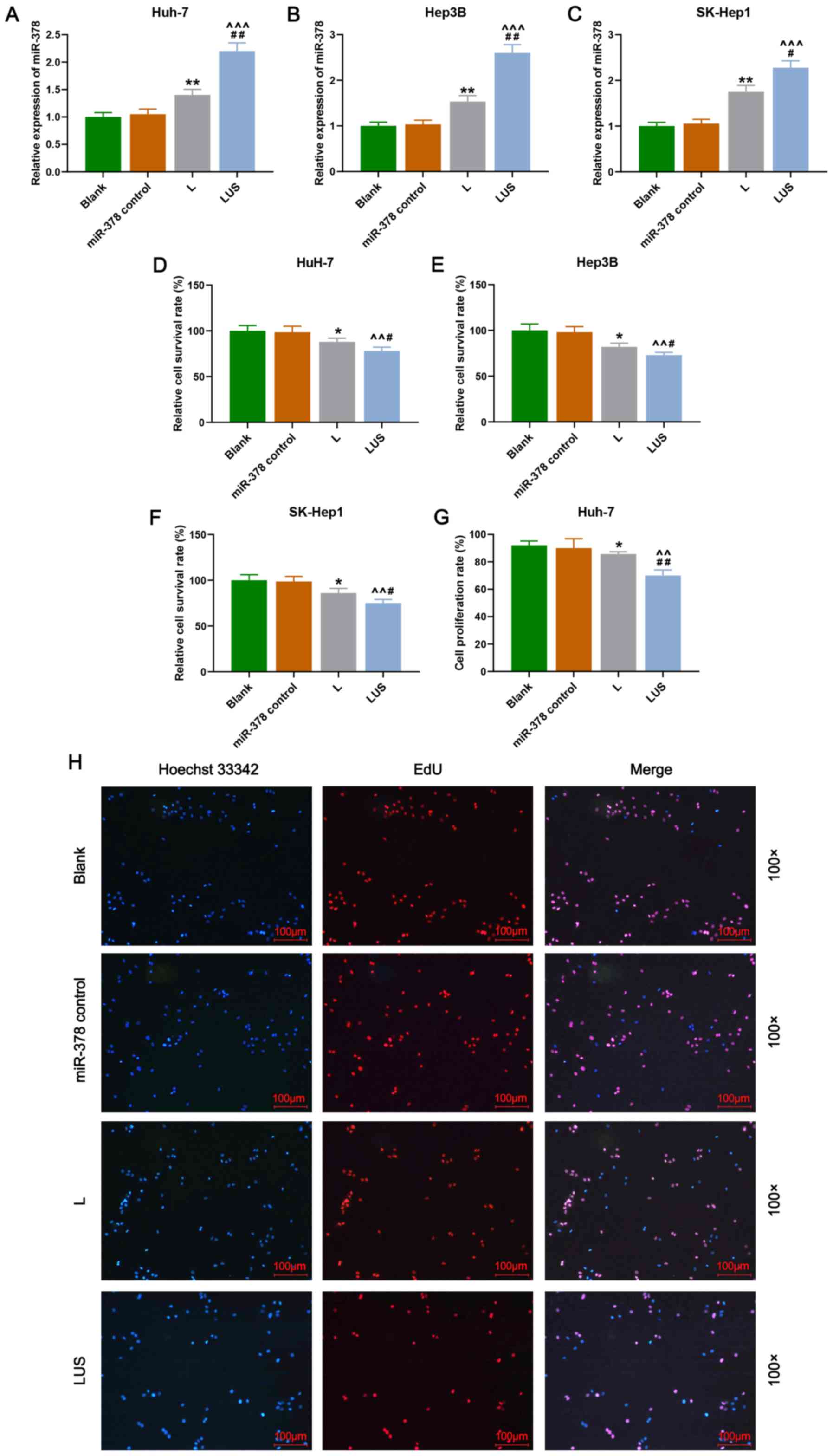
Results
miR-378 inhibits the proliferation of
HuH-7 cells more efficiently using a combination of ultrasonic
irradiation and SonoVue microbubbles method
To investigate the role of miR-378 on the
proliferation of HuH-7, Hep3B and SK-Hep1 cells, and the efficiency
of transfection using ultrasonic irradiation in combination with
SonoVue microbubbles, cell survival rate was detected by performing
CCK-8 and cell proliferation rate was measured using double
cytochemical staining. In the present study, the results revealed
that the relative expression of miR-378 in the L group was
significantly increased compared with that in the Blank group
(P<0.01), and in the LUS group this expression was significantly
increased compared with that in the miR-378 control and L groups
(P<0.001; Fig. 1A-C). Moreover,
the relative cell survival rate (%) of HuH-7, Hep3B and SK-Hep1
cells in the L group was reduced compared with that in the Blank
group, and was also reduced in the LUS group compared with that in
the miR-378 control and L groups (P<0.05 and P<0.01,
respectively; Fig. 1D-F).
Furthermore, the cell proliferation rate (%) of Huh-7 cells in the
L and LUS groups demonstrated a similar trend to that of the cell
survival rate (P<0.05 and P<0.01, respectively; Fig. 1G and H). Thus, these data suggested
that miR-378 expression contributed to the suppression of the
proliferation of HuH-7 cells and that the combination of ultrasonic
irradiation and SonoVue microbubbles method was more effective in
the transfection of miRNA.
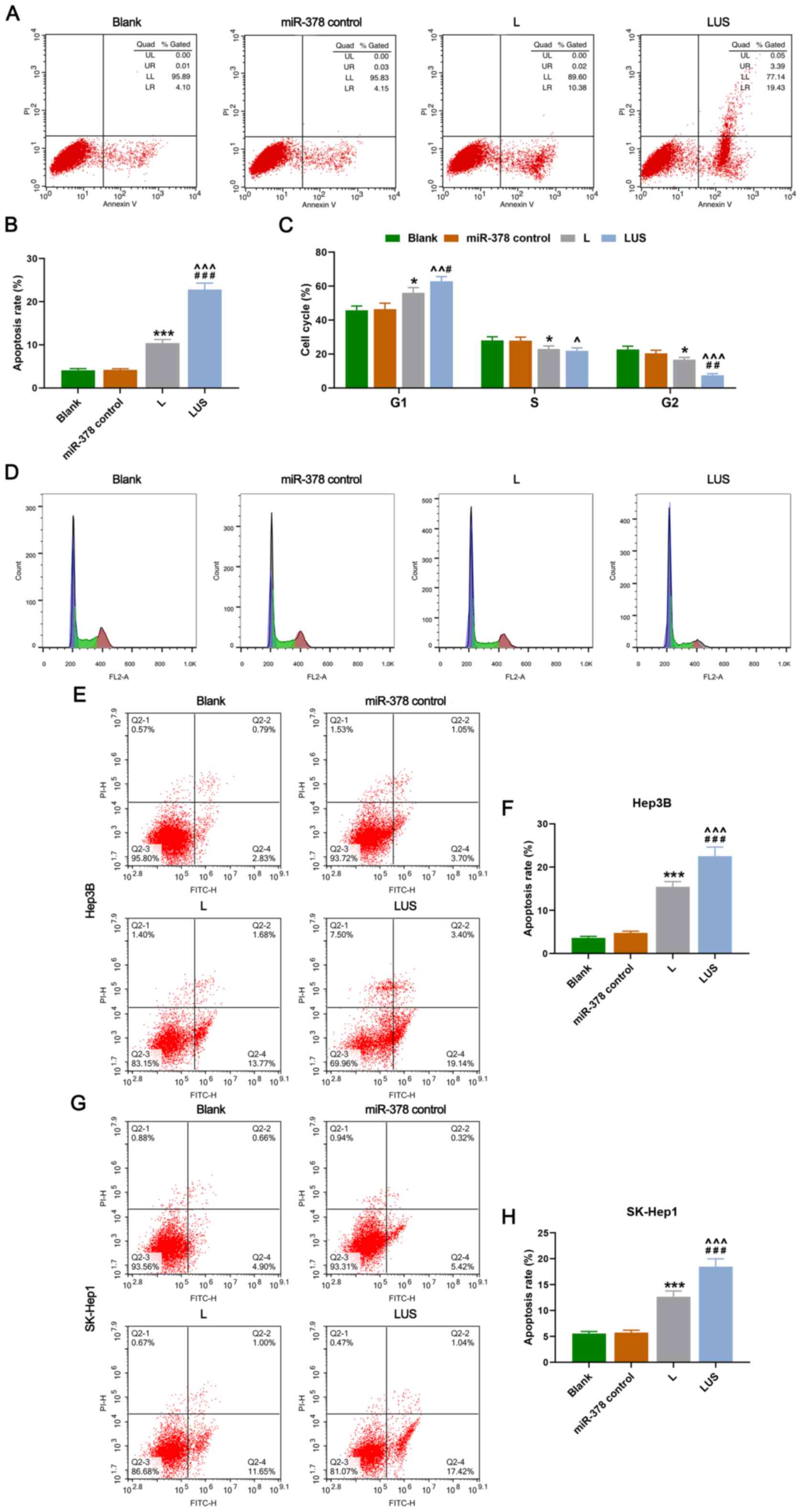
miR-378 increases the apoptosis rate
and arrests the cell cycle of HuH-7 cells more efficiently using a
combined method of ultrasonic irradiation and SonoVue
microbubbles
Cell apoptosis and cell cycle of each phase were
then evaluated. As expected, the apoptosis rate (%) of HuH-7 cells
in the L group was increased compared with that in the Blank group,
moreover, it was significantly elevated in the LUS group compared
with that in the miR-378 control and L groups (P<0.001; Fig. 2A and B). Furthermore, the
percentage of HuH-7 cells in the G1 phase in the L group was
increased compared with that in the Blank group, but it was much
higher in the LUS group than that in the miR-378 control and L
groups (P<0.05 and P<0.01, respectively; Fig. 2C and D). However, the S and G2
phase in the L group were significantly lower than that in the
miR-378 control group, while the two phases in the LUS group were
reduced compared with those in the miR-378 control and L groups,
and no significant difference between the L and LUS groups on S
phase was observed (P<0.05, P<0.01 and P<0.001; Fig. 2C and D). Thus, miR-378 expression
could increase the rate of apoptosis and arrest the cell cycle of
HuH-7 cells in G1 phase more efficiently using a combination of
ultrasonic irradiation and SonoVue microbubbles method. Similarly,
the apoptosis rate (%) of Hep3B cells in the L group was increased
compared with that in the Blank group and it was significantly
higher in the LUS group compared with that in the miR-378 control
and L groups (P<0.001; Fig. 2E and
F). Moreover, the apoptosis rate (%) of SK-Hep1 cells in the L
group was higher than that in the Blank group and it was
significantly increased in the LUS group compared with that in the
miR-378 control and L groups (P<0.001; Fig. 2G and H).
miR-378 regulates genes related to
apoptosis and proliferation of HuH-7 cells
The present study also explored whether genes
related to apoptosis and the proliferation of HuH-7 cells could be
affected by miR-378 expression, and the efficiency of the combined
transfection method. In the current study, the data revealed that
the protein and mRNA expression levels of Cyclin D1, Bcl-2, Akt and
Survivin in the L group were lower than those in the miR-378
control group, and the levels were significantly decreased in the
LUS group compared with those in the L and miR-378 control groups
(P<0.01 and P<0.001; Fig.
3A-C). On the other hand, the expression of Bax and p53
exhibited a different trend, expression increased in the L and LUS
groups compared with the miR-378 control and Blank groups, with the
highest expression found in the LUS group (P<0.05 and
P<0.001; Fig. 3A-C). In
addition, β-catenin expression was significantly reduced in the LUS
group compared with that in the miR-378 control and L groups
(P<0.05 and P<0.001; Fig.
3D-E). Taken together, these results indicated that miR-378
expression led to a decrease in the expression of
proliferation-related genes and increased the expression of
apoptosis-related genes. Additionally, it was demonstrated that the
use of combined ultrasonic irradiation and SonoVue microbubbles
method for the transfection of miR-378 was more effective.
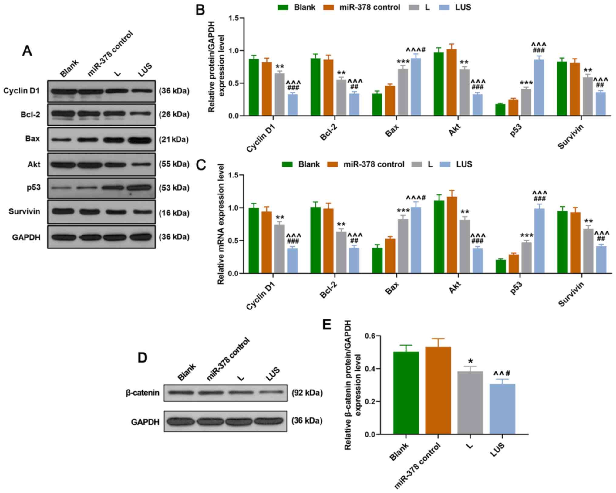 | Figure 3.miR-378 regulates genes related to
apoptosis and proliferation of HuH-7 cells more efficiently. (A)
Western blot images showing protein expression of Cyclin D1, Bcl-2,
Bax, Akt, p53 and Survivin in Blank, miR-378 control, L and LUS
groups. Histograms showing (B) relative protein and (C) mRNA
expression levels in each group. (D) Western blotting was used to
measure the expression of β-catenin. (E) Relative expression of
β-catenin was quantified via western blotting. Bars indicate mean ±
SD. *P<0.05, **P<0.01 and ***P<0.001 vs. Blank;
^^P<0.01 and ^^^P<0.001 vs. miR-378
control; #P<0.05, ##P<0.01 and
###P<0.001 vs. L. L group, the hepatocellular
carcinoma cells were transfected with miR-378; LUS group, the
hepatocellular carcinoma cells were treated with miR-378 mimic
combined with Ultrasonic irradiation and SonoVue microbubbles. miR,
microRNA. |
Discussion
At present, miRNAs have been increasingly found to
have important roles in the development of cancer. It has been
shown that miR-146a not only has a tumor-suppressive effect, but
also plays a critical role in the growth of HCC cells and that
genetic variation of miR-146a may be a risk factor for developing
HCC (21). The expression of
miR143HG was significantly downregulated in HCC cells and tissues,
and was associated with the staging and prognosis of patients with
HCC (22). Furthermore, previous
research has also suggested that the upregulation of miR-21-5p may
be a functional regulator of HCC apoptosis and could be a new tumor
marker for early diagnosis of HCC (23). The current study examined the
effects of miR-378 overexpression on HuH-7 cells and explored the
efficiency of the combined transfection method of ultrasonic
irradiation and SonoVue microbubbles. The results demonstrated that
miR-378 functioned as a suppressor in the proliferation of HuH-7,
Hep3B and SK-Hep1 cells, and this effect of miR-378 could be
enhanced by using the combined method. Thus, these results
indicated that a combined transfection approach is useful and may
have potential in gene therapy.
Overexpressed miR-378 could be transfected into
HuH-7, Hep3B and SK-Hep1 cells, which are representative of
hepatoma cells. Furthermore, it was found that miR-378
overexpression reduced the proliferation ability and increased the
apoptosis rate of HuH-7, Hep3B and SK-Hep1 cells. The cell cycle is
a process during which cells undergo a series of controls and
regulations to maintain an orderly progression of cell growth and
proliferation (24–26). Cyclin/CDK is a promoter of the cell
cycle (24–26). Cyclin D1, a member of the Cyclin
family and also a proto-oncogene, promotes cell G1/S phase via CDK,
thereby promoting cancer development (24–26).
p53 is a tumor suppressor gene located at 17p in human chromosomes
and is normally expressed at low levels in the nucleus (27–30).
Mutated p53 genes have been found in various human tumors,
including in HCC (27–30). p53 proteins are involved in several
cellular processes, including gene transcription, DNA repair, cell
cycle, genome stabilization, chromosome segregation, senescence,
apoptosis and angiogenesis (27–30).
The p53 signal transduction pathway is an important signaling
pathway regulating G1/S phase (27–30).
The role of the Bcl-2 family in apoptosis has received much
attention (31), Bcl-2 is an
inhibitor of apoptosis protein, while Bax is an apoptosis-promoting
protein, these two factors are closely related to the regulation of
apoptosis (31–34). Specifically, Bax forms a
heterodimer with Bcl-2, thereby inhibiting the function of Bcl-2
(35–37), moreover, the susceptibility of cell
apoptosis depends on the ratio of Bax/Bcl-2, which also determines
cell survival after receiving apoptosis signals, thus, the ratio
plays an important role in tumor occurrence (35–37).
Akt is a serine/threonine protein kinase (38–40)
and when cells are stimulated by growth factors, PI3K activates and
produces PIP3 that binds to the PH domain of Akt to trigger the
recruitment of Akt to the cell membrane (38–40).
This process is involved in resistance to apoptosis, glucose
metabolism, protein synthesis and could promote cell growth and
proliferation (38–40). Survivin is one of the members of
the IAP family with the lightest molecular weight (41,42)
and it specifically binds to mitotic spindles during G2/M phase and
can inhibit caspase-3, caspase-7, caspase-8 and cytochrome C, which
are the key factors in apoptosis signaling pathways, therefore it
can promote cancer development (41,42).
In the present study, expression of Cyclin D1, Bcl-2, Akt and
Survivin were reduced in HuH-7 cells transfected with miR-378,
while Bax and p53 expression was elevated, suggesting that the
proliferation of HuH-7 cells was downregulated and apoptosis was
increased by the overexpression of miR-378.
Several researchers have investigated the role of
miR-378 in hepatoma. An et al (43) reported that in a Chinese
population, mutant miR-378 was related to the prognosis of HCC.
Similarly, Hyun et al (44)
revealed that the activation of hepatic stellate cells and the
liver fibrosis process could be suppressed by miR-378. In addition,
Zhou et al (45) found that
miR-378 could be overexpressed by metformin to reduce proliferation
of HCC. Thus, miR-378 may be a suppressor in the process of
hepatoma.
Interestingly, in the present study it was found
that when miR-378 was transfected into HuH-7, Hep3B and SK-Hep1
cells by the combined method of ultrasonic irradiation and SonoVue
microbubbles, the suppressive effect of miR-378 was more
significant. Ran et al (19) indicated that interference of the
target gene by short interfering RNA using the combined method of
ultrasonic irradiation and SonoVue microbubbles was more efficient
than using the lipidosome method for cell transfection. Li et
al (46) also showed that
SonoVue and ultrasound irradiation may have promising effects in
breast cancer therapy by delivering the target genes
effectively.
Generally, traditional viral transfection and
lipofection methods limit the functionality of target genes in
target cells, due to their low safety and poor transfection
efficiency (47,48). However, SonoVue differs from
traditional gene vector microbubbles and can overcome such
limitations (49). When SonoVue
microbubbles are irradiated by ultrasound, they can produce
continuous compression or expansion, leading to the rupture of
microbubbles with accompanied cavitation effect (50). Thus, the permeability of the target
tissue or cell membrane is increased, which promotes the entry of
the gene into the target cells (50).
In conclusion, miR-378 is shown to be a suppressive
factor in HCC, as it suppresses the proliferation and increases the
apoptosis of HCC cells. Moreover, the combination of ultrasonic
irradiation and SonoVue microbubbles method is more efficient in
the transfection of miRNA. Therefore, the present findings
contribute to the current gene therapy for hepatoma, however, the
feasibility of such a method should be further explored.
Acknowledgements
Not applicable.
Funding
No funding was received.
Availability of data and materials
The datasets used and/or analyzed during the current
study are available from the corresponding author on reasonable
request.
Authors' contributions
JW and YL made substantial contributions to study
conception and design, drafted the article and critically revised
it for important intellectual content. QM and JH were involved in
data acquisition, analysis and interpretation. All authors gave
final approval of the version to be published and agree to be
accountable for all aspects of the work in ensuring that questions
related to the accuracy or integrity of the work are appropriately
investigated and resolved. All authors read and approved the final
manuscript.
Ethics approval and consent to
participate
Not applicable.
Patient consent for publication
Not applicable.
Competing interests
The authors declare that they have no competing
interests.
References
|
1
|
Cheng AL, Kang YK, Chen Z, Tsao CJ, Qin S,
Kim JS, Luo R, Feng J, Ye S, Yang TS, et al: Efficacy and safety of
sorafenib in patients in the Asia-Pacific region with advanced
hepatocellular carcinoma: A phase III randomised, double-blind,
placebo-controlled trial. Lancet Oncol. 10:25–34. 2009. View Article : Google Scholar : PubMed/NCBI
|
|
2
|
Llovet JM, Ricci S, Mazzaferro V, Hilgard
P, Gane E, Blanc JF, de Oliveira AC, Santoro A, Raoul JL, Forner A,
et al: Sorafenib in advanced hepatocellular carcinoma. N Engl L
Med. 359:378–390. 2008. View Article : Google Scholar
|
|
3
|
Bruix J and Sherman M; American
Association for the Study of Liver Diseases, : Management of
hepatocellular carcinoma: An update. Hepatology. 53:1020–1022.
2011. View Article : Google Scholar : PubMed/NCBI
|
|
4
|
Llovet JM and Bruix J: Systematic review
of randomized trials for unresectable hepatocellular carcinoma:
Chemoembolization improves survival. Hepatology. 37:429–442. 2003.
View Article : Google Scholar : PubMed/NCBI
|
|
5
|
Nam SW, Park JY, Ramasamy A, Shevade S,
Islam A, Long PM, Park CK, Park SE, Kim SY, Lee SH, et al:
Molecular changes from dysplastic nodule to hepatocellular
carcinoma through gene expression profiling. Hepatology.
42:809–818. 2005. View Article : Google Scholar : PubMed/NCBI
|
|
6
|
Hoshida Y, Villanueva A, Kobayashi M, Peix
J, Chiang DY, Camargo A, Gupta S, Moore J, Wrobel MJ, Lerner J, et
al: Gene expression in fixed tissues and outcome in hepatocellular
carcinoma. N Engl J Med. 359:1995–2004. 2008. View Article : Google Scholar : PubMed/NCBI
|
|
7
|
Van Roosbroeck K and Calin GA: Cancer
hallmarks and MicroRNAs: The therapeutic connection. Adv Cancer
Res. 135:119–149. 2017. View Article : Google Scholar : PubMed/NCBI
|
|
8
|
Wang G, Song L, Hou X, Kala S, Wong KF,
Tang L, Dai Y and Sun L: Surface-modified GVs as nanosized contrast
agents for molecular ultrasound imaging of tumor. Biomaterials.
236:1198032020. View Article : Google Scholar : PubMed/NCBI
|
|
9
|
Qiu L, Leng QY and Luo Y: Progress of
ultrasound microbubble contrast technology in the diagnosis and
treatment of clinical diseases. Sichuan Da Xue Xue Bao Yi Xue Ban.
45:974–978. 2014.(In Chinese). PubMed/NCBI
|
|
10
|
Liu Y, Miyoshi H and Nakamura M:
Encapsulated ultrasound microbubbles: Therapeutic application in
drug/gene delivery. J Control Release. 114:89–99. 2006. View Article : Google Scholar : PubMed/NCBI
|
|
11
|
Price RJ and Kaul S: Contrast ultrasound
targeted drug and gene delivery: An update on a new therapeutic
modality. J Cardiovasc Pharmacol Ther. 7:171–180. 2002. View Article : Google Scholar : PubMed/NCBI
|
|
12
|
Wallace N and Wrenn SP: Ultrasound
triggered drug delivery with liposomal nested microbubbles.
Ultrasonics. 63:31–38. 2015. View Article : Google Scholar : PubMed/NCBI
|
|
13
|
Mountford PA, Sirsi SR and Borden MA:
Condensation phase diagrams for lipid-coated perfluorobutane
microbubbles. Langmuir. 30:6209–6218. 2014. View Article : Google Scholar : PubMed/NCBI
|
|
14
|
Kooiman K, van Rooij T, Qin B, Mastik F,
Vos HJ, Versluis M, Klibanov AL, de Jong N, Villanueva FS and Chen
X: Focal areas of increased lipid concentration on the coating of
microbubbles during short tone-burst ultrasound insonification.
PLoS One. 12:e01807472017. View Article : Google Scholar : PubMed/NCBI
|
|
15
|
Myrset AH, Fjerdingstad HB, Bendiksen R,
Arbo BE, Bjerke RM, Johansen JH, Kulseth MA and Skurtveit R: Design
and characterization of targeted ultrasound microbubbles for
diagnostic use. Ultrasound Med Biol. 37:136–150. 2011. View Article : Google Scholar : PubMed/NCBI
|
|
16
|
Lammertink B, Deckers R, Storm G, Moonen C
and Bos C: Duration of ultrasound-mediated enhanced plasma membrane
permeability. Int J Pharm. 482:92–98. 2015. View Article : Google Scholar : PubMed/NCBI
|
|
17
|
Yang PS, Tung FI, Chen HP, Liu TY and Lin
YY: A novel bubble-forming material for preparing
hydrophobic-agent-loaded bubbles with theranostic functionality.
Acta Biomater. 10:3762–3774. 2014. View Article : Google Scholar : PubMed/NCBI
|
|
18
|
Rychak JJ and Klibanov AL: Nucleic acid
delivery with microbubbles and ultrasound. Adv Drug Deliv Rev.
72:82–93. 2014. View Article : Google Scholar : PubMed/NCBI
|
|
19
|
Ran LW, Wang H, Lan D, Jia HX and Yu SS:
Effect of RNA interference targeting STAT3 gene combined with
ultrasonic irradiation and SonoVue microbubbles on proliferation
and apoptosis in keratinocytes of psoriatic lesions. Chin Med J
(Engl). 131:2097–2104. 2018. View Article : Google Scholar : PubMed/NCBI
|
|
20
|
Livak KJ and Schmittgen TD: Analysis of
relative gene expression data using real-time quantitative PCR and
the 2(-Delta Delta C(T)) method. Methods. 25:402–408. 2001.
View Article : Google Scholar : PubMed/NCBI
|
|
21
|
Wang H, Li X, Li T, Wang L, Wu X, Liu J,
Xu Y and Wei W: Multiple roles of microRNA-146a in immune responses
and hepatocellular carcinoma. Oncol Lett. 18:5033–5042.
2019.PubMed/NCBI
|
|
22
|
Lin X, Xiaoqin H, Jiayu C, Li F, Yue L and
Ximing X: Long non-coding RNA miR143HG predicts good prognosis and
inhibits tumor multiplication and metastasis by suppressing
mitogen-activated protein kinase and Wnt signaling pathways in
hepatocellular carcinoma. Hepatol Res. 49:902–918. 2019. View Article : Google Scholar : PubMed/NCBI
|
|
23
|
Zhong XZ, Deng Y, Chen G and Yang H:
Investigation of the clinical significance and molecular mechanism
of miR-21-5p in hepatocellular carcinoma: A systematic review based
on 24 studies and bioinformatics investigation. Oncol Lett.
17:230–246. 2019.PubMed/NCBI
|
|
24
|
Kim JK and Diehl JA: Nuclear cyclin D1: An
oncogenic driver in human cancer. J Cell Physiol. 220:292–296.
2009. View Article : Google Scholar : PubMed/NCBI
|
|
25
|
Qie S and Diehl JA: Cyclin D1, cancer
progression, and opportunities in cancer treatment. J Mol Med
(Berl). 94:1313–1326. 2016. View Article : Google Scholar : PubMed/NCBI
|
|
26
|
Witzel II, Koh LF and Perkins ND:
Regulation of cyclin D1 gene expression. Biochem Soc Trans.
38:217–222. 2010. View Article : Google Scholar : PubMed/NCBI
|
|
27
|
Hayman L, Chaudhry WR, Revin VV, Zhelev N
and Bourdon JC: What is the potential of p53 isoforms as a
predictive biomarker in the treatment of cancer? Expert Rev Mol
Diagn. 19:149–159. 2019. View Article : Google Scholar : PubMed/NCBI
|
|
28
|
Tiwari B, Jones AE and Abrams JM:
Transposons, p53 and genome security. Trends Genet. 34:846–855.
2018. View Article : Google Scholar : PubMed/NCBI
|
|
29
|
Xue Y, San Luis B and Lane DP: Intratumour
heterogeneity of p53 expression; causes and consequences. J Pathol.
249:274–285. 2019. View Article : Google Scholar : PubMed/NCBI
|
|
30
|
Chabeda A, Yanez RJR, Lamprecht R, Meyers
AE, Rybicki EP and Hitzeroth II: Therapeutic vaccines for high-risk
HPV-associated diseases. Papillomavirus Res. 5:46–58. 2018.
View Article : Google Scholar : PubMed/NCBI
|
|
31
|
O'Neill JW and Hockenbery DM:
Bcl-2-related proteins as drug targets. Curr Med Chem.
10:1553–1562. 2003. View Article : Google Scholar : PubMed/NCBI
|
|
32
|
Brown LM, Hanna DT, Khaw SL and Ekert PG:
Dysregulation of BCL-2 family proteins by leukemia fusion genes. J
Biol Chem. 292:14325–14333. 2017. View Article : Google Scholar : PubMed/NCBI
|
|
33
|
Peña-Blanco A and García-Sáez AJ: Bax, Bak
and beyond-mitochondrial performance in apoptosis. FEBS J.
285:416–431. 2018. View Article : Google Scholar : PubMed/NCBI
|
|
34
|
Renault TT, Dejean LM and Manon S: A
brewing understanding of the regulation of Bax function by Bcl-xL
and Bcl-2. Mech Ageing Dev. 161:201–210. 2017. View Article : Google Scholar : PubMed/NCBI
|
|
35
|
Stefanaki C, Antoniou C, Stefanaki K,
Petrikos G, Argyrakos T, Constantinidou CV, Karentzou O, Stratigos
A and Katsambas A: Bcl-2 and Bax in congenital naevi. Br J
Dermatol. 154:1175–1179. 2006. View Article : Google Scholar : PubMed/NCBI
|
|
36
|
Abu Zeid EH, Hussein MMA and Ali H:
Ascorbic acid protects male rat brain from oral potassium
dichromate-induced oxdative DNA damage and apoptotic changes: The
expression patterns of caspase-3, P 53, Bax, and Bcl-2 genes.
Environ Sci Pollut Res Int. 25:13056–13066. 2018. View Article : Google Scholar : PubMed/NCBI
|
|
37
|
Zhang Y, Yang X, Ge X and Zhang F:
Puerarin attenuates neurological deficits via Bcl-2/Bax/cleaved
caspase-3 and Sirt3/SOD2 apoptotic pathways in subarachnoid
hemorrhage mice. Biomed Pharmacother. 109:726–733. 2019. View Article : Google Scholar : PubMed/NCBI
|
|
38
|
Lien EC, Dibble CC and Toker A: PI3K
signaling in cancer: Beyond AKT. Curr Opin Cell Biol. 45:62–71.
2017. View Article : Google Scholar : PubMed/NCBI
|
|
39
|
Driessen GJ, IJspeert H, Wentink M, Yntema
HG, van Hagen PM, van Strien A, Bucciol G, Cogulu O, Trip M,
Nillesen W, et al: Increased PI3K/Akt activity and deregulated
humoral immune response in human PTEN deficiency. J Allergy Clin
Immunol. 138:1744–1747.e1745. 2016. View Article : Google Scholar : PubMed/NCBI
|
|
40
|
Li Y, Fu LX, Zhu WL, Shi H, Chen LJ and Ye
B: Blockade of CXCR6 reduces invasive potential of gastric cancer
cells through inhibition of AKT signaling. Int J Immunopathol
Pharmacol. 28:194–200. 2015. View Article : Google Scholar : PubMed/NCBI
|
|
41
|
Han G, Gong H, Wang Y, Guo S and Liu K:
AMPK/mTOR-mediated inhibition of survivin partly contributes to
metformin-induced apoptosis in human gastric cancer cell. Cancer
Biol Ther. 16:77–87. 2015. View Article : Google Scholar : PubMed/NCBI
|
|
42
|
Liu JL, Gao W, Kang QM, Zhang XJ and Yang
SG: Prognostic value of survivin in patients with gastric cancer: A
systematic review with meta-analysis. PLoS One. 8:e719302013.
View Article : Google Scholar : PubMed/NCBI
|
|
43
|
An J, Liu J, Liu L, Liu Y, Pan Y, Huang M,
Qi F, Wen J, Xie K, Ma H, et al: A genetic variant in primary
miR-378 is associated with risk and prognosis of hepatocellular
carcinoma in a Chinese population. PLoS One. 9:e937072014.
View Article : Google Scholar : PubMed/NCBI
|
|
44
|
Hyun J, Wang S, Kim J, Rao KM, Park SY,
Chung I, Ha CS, Kim SW, Yun YH and Jung Y: MicroRNA-378 limits
activation of hepatic stellate cells and liver fibrosis by
suppressing Gli3 expression. Nat Commun. 7:109932016. View Article : Google Scholar : PubMed/NCBI
|
|
45
|
Zhou J, Han S, Qian W, Gu Y, Li X and Yang
K: Metformin induces miR-378 to downregulate the CDK1, leading to
suppression of cell proliferation in hepatocellular carcinoma.
OncoTargets Ther. 11:4451–4459. 2018. View Article : Google Scholar
|
|
46
|
Li XH, Zhou P, Wang LH, Tian SM, Qian Y,
Chen LR and Zhang P: The targeted gene (KDRP-CD/TK) therapy of
breast cancer mediated by SonoVue and ultrasound irradiation in
vitro. Ultrasonics. 52:186–191. 2012. View Article : Google Scholar : PubMed/NCBI
|
|
47
|
Thomas CE, Ehrhardt A and Kay MA: Progress
and problems with the use of viral vectors for gene therapy. Nat
Rev Genet. 4:346–358. 2003. View Article : Google Scholar : PubMed/NCBI
|
|
48
|
Ding B, Li T, Zhang J, Zhao L and Zhai G:
Advances in liver-directed gene therapy for hepatocellular
carcinoma by non-viral delivery systems. Curr Gene Ther. 12:92–102.
2012. View Article : Google Scholar : PubMed/NCBI
|
|
49
|
Delalande A, Bastié C, Pigeon L, Manta S,
Lebertre M, Mignet N, Midoux P and Pichon C: Cationic gas-filled
microbubbles for ultrasound-based nucleic acids delivery. Biosci
Rep. 37:BSR201606192017. View Article : Google Scholar : PubMed/NCBI
|
|
50
|
Qin P, Han T, Yu ACH and Xu L: Mechanistic
understanding the bioeffects of ultrasound-driven microbubbles to
enhance macromolecule delivery. J Control Release. 272:169–181.
2018. View Article : Google Scholar : PubMed/NCBI
|