Introduction
In China, lung cancer is the leading cause of cancer
death in males and the second leading cause of death in females
(1). Lung cancer is also one of
the leading causes of cancer-associated mortality globally
(2). Lung cancer can be classified
as small cell lung cancer and non-small cell lung cancer (NSCLC)
(2). The standard first-line
therapies for lung cancer include surgery, radiotherapy,
chemotherapy, targeted therapy and immunotherapy (2); however, NSCLC is characterised by
both aggressive behaviour and a poor response to chemotherapy
(3). Moreover, secondary
resistance often occurs as a result of exposure to chemotherapeutic
agents (4). Therefore, it is
important to explore new strategies to treat NSCLC.
Gonzalez et al (5), reported that mannose, a type of
monosaccharide, induced growth retardation in various tumour types
in vitro and enhanced cell death in response to major forms of
chemotherapy in several cancers, including osteogenic sarcoma,
ovarian cancer and pancreatic cancer. Whether mannose exerts
antigrowth effects on NSCLC remains to be elucidated. The present
study aimed to identify the effects of mannose on the
proliferation, cell cycle progression, cisplatin-mediated apoptosis
and metastasis of A549 and H1299 cells, in order to determine the
mechanisms responsible for the mannose-induced anticancer
effects.
Materials and methods
Cancer cell lines
A549 and H1299 lung adenocarcinoma cells were
purchased from the American Type Culture Collection. 293FT cells
were purchased from the Shanghai Cell Bank of the Chinese Academy
of Sciences. Cells were cultured in Dulbeccos modified Eagles
medium (DMEM; Thermo Fisher Scientific, Inc.) supplemented with 10%
FBS (Biological Industries), streptomycin (100 U/ml) and penicillin
(100 U/ml). All cells were incubated at 37°C in an incubator
containing 5% CO2. Mannose was dissolved in water, and
the concentration of the storage stock was 500 mM (stored at
−20°C).
Cell Counting Kit-8 (CCK-8) assay
Cell viability was detected with a CCK-8 assay
(Dojindo Molecular Technologies, Inc.) according to the
manufacturers protocol. Cancer cells (5,000 cells/well) were seeded
in 96-well plates in complete culture medium for 24 h. To evaluate
the antitumour effects of mannose on A549 and H1299 cells in vitro,
different concentrations (0, 15, 30 and 60 mM) of mannose were used
to treat cancer cells for 24 h at 37°C. The IC50 values
of cisplatin against A549 and H1299 cells were 34.08 and 23.12 µM,
respectively, which were calculated using GraphPad Prism v6
(GraphPad Software, Inc.). In addition, 30 mM mannose was used to
treat cancer cells for 12, 24 or 48 h. To determine whether mannose
could augment cisplatin-mediated apoptosis in lung cancer, 30 mM
mannose, 30 µM cisplatin or mannose + cisplatin was used to treat
cancer cells for 24 h at 37°C. Then flow cytometry and western
blotting were performed to evaluate the effects of mannose on
cisplatin-mediated apoptosis of lung cancer cells.
Flow cytometry-based method for
evaluating the cell cycle distribution
To assess the influence of mannose on the cell cycle
progression of lung cancer cells, after being treated with 30 mM
mannose for 24 h, cancer cells were harvested and fixed with 70%
ethanol at −20°C for 24 h. Then, cells were stained with 0.1 mg/ml
propidium iodide (PI; Sigma-Aldrich; Merck KGaA) for 15 min at room
temperature in the dark, and the cell cycle distribution was
evaluated and analysed using an Accuri C6 flow cytometer (BD
Biosciences).
Flow cytometry-based method for
evaluating apoptosis
To evaluate the effects of mannose on
cisplatin-induced apoptosis, cell samples were grouped as follows:
Control, 30 mM mannose, 30 µM cisplatin and 30 mM mannose + 30 µM
cisplatin. After 24 h, cells were collected for cell apoptosis
analysis. Apoptotic cells were identified using flow cytometric
analysis with an Annexin V-FITC kit (Invitrogen; Thermo Fisher
Scientific, Inc.) according to the manufacturers instructions.
Samples were stained with annexin-V-FITC and PI for 15 min at room
temperature in the dark. In the four fields on the original images,
the dots indicate the number of Annexin V-/PI- (the bottom left
field, indicating live cells), Annexin V+/PI- (the bottom right
field, indicating early apoptotic cells), Annexin V+/PI+ (the top
right field, indicating late apoptotic cells), and Annexin V-/PI+
(the top left field, indicating dead cells) cells. The percent of
apoptotic cells was calculated as follows: Apoptosis rate (%) =
rate of early apoptotic cells (%) + rate of late apoptotic cells
(%). The apoptotic rate was evaluated and analysed using an Accuri
C6 flow cytometer (BD Biosciences).
Transwell migration and invasion
assays
To detect the invasive ability of cancer cells in
vitro, 5 µl of Matrigel (BD Biosciences) was spread in the
upper chamber of a 24-well Transwell plate (BD Biosciences). After
being treated with or without 30 mM mannose for 24 h, cancer cells
were resuspended at a density of 1×10⁵ cells/ml in DMEM
without FBS. Then, 0.5 ml of the cell suspension was added to each
upper chamber of the 24-well plate, and 0.5 ml of DMEM containing
20% FBS to the lower chambers. After 24 h, the Matrigel and NSCLC
cells in the upper chambers were cleaned carefully and thoroughly.
The upper and lower chambers were washed once with 1X PBS, and
cells were fixed with 4% paraformaldehyde for 20 min at room
temperature. NSCLC cells on the lower surface of the upper chambers
were stained with 0.25% crystal violet, which was dissolved in 20%
methanol, for 30 min at room temperature. The samples were washed
twice with PBS, and the membranes of the chambers were sealed with
cover glue (Leica Microsystems GmbH). Membranes were wetted with
PBS and observed under a microscope (Nikon Corporation); 5 random
fields (magnification, ×20) were selected, the NSCLC cells were
counted, and the average numbers were analysed and compared.
For detection of the migration ability of cancer
cells in vitro, the protocol was the same as that used for the
invasion assays, but Matrigel was not used.
Western blot assay
For western blotting, cancer cells were treated with
30 and 60 mM mannose for 24 h. Then, total protein lysate from
cancer cells was extracted with RIPA lysis buffer (Shanghai Yeasen
Biotechnology Co.) and the protein concentration was evaluated by
bicinchoninic acid assay (Thermo Fisher Scientific, Inc.). A total
of 10–20 µg protein lysate per lane was separated by 10% SDS-PAGE
and transferred to polyvinylidene difluoride membranes (EMD
Millipore). Membranes were blocked with 5% non-fat milk in Tris-HCl
buffer solution containing 0.1% Tween 20 (TBST) for 1 h at room
temperature and incubated separately with primary antibodies
against the following proteins at 4°C overnight: p21 (1:1,000; cat.
no. ab218311; Abcam), proliferating cell nuclear antigen (PCNA;
1:1,000; cat. no. ab92552; Abcam), Bcl-2 (1:1,000; cat. no.
ab185002; Abcam), Bax (1:1,000; cat. no. ab32503; Abcam), cleaved
caspase-3 (1:1,000; cat. no. ab2302; Abcam), GAPDH (1:10,000; cat.
no. ab181602; Abcam), matrix metalloproteinase 2 (MMP2; 1:1,000;
cat. no. ab97779; Abcam), E-cadherin (1:1,000; cat. no. ab1416;
Abcam), N-cadherin (1:1,000; cat. no. ab18203; Abcam),
phosphorylated (p)-AKT (Ser473; 1:1,000; cat. no. ab81283; Abcam),
total AKT (1:1,000; cat. no. ab8805; Abcam), p-ERK1/2 (1:1,000;
cat. no. ab223500; Abcam), and total ERK (1:1,000; cat. no.
ab17942; Abcam). After washing with TBST, membranes were incubated
with horseradish peroxidase-conjugated anti-rabbit IgG (1:2,500;
cat. nos. ab6721 and ab6728; Abcam) for 1 h at room temperature.
Visualization of blots was performed using a standard protocol for
electro-chemiluminescence (cat. no. ab133406; Abcam). The levels of
GAPDH were used as the internal standards. Densitometry was
performed using Image J software (v1.47; National Institutes of
Health).
Statistical analysis
Data are presented as the mean ± SD. Students t-test
or one-way analysis of variance (ANOVA) was used to evaluate
statistical differences via GraphPad Prism v6 (GraphPad Software,
Inc.). Tukeys multiple comparisons test was used to analyze data
following ANOVA. P<0.05 was considered to indicate a
statistically significant difference.
Results
Mannose inhibits the viability of lung
cancer cells in vitro
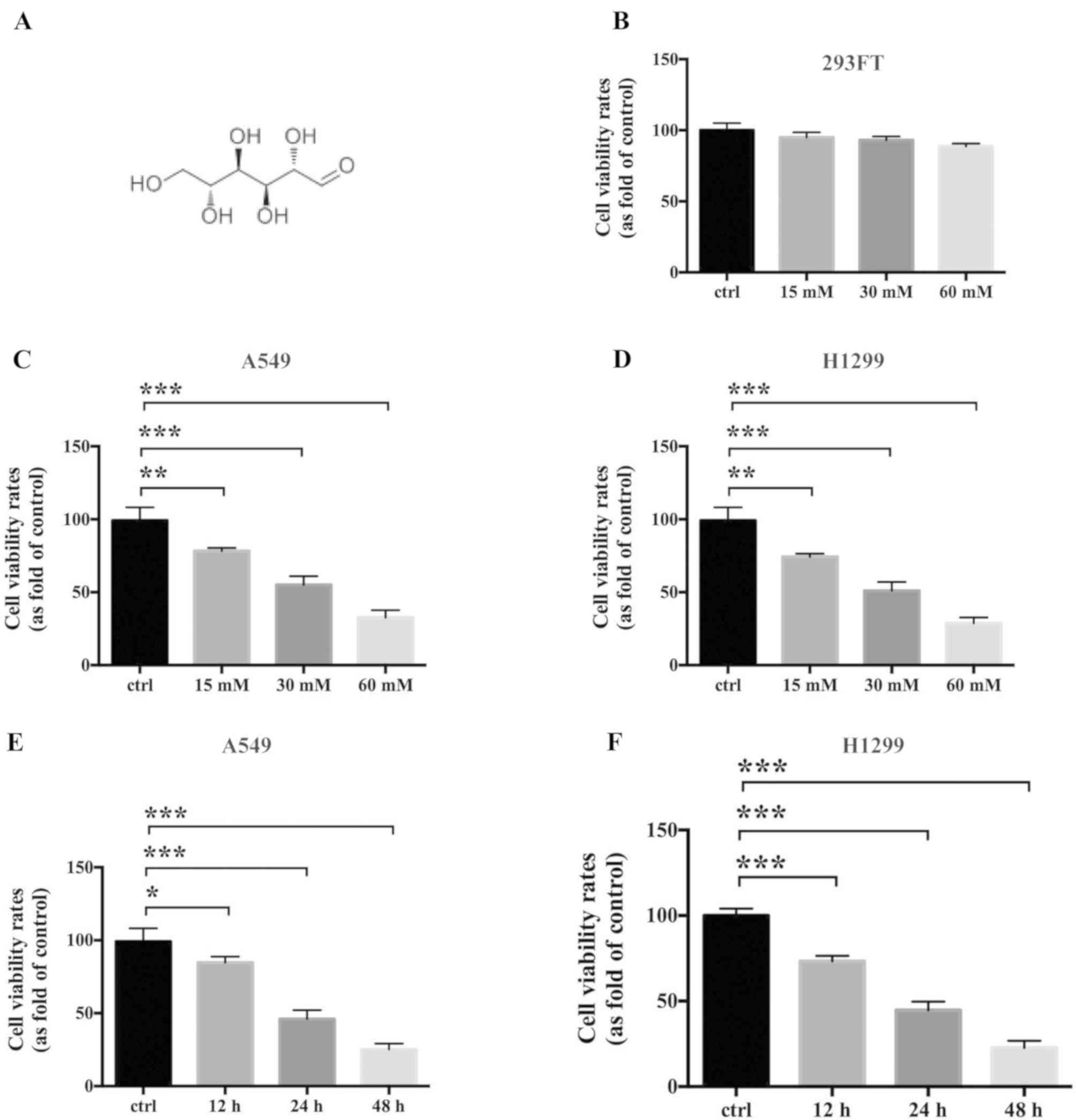
Mannose is a carbohydrate; the structure of mannose
is shown in Fig. 1A. A549, H1299
and 293FT cells were used in the present study to evaluate the
antigrowth effects of mannose on NSCLC and non-cancer cell lines in
vitro. A CCK-8 assay showed that mannose does not significantly
affect the viability of 293FT cells (n=3; Fig. 1B). Conversely, mannose
significantly inhibited the viability of A549 and H1299 cells
dose-dependently at 24 h in vitro (n=3; Fig. 1C and D). The IC50 of
mannose against A549 and H1299 cells at 24 h was ~30 mM, which was
used as the experimental concentration in subsequent assays. In
addition, 30 mM mannose significantly reduced the viability of A549
and H1299 cells in a time-dependent manner in vitro (n=3; Fig. 1E and F).
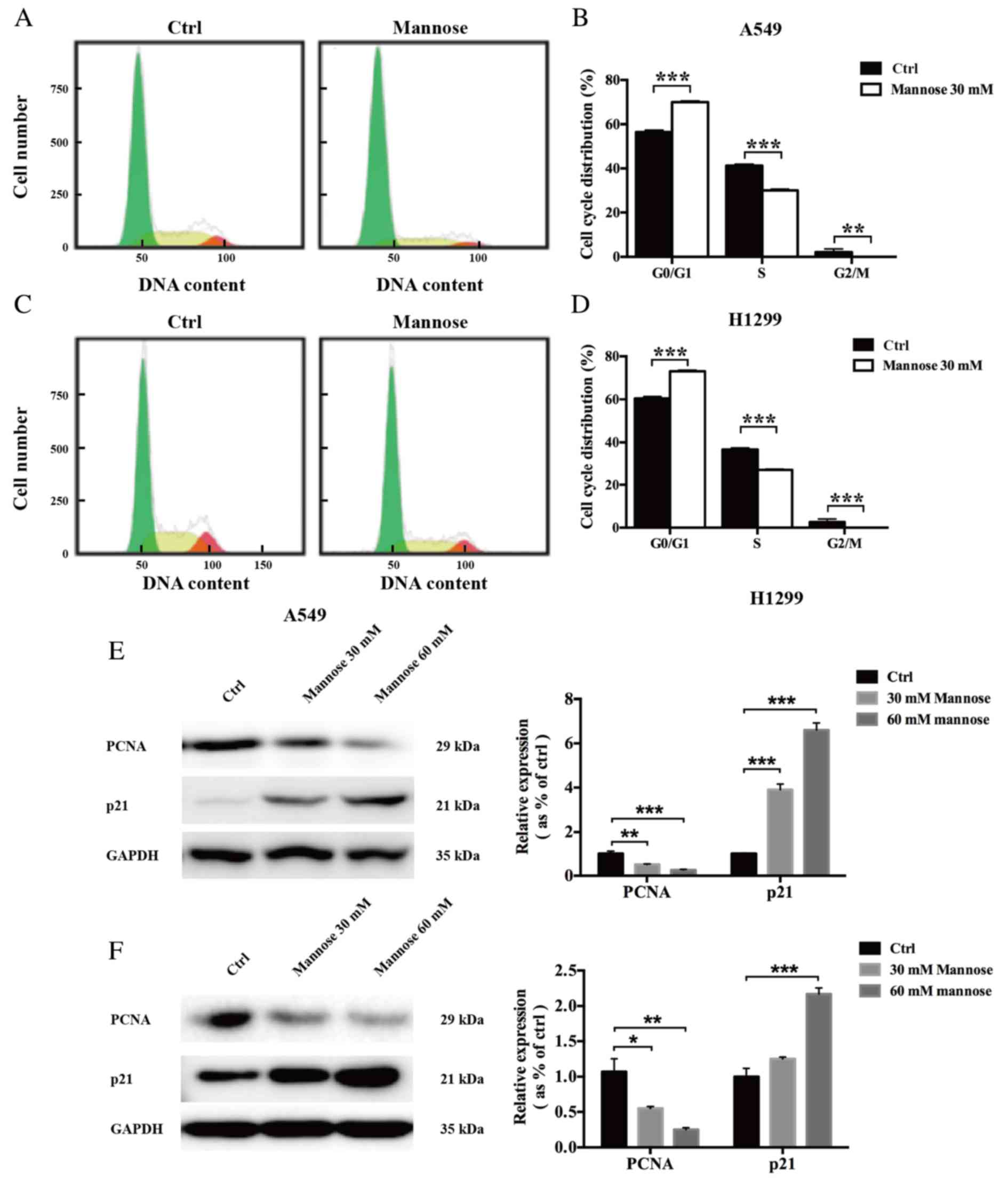
Mannose induces G0/G1 phase arrest in
lung cancer cells in vitro
To further examine whether mannose inhibits the
proliferation of NSCLC cells by regulating cell cycle progression,
a flow cytometry-based method was used. The results showed that
after treatment with 30 mM mannose for 24 h, the cell cycle
distribution of A549 and H1299 cells was significantly altered
compared with the control group (Fig.
2A-D). It was revealed that mannose increased the population of
G0/G1-phase and reduced the population of S/G2/M-phase A549 (n=3;
Fig. 2A and B) and H1299 cells
in vitro (n=3; Fig. 2C and
D). Next, the effects of mannose on the expression levels of
two regulators of the cell cycle, PCNA and p21, were further
investigated via western blotting. The western blot assay results
showed that treatment with 30 or 60 mM mannose for 24 h
downregulated PCNA expression in both A549 and H1299 cells in
vitro; in contrast, mannose treatment significantly upregulated
p21 expression in NSCLC cells (Fig. 2E
and F). Furthermore, the effects of mannose on the expression
levels of PCNA and p21 increased in a concentration-dependent
manner. Mannose may induce G0/G1 cell cycle arrest in A549 and
H1299 cells by affecting the expression of PCNA and p21.
Mannose increases cisplatin-mediated
apoptosis of lung cancer cells in vitro
Cisplatin resistance in NSCLC is an urgent problem
that requires overcoming to improve the outcomes of lung cancer
therapy (6). In the present study,
flow cytometric and western blot analyses were conducted to
evaluate whether mannose could induce apoptosis in A549 and H1299
cells in vitro, and whether it could augment
cisplatin-mediated cellular death. First, a CCK-8 cell viability
assay was used to determine the IC50 of cisplatin
against both A549 and H1299 cells. The IC50 values of
cisplatin against A549 and H1299 cells were 34.08 µM and 23.12 µM,
respectively (Fig. 3A), so 30 µM
cisplatin was selected for use in subsequent experiments. Then, a
flow cytometry-based method was used to evaluate the effects of
mannose, cisplatin and mannose + cisplatin on apoptosis induction
in A549 and H1299 cells in vitro. Compared with the control
treatment, both 30 mM mannose and 30 µM cisplatin could
significantly induce apoptosis in NSCLC cells (Fig. 3B). Furthermore, under treatment
with the combination of 30 mM mannose + 30 µM cisplatin, the
percentage of apoptotic cancer cells was further increased compared
with single-agent mannose or cisplatin treatment (Fig. 3B). To further explore the molecular
changes responsible for this phenomenon, western blotting was
conducted to measure the expression levels of apoptosis-related
proteins, including cleaved caspase-3, Bax and Bcl-2. The results
showed that treatment with 30 mM mannose or 30 µM cisplatin for 24
h downregulated Bcl-2 expression in both A549 and H1299 cells in
vitro. In contrast, the expression of both cleaved caspase-3 and
Bax in NSCLC cells was upregulated (Fig. 3C and E). Furthermore, mannose
significantly augmented the effects of cisplatin on the expression
of cleaved caspase-3, Bax and Bcl-2. Mannose may thus enhance the
cisplatin-mediated apoptosis of A549 and H1299 cells by affecting
the expression of Bax/Bcl-2 and caspase-3.
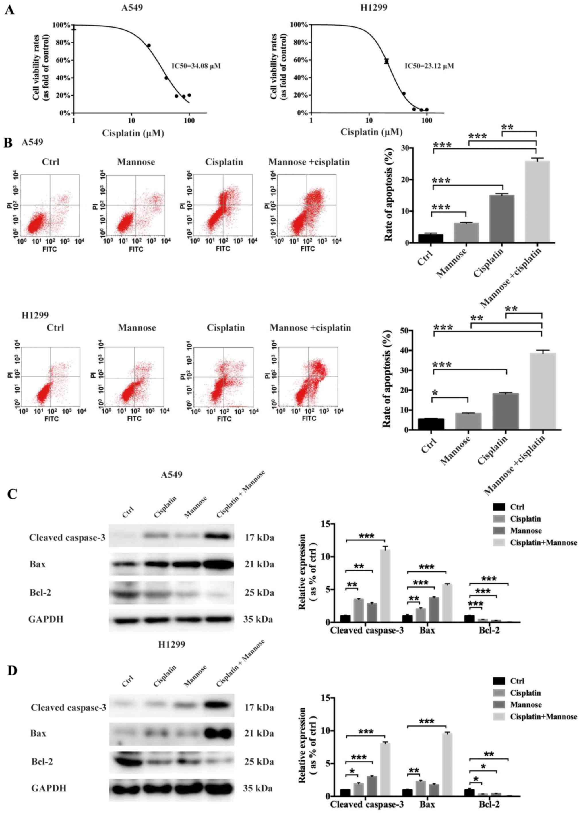 | Figure 3.Mannose increases the
cisplatin-mediated apoptosis of NSCLC cells in vitro. (A)
CCK-8 cell viability assays were conducted to determine the
IC50 of cisplatin against A549 and H1299 cells at 24 h.
(B) Flow cytometry was performed to evaluate the effects of
mannose, cisplatin, or a combination of cisplatin and mannose on
the apoptosis of NSCLC cells in vitro. Early apoptotic cells
are represented in the bottom right field (Annexin V+/PI-). Late
apoptotic cells are represented in the top right field (Annexin
V+/PI+). (C and D) Western blotting was conducted to evaluate the
effects of mannose, cisplatin, or a combination of cisplatin and
mannose on the expression of apoptosis-related proteins, including
cleaved caspase-3, Bax and Bcl-2. Data are presented as the mean ±
standard deviation. *P<0.05, **P<0.01, ***P<0.001. NSCLC,
non-small cell lung cancer; ctrl, control; PI, propidium
iodide. |
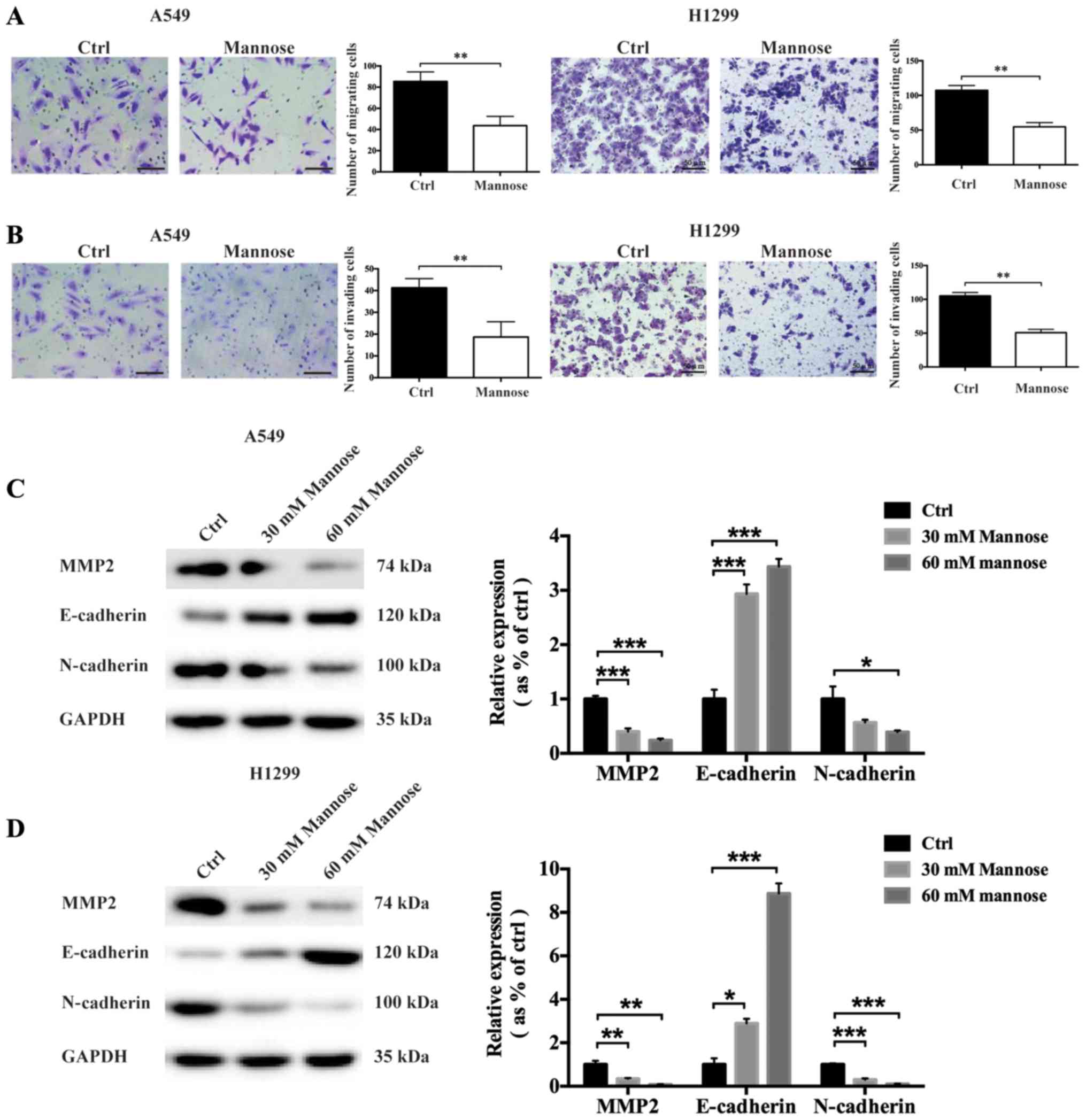
Mannose reduces the migration and
invasive abilities of lung cancer cells in vitro
The metastasis of malignant tumours is one of the
primary complications of cancers, including NSCLC (7). To determine whether mannose could
inhibit the invasive abilities of NSCLC cells, Transwell migration
and invasion, and western blot assays were used to evaluate the
anti-invasion effects of mannose. Following treatment with 30 mM
mannose for 24 h, the migration of A549 and H1299 cells was
significantly reduced in vitro (Fig.
4A). The Transwell invasion assays showed that mannose also
reduced the invasive abilities of NSCLC cells in vitro (Fig. 4B). To further explore the effects
of mannose on epithelial-mesenchymal transition (EMT) in A549 and
H1299 cells, western blotting was used to assess the expression of
MMP2, E-cadherin and N-cadherin. It was observed that treatment
with 30 or 60 mM mannose for 24 h downregulated N-cadherin and MMP2
expression in both A549 and H1299 cells in vitro (Fig. 4C and D). In contrast, mannose
treatment significantly upregulated E-cadherin expression in NSCLC
cells. Furthermore, the effects of mannose on the expression of
MMP2, E-cadherin and N-cadherin significantly increased in a
concentration-dependent manner. Mannose may inhibit the metastatic
ability of NSCLC cells by inhibiting EMT.
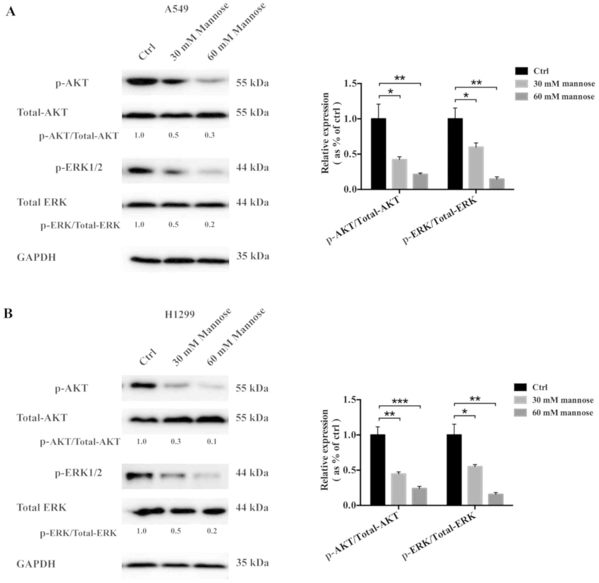
Mannose inhibits the PI3K/AKT and ERK
signalling pathways in lung cancer
A number of studies have reported the contribution
of the PI3K/AKT signalling pathway (8) and mitogen-activated protein kinase
(MAPK/ERK) signalling pathway (9,10) to
the progression of lung cancer, including the promotion of
proliferation, reductions in apoptosis and the facilitation of
metastasis. In the current study, western blotting was conducted to
examine the effects of mannose on the activation of the PI3K/AKT
and ERK signalling pathways in A549 and H1299 cells in
vitro, including the levels of p-AKT (Ser473) and p-ERK1/2. It
was found that treatment with 30 or 60 mM mannose for 24 h
decreased the levels of p-AKT (Ser473) and p-ERK1/2 in both A549
and H1299 cells in vitro (Fig.
5A and B). The ratios of p-AKT/total AKT and p-ERK1/2/total ERK
were significantly decreased in A549 and H1299 cells. The
inhibitory regulation of the AKT and ERK signalling pathways by
mannose increased in a concentration-dependent manner. Mannose may
exert anticancer effects on A549 and H1299 cells by inhibiting the
PI3K/AKT and ERK signalling pathways.
Discussion
The present study identified that mannose exerted
anticancer effects on NSCLC cell lines (A549 and H1299) in
vitro by inhibiting proliferation, inducing G0/G1 cell cycle
arrest, promoting cisplatin-induced apoptosis and decreasing
invasion. Li et al (11),
observed that Pseudomonas aeruginosa mannose-sensitive
haemagglutinin (PA-MSHA) exhibited antitumour properties against
hepatocellular carcinoma (HCC) through mannose-binding activity and
downregulation of the epidermal growth factor receptor
(EGFR)/Akt/IκBβ/NF-κB pathway. PA-MSHA induced significant cell
proliferation inhibition and cell cycle arrest by decreasing the
expression levels of PCNA, cyclin-D1/E, cyclin-dependent kinases
2/4 and increasing the levels of p21 and p27 (11). PA-MSHA also suppressed the
migration, invasion and adhesion of HCC cells by inhibiting EMT,
consistent with the present study (11). Sturge et al (12), reported that the mannose receptor
(MR) was important for macrophage migration; bone marrow-derived
macrophages obtained from MR-deficient mice exhibited an increase
in random cell migration. Ding et al (13), hypothesised that MR could be a
novel biomarker for colorectal cancer (CRC), reporting that the
expression of MR in CRC tissues was significantly upregulated
compared with in paracancerous tissues. Furthermore, high
expression of MR in CRC tissues was also associated with reduced
overall survival and an increased degree of lymphatic metastasis.
These findings highlight the potential value of exploring the
effects of mannose and its receptor on cancer metastasis.
It has been reported that the PI3K-AKT and ERK1/2
signalling pathways, and the crosstalk between them, can facilitate
cancer progression (14). Aberrant
activation of the PI3K-AKT and ERK signalling pathways contributes
to cancer growth, metastasis (15)
and chemoresistance (16,17). Furthermore, overactivation of the
PI3K/AKT/mTOR pathway is one of the mechanisms of acquired
resistance to EGFR-tyrosine kinase (EGFR-TK) inhibitors in patients
with NSCLC carrying EGFR-activating mutations (18). The treatment of NSCLC has been
modified due to emerging novel strategies against certain molecular
targets, including PI3K (19), AKT
and MAPK/ERK kinase (MEK) (20),
mTOR (21) and ERK (22). The present study identified that
mannose may decrease the activation of both AKT and ERK1/2,
potentially contributing to its anticancer effects on NSCLC cells
in vitro. These data indicated that mannose could be combined with
AKT or ERK-targeted therapies in lung cancer. However, additional
experiments should be conducted to explore the molecular mechanisms
underlying the anticancer effects of mannose against NSCLC in
vitro and in vivo.
Abnormal metabolism is involved in various malignant
behaviours of NSCLC, including cell survival, metastatic potential
and disease progression (23).
Mannose in animal cells is phosphorylated by hexokinase and
subsequently isomerised by mannose phosphate isomerase (MPI) to
fructose-6-P, which is used in the glycolysis pathway (24). de la Fuente and Hernanz (24), reported a significant decrease in
MPI activity in splenic lymphoid cells from a leukaemia-bearing
mouse model, which enhanced the toxicity of mannose towards splenic
lymphoid cells. These authors also observed that MPI activity was
downregulated in peripheral blood lymphocytes from patients with
chronic lymphocytic leukaemia compared with in lymphocytes from
healthy donors. The present study identified that mannose augmented
the cisplatin-induced apoptosis of NSCLC cells in vitro,
potentially via the Bcl-2/Bax/caspase-3 pathway. However, the
association between MPI and NSCLC is yet to be reported, to the
best of our knowledge. In addition, whether MPI contributes to the
chemoresistance of NSCLC remains unclear. Cazet et al
(25), reported that MPI was a
potential enzymatic target for glioma therapy; MPI knockdown
significantly reduced glioma survival and increased
radiosensitivity, indicating the potential value of combining
mannose with MPI-targeted therapy for cancer treatment.
In summary, the present study revealed the
anticancer activity of mannose against NSCLC cells in vitro
via the inhibition of the AKT/ERK signalling pathway. Further
experiments are required to explore the mechanisms underlying the
anticancer properties of mannose, including its effects on mice
bearing tumours. The present study provides a rationale for the
potential application of mannose-based strategies against
NSCLC.
Acknowledgements
Not applicable.
Funding
The study was funded by Science and Technology
Research Program of Sichuan (grant no. 2016FX0092).
Availability of data and materials
The datasets and supporting materials generated
during and/or analysed during the present study are available from
the corresponding author on reasonable request.
Authors contributions
YW participated in the design of the study and
drafted the manuscript. SX collected and analyzed the data. BH
designed the study, revised the manuscript and is responsible for
authenticity of data. All authors read and approved the final
manuscript.
Ethics approval and consent to
participate
Not applicable.
Patient consent for publication
Not applicable.
Competing interests
The authors declare that they have no competing
interests.
Glossary
Abbreviations
Abbreviations:
|
NSCLC
|
non-small cell lung cancer
|
|
CCK-8
|
Cell Counting Kit-8
|
|
PA-MSHA
|
pseudomonas
aeruginosa-mannosesensitive haemagglutinin
|
|
HCC
|
hepatocellular carcinoma
|
|
MR
|
mannose receptor
|
|
CRC
|
colorectal cancer
|
|
EGFR-TK
|
epidermal growth factor
receptor-tyrosine kinase
|
References
|
1
|
Hong QY, Wu GM, Qian GS, Hu CP, Zhou JY,
Chen LA, Li WM, Li SY, Wang K, Wang Q, et al Lung Cancer Group of
Chinese Thoracic Society; Chinese Alliance Against Lung Cancer, :
Prevention and management of lung cancer in China. Cancer. 121
(Suppl 17):2957–3088. 2015. View Article : Google Scholar
|
|
2
|
Torre LA, Siegel RL and Jemal A: Lung
Cancer Statistics. Adv Exp Med Biol. 893:1–19. 2016. View Article : Google Scholar : PubMed/NCBI
|
|
3
|
Zhao Y and Adjei AA: New strategies to
develop new medications for lung cancer and metastasis. Cancer
Metastasis Rev. 34:265–275. 2015. View Article : Google Scholar : PubMed/NCBI
|
|
4
|
Chang A: Chemotherapy, chemoresistance and
the changing treatment landscape for NSCLC. Lung Cancer. 71:3–10.
2011. View Article : Google Scholar : PubMed/NCBI
|
|
5
|
Gonzalez PS, OPrey J, Cardaci S, Barthet
VJA, Sakamaki JI, Beaumatin F, Roseweir A, Gay DM, Mackay G,
Malviya G, et al: Mannose impairs tumour growth and enhances
chemotherapy. Nature. 563:719–723. 2018. View Article : Google Scholar : PubMed/NCBI
|
|
6
|
Hu C, Zhang M, Moses N, Hu CL, Polin L,
Chen W, Jang H, Heyza J, Malysa A, Caruso JA, et al: The
USP10-HDAC6 axis confers cisplatin resistance in non-small cell
lung cancer lacking wild-type p53. Cell Death Dis. 11:3282020.
View Article : Google Scholar : PubMed/NCBI
|
|
7
|
Nevel KS, DiStefano N, Lin X, Skakodub A,
Ogilvie SQ, Reiner AS, Pentsova E and Boire A: A retrospective,
quantitative assessment of disease burden in patients with
leptomeningeal metastases from non-small-cell lung cancer. Neuro
Oncol. 22:675–683. 2019. View Article : Google Scholar
|
|
8
|
Fu QF, Liu Y, Fan Y, Hua SN, Qu HY, Dong
SW, Li RL, Zhao MY, Zhen Y, Yu XL, et al: Alpha-enolase promotes
cell glycolysis, growth, migration, and invasion in non-small cell
lung cancer through FAK-mediated PI3K/AKT pathway. J Hematol Oncol.
8:222015. View Article : Google Scholar : PubMed/NCBI
|
|
9
|
Fong Y, Wu CY, Chang KF, Chen BH, Chou WJ,
Tseng CH, Chen YC, Wang HD, Chen YL and Chiu CC: Dual roles of
extracellular signal-regulated kinase (ERK) in quinoline compound
BPIQ-induced apoptosis and anti-migration of human non-small cell
lung cancer cells. Cancer Cell Int. 17:372017. View Article : Google Scholar : PubMed/NCBI
|
|
10
|
Wang H, Yu Z, Huo S, Chen Z, Ou Z, Mai J,
Ding S and Zhang J: Overexpression of ELF3 facilitates cell growth
and metastasis through PI3K/Akt and ERK signaling pathways in
non-small cell lung cancer. Int J Biochem Cell Biol. 94:98–106.
2018. View Article : Google Scholar : PubMed/NCBI
|
|
11
|
Li T, Dong ZR, Guo ZY, Wang CH, Zhi XT,
Zhou JW, Li DK, Chen ZT, Chen ZQ and Hu SY: Mannose-mediated
inhibitory effects of PA-MSHA on invasion and metastasis of
hepatocellular carcinoma via EGFR/Akt/IκBβ/NF-κB pathway. Liver
Int. 35:1416–1429. 2015. View Article : Google Scholar : PubMed/NCBI
|
|
12
|
Sturge J, Todd SK, Kogianni G, McCarthy A
and Isacke CM: Mannose receptor regulation of macrophage cell
migration. J Leukoc Biol. 82:585–593. 2007. View Article : Google Scholar : PubMed/NCBI
|
|
13
|
Ding D, Yao Y, Yang C and Zhang S:
Identification of mannose receptor and CD163 as novel biomarkers
for colorectal cancer. Cancer Biomark. 21:689–700. 2018. View Article : Google Scholar : PubMed/NCBI
|
|
14
|
Dent P: Crosstalk between ERK, AKT, and
cell survival. Cancer Biol Ther. 15:245–246. 2014. View Article : Google Scholar : PubMed/NCBI
|
|
15
|
Nathanson L: Malignant Melanoma: Biology
Diagnosis, and Therapy. Springer US; Boston, MA: 1988, View Article : Google Scholar
|
|
16
|
Chung LY, Tang SJ, Sun GH, Chou TY, Yeh
TS, Yu SL and Sun KH: Galectin-1 promotes lung cancer progression
and chemoresistance by upregulating p38 MAPK, ERK, and
cyclooxygenase-2. Clin Cancer Res. 18:4037–4047. 2012. View Article : Google Scholar : PubMed/NCBI
|
|
17
|
Wang H, Wang D, Li C, Zhang X, Zhou X and
Huang J: High Kpnβ1 expression promotes non-small cell lung cancer
proliferation and chemoresistance via the PI3-kinase/AKT pathway.
Tissue Cell. 51:39–48. 2018. View Article : Google Scholar : PubMed/NCBI
|
|
18
|
Fumarola C, Bonelli MA, Petronini PG and
Alfieri RR: Targeting PI3K/AKT/mTOR pathway in non small cell lung
cancer. Biochem Pharmacol. 90:197–207. 2014. View Article : Google Scholar : PubMed/NCBI
|
|
19
|
Vansteenkiste JF, Canon JL, De Braud F,
Grossi F, De Pas T, Gray JE, Su WC, Felip E, Yoshioka H, Gridelli
C, et al: Safety and efficacy of buparlisib (BKM120) in patients
with PI3K pathway-activated non-small cell lung cancer: Results
from the phase II BASALT-1 study. J Thorac Oncol. 10:1319–1327.
2015. View Article : Google Scholar : PubMed/NCBI
|
|
20
|
Tolcher AW, Khan K, Ong M, Banerji U,
Papadimitrakopoulou V, Gandara DR, Patnaik A, Baird RD, Olmos D,
Garrett CR, et al: Antitumor activity in RAS-driven tumors by
blocking AKT and MEK. Clin Cancer Res. 21:739–748. 2015. View Article : Google Scholar : PubMed/NCBI
|
|
21
|
Deutsch E, Le Péchoux C, Faivre L, Rivera
S, Tao Y, Pignon JP, Angokai M, Bahleda R, Deandreis D, Angevin E,
et al: Phase I trial of everolimus in combination with thoracic
radiotherapy in non-small-cell lung cancer. Ann Oncol.
26:1223–1229. 2015. View Article : Google Scholar : PubMed/NCBI
|
|
22
|
Blumenschein GR Jr, Gatzemeier U, Fossella
F, Stewart DJ, Cupit L, Cihon F, OLeary J and Reck M: Phase II,
multicenter, uncontrolled trial of single-agent sorafenib in
patients with relapsed or refractory, advanced non-small-cell lung
cancer. J Clin Oncol. 27:4274–4280. 2009. View Article : Google Scholar : PubMed/NCBI
|
|
23
|
Cairns RA, Harris IS and Mak TW:
Regulation of cancer cell metabolism. Nat Rev Cancer. 11:85–95.
2011. View
Article : Google Scholar : PubMed/NCBI
|
|
24
|
de la Fuente M and Hernanz A: Enzymes of
mannose metabolism in murine and human lymphocytic leukaemia. Br J
Cancer. 58:567–569. 1988. View Article : Google Scholar : PubMed/NCBI
|
|
25
|
Cazet A, Charest J, Bennett DC, Sambrooks
CL and Contessa JN: Mannose phosphate isomerase regulates
fibroblast growth factor receptor family signaling and glioma
radiosensitivity. PLoS One. 9:e1103452014. View Article : Google Scholar : PubMed/NCBI
|